

The effect of shear on the cytoskeleton remodeling and physiological performance of myocardium cells through Tmod1
- PMID: 35548140
- PMCID: PMC9086437
- DOI: 10.1039/c8ra05982a
The effect of shear on the cytoskeleton remodeling and physiological performance of myocardium cells through Tmod1
Abstract
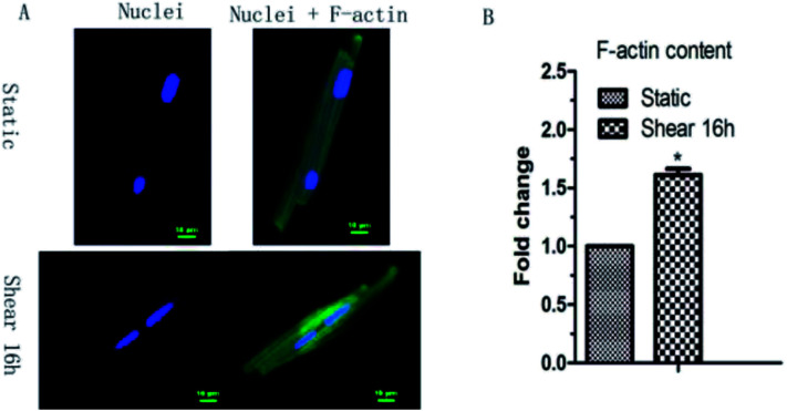
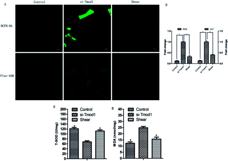
Objective: mechanical stimulation alters cell metabolism, but little is known about the effects of mechanical strain on the cytoskeleton of myocardium cells. This study was to investigate the changes of F-actin, a cytoskeleton protein of myocardium cells, and to provide a theoretical basis for further investigation of the mechanism of myocardium-remodeling. Methods: we examined the effects of fluid shear stress on the Tmod1 expression and F-actin cytoskeleton remodeling. Then, after myocardial cells, silenced by si-Tmod1, were treated by fluid shear stress, the change of intracellular calcium ion concentration, ROS in myocardial cells, cytochrome C, and the amount of F-actin, LDH and T-SOD MDA were evaluated with laser light confocal microscopy, western blot, and ELISA, respectively. Results: fluid shear stress can induce F-actin cytoskeleton remodeling and upregulate Tmod1 expression. After myocardial cells were under the conditions of Tmod1 inhibition, shear stress can significantly reduce the increase of ROS levels and calcium content, decrease the release of cells cytochrome C and LDH, decrease the MDA content, and increase the level of T-SOD. Conclusion: in conclusion, shear treatment can remodel the cytoskeleton through Tmod1, and its mechanism may be related to scavenging oxidative stress products, ROS and MDA, the increase of intracellular antioxidant enzyme activity of SOD and improvement in mitochondrial dysfunction.
This journal is © The Royal Society of Chemistry.
Conflict of interest statement
The authors declared that they have no conflicts of interest to this work. We declare that we do not have any commercial or associative interest that represents a conflict of interest in connection with the work submitted.
Figures


References
-
- Wang F. W. Zhao F. Qian X. Y. Yu Z. Z. Zhao J. Su L. Zhang Y. Zhang S. L. Zhao B. X. Miao J. Y. RSC Adv. 2014;4:56722–56730. doi: 10.1039/C4RA10404H. - DOI
LinkOut - more resources
Full Text Sources