

Fabrication of a blood compatible composite membrane from chitosan nanoparticles, ethyl cellulose and bacterial cellulose sulfate
- PMID: 35548235
- PMCID: PMC9085638
- DOI: 10.1039/c8ra05536j
Fabrication of a blood compatible composite membrane from chitosan nanoparticles, ethyl cellulose and bacterial cellulose sulfate
Abstract
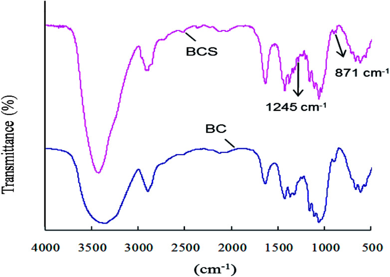
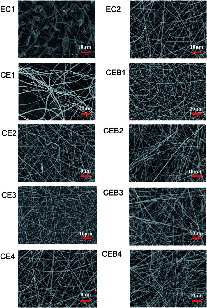
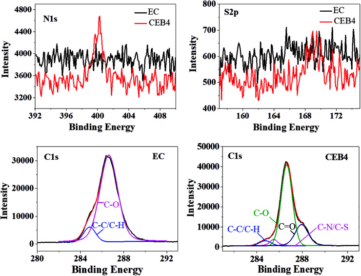
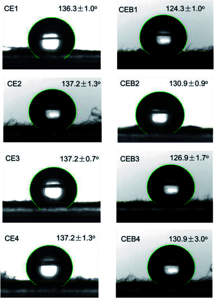
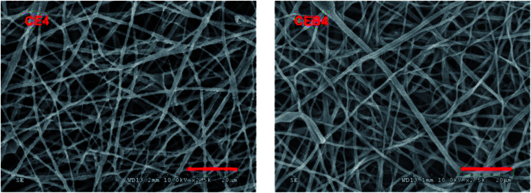
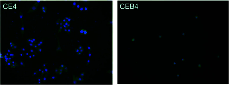
A heparin-like composite membrane was fabricated through electrospinning chitosan nanoparticles (CN) together with an ethylcellulose (EC) ethanol solution onto a bacterial cellulose sulfate membrane (BCS). Scanning electron microscopy images revealed that there were no chitosan particles in the obtained composite CN-EC/BCS membranes (CEB), indicating CN had been stretched to nanofibers. X-ray photoelectron spectroscopy verified the existence of -NH2 from chitosan and -SO3 - from BCS on the surface of CEB membranes. Positively charged CN in the electrospinning solution and negatively charged BCS on the collector increased the electrostatic force and the electrospinning ability of the EC was increased. The membrane was hydrophobic, with a water contact angle higher than 120°. CEB membranes expressed good blood compatibility according to the results of coagulation time and platelet adhesion experiments. No platelets adhered on the surface of the CEB membranes. An inflammatory response was investigated according to activation of the macrophages seeded onto the membranes. Macrophages seeded on CEB membranes are not activated after 24 h incubation.
This journal is © The Royal Society of Chemistry.
Conflict of interest statement
There are no conflicts to decalre.
Figures
Similar articles
-
Preparation and anticoagulant properties of heparin-like electrospun membranes from carboxymethyl chitosan and bacterial cellulose sulfate.Int J Biol Macromol. 2018 Dec;120(Pt B):1396-1405. doi: 10.1016/j.ijbiomac.2018.09.133. Epub 2018 Sep 25. Int J Biol Macromol. 2018. PMID: 30266642
-
Tunable Physical Properties of Ethylcellulose/Gelatin Composite Nanofibers by Electrospinning.J Agric Food Chem. 2018 Feb 28;66(8):1907-1915. doi: 10.1021/acs.jafc.7b06038. Epub 2018 Feb 15. J Agric Food Chem. 2018. PMID: 29425459
-
Nanoporous membranes with cellulose nanocrystals as functional entity in chitosan: removal of dyes from water.Carbohydr Polym. 2014 Nov 4;112:668-76. doi: 10.1016/j.carbpol.2014.06.048. Epub 2014 Jun 25. Carbohydr Polym. 2014. PMID: 25129796
-
In Situ Generation of Cellulose Nanocrystals in Polycaprolactone Nanofibers: Effects on Crystallinity, Mechanical Strength, Biocompatibility, and Biomimetic Mineralization.ACS Appl Mater Interfaces. 2015 Sep 9;7(35):19672-83. doi: 10.1021/acsami.5b04682. Epub 2015 Aug 26. ACS Appl Mater Interfaces. 2015. PMID: 26295953
-
Hemocompatible and antibacterial porous membranes with heparinized copper hydroxide nanofibers as separation layer.Colloids Surf B Biointerfaces. 2013 Oct 1;110:36-44. doi: 10.1016/j.colsurfb.2013.04.020. Epub 2013 Apr 28. Colloids Surf B Biointerfaces. 2013. PMID: 23707848
Cited by
-
Shrimp Waste Upcycling: Unveiling the Potential of Polysaccharides, Proteins, Carotenoids, and Fatty Acids with Emphasis on Extraction Techniques and Bioactive Properties.Mar Drugs. 2024 Mar 28;22(4):153. doi: 10.3390/md22040153. Mar Drugs. 2024. PMID: 38667770 Free PMC article. Review.
-
Fungal Carboxymethyl Chitosan-Impregnated Bacterial Cellulose Hydrogel as Wound-Dressing Agent.Gels. 2023 Feb 27;9(3):184. doi: 10.3390/gels9030184. Gels. 2023. PMID: 36975633 Free PMC article.
-
Leaftronics: Natural lignocellulose scaffolds for sustainable electronics.Sci Adv. 2024 Nov 8;10(45):eadq3276. doi: 10.1126/sciadv.adq3276. Epub 2024 Nov 8. Sci Adv. 2024. PMID: 39514653 Free PMC article.
-
Synthesis and characterization of a magnetic bacterial cellulose-chitosan nanocomposite and evaluation of its applicability for osteogenesis.Bioimpacts. 2024;14(6):30159. doi: 10.34172/bi.2024.30159. Epub 2024 Mar 24. Bioimpacts. 2024. PMID: 39493895 Free PMC article.
-
Mechanical and Water-Resistant Properties of Eco-Friendly Chitosan Membrane Reinforced with Cellulose Nanocrystals.Polymers (Basel). 2019 Jan 18;11(1):166. doi: 10.3390/polym11010166. Polymers (Basel). 2019. PMID: 30960152 Free PMC article.
References
-
- Müller U., Medical. Device. Technology., 2008, vol. 19, pp. 32–34 - PubMed
LinkOut - more resources
Full Text Sources