. 2019 Jul 25;9(40):22966-22979.
doi: 10.1039/c9ra04026a. eCollection 2019 Jul 23.

Hybrid cellulose nanocrystal/alginate/gelatin scaffold with improved mechanical properties and guided wound healing

Affiliations

Hybrid cellulose nanocrystal/alginate/gelatin scaffold with improved mechanical properties and guided wound healing

Yue Shan et al. RSC Adv. .

Erratum in

Abstract

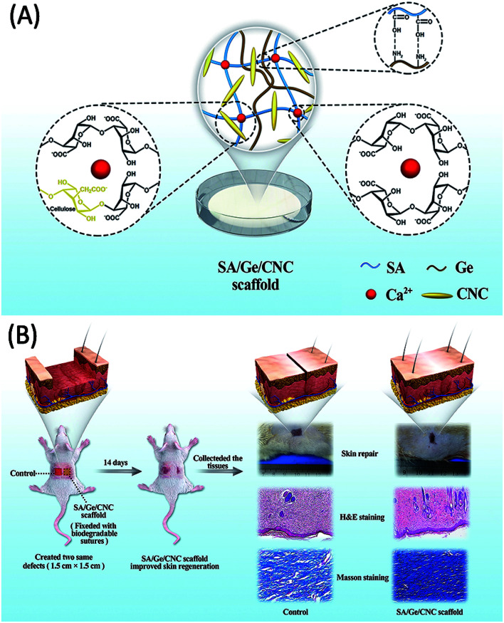
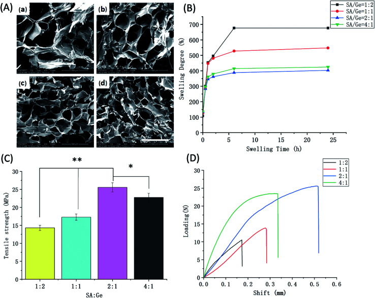
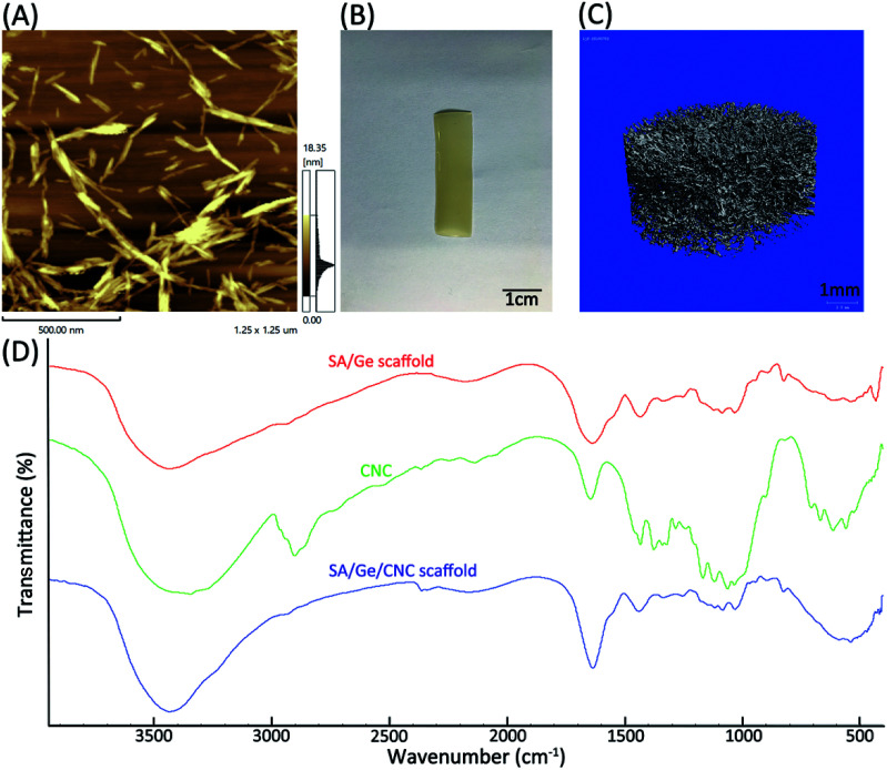
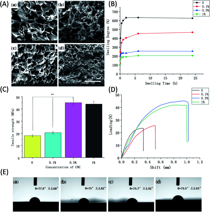
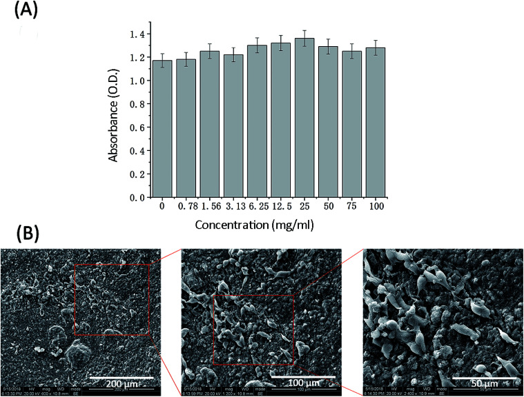
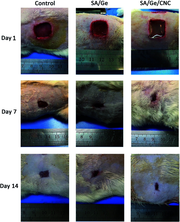
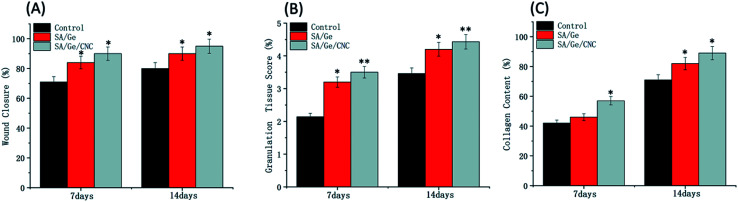
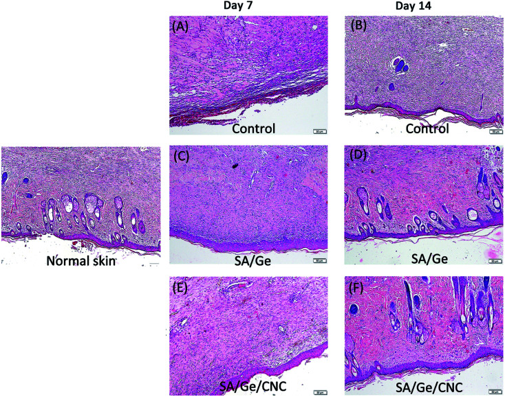
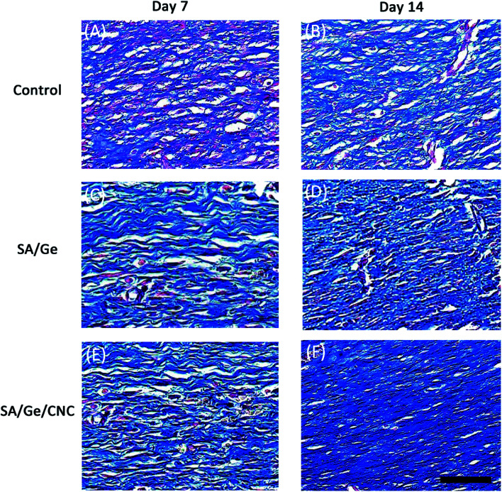
Nature derived biopolymers such as polysaccharides and collagen have attracted considerable attention in biomedical applications. Despite excellent biocompatibility and bioactivity, their poor mechanical properties could not meet the requirement for skin regeneration. In this study, cellulose nanocrystal (CNC) was incorporated into the calcium cross-linked sodium alginate/gelatin (SA/Ge) scaffold to reinforce its physicochemical properties. A novel sodium alginate/gelatin/cellulose nanocrystal (SA/Ge/CNC) scaffold was successfully prepared through electrostatic interaction of sodium alginate and gelatin, ionic cross-linking of calcium ions with sodium alginate, and incorporation of CNC. Afterwards, the SA/Ge and SA/Ge/CNC scaffolds were fully characterized and compared with scanning electron microscopy images, swelling behaviors, tensile strengths and contact angles. The involvement of CNC produces a hybrid SA/Ge/CNC scaffold with desired porous network, moderate swelling behavior, and superior mechanical strength (from 18 MPa to 45 MPa). Furthermore, in vitro cytotoxicity and cell growth assay using mouse embryonic fibroblast cells validated that SA/Ge/CNC scaffold was non-toxic and can prompt cell adhesion and proliferation. The in vivo skin regeneration experiments using the SA/Ge/CNC scaffold group showed an improved skin wound healing process with accelerated re-epithelialization, increased collagen deposition and faster extracellular matrix remodeling. Overall, the results suggested that the SA/Ge/CNC hybrid scaffold with enhanced mechanical performance and wound healing efficacy was a promising biomaterial for skin defect regeneration.


Conflict of interest statement

The authors declare no conflicts of interest.

Figures

Scheme 1
Scheme 1. (A) The illustration of the SA/Ge/CNC scaffold. (B) The skin regeneration improved by the SA/Ge/CNC scaffold in SD rats.
Fig. 1
Fig. 1. Physical characteristics of SA/Ge scaffolds (A) SEM images of SA/Ge scaffolds with different SA/Ge ratios: (a) SA/Ge = 1 : 2, (b) SA/Ge = 1 : 1, (c) SA/Ge = 2 : 1, (d) SA/Ge = 4 : 1. The bar corresponds to 500 μm. (B) Swelling degree, (C) maximum tensile strength at breaking points, and (D) representative shift–loading curves of SA/Ge scaffolds in different ratios. * and ** denote significant difference at p < 0.05 and p < 0.01 levels, respectively.
Fig. 2
Fig. 2. (A) Atomic force microscopy image of 0.01 wt% CNC dispersed in aqueous solution. (B) Visual appearance of SA/Ge/CNC (SA : Ge = 2 : 1, CNC = 0.5%) scaffold (length = 3 cm, width = 1 cm). (C) Micro-CT image of SA/Ge/CNC scaffold (SA : Ge = 2 : 1, CNC = 0.5%) (diameter = 10 mm). (D) FTIR spectra of SA/Ge scaffold, CNC, and SA/Ge/CNC scaffold.
Fig. 3
Fig. 3. Physical characteristics of SA/Ge/CNC (SA/Ge = 2 : 1) scaffolds with different concentrations of CNC (A) SEM images of SA/Ge/CNC scaffolds: (a) without CNC, (b) 0.1% CNC, (c) 0.5% CNC, (d) 1% CNC. The bar corresponds to 500 μm. (B) Swelling degree, (C) maximum tensile strength at breaking points, (D) representative shift–loading curves, and (E) contact angles of SA/Ge/CNC scaffolds with different CNC concentration: (a) without CNC, (b) 0.1% CNC, (c) 0.5% CNC, (d) 1% CNC. ** denote significant difference at p < 0.01 level.
Fig. 4
Fig. 4. Cell study on SA/Ge/CNC (SA/Ge = 2 : 1, CNC = 0.5%) scaffold. (A) Cytotoxicity of 3T3 cells after 24 hour culture with scaffold leachate (MTT assay). (B) SEM images of 3T3 cells cultured on SA/Ge/CNC scaffold for 3 days.
Fig. 5
Fig. 5. Macroscopic observation of skin wounds in control, SA/Ge, and SA/Ge/CNC groups at 1 days, 7 days, and 14 days after surgery.
Fig. 6
Fig. 6. (A) The wound closure, (B) granulation tissue score, (C) collagen content in control, SA/Ge, and SA/Ge/CNC groups at 7 days and 14 days after surgery.
Fig. 7
Fig. 7. H&E staining images in control, SA/Ge, and SA/Ge/CNC groups at 7 days and 14 days after surgery. The bar corresponds to 50 μm.
Fig. 8
Fig. 8. Masson's trichrome staining images in control, SA/Ge, and SA/Ge/CNC groups at 7 and 14 days after surgery. The bar corresponds to 50 μm.

References

    1. Sousa M. P. Neto A. I. Correia T. R. Miguel S. P. Matsusaki M. Correia I. J. Mano J. F. Biomater. Sci. 2018;6:1962–1975. doi: 10.1039/C8BM00319J. - DOI - PMC - PubMed
    1. Qi C. Xu L. Deng Y. Wang G. Wang Z. Wang L. Biomater. Sci. 2018;6:2859–2870. doi: 10.1039/C8BM00934A. - DOI - PubMed
    1. Liao J. Jia Y. Wang B. Shi K. Qian Z. ACS Biomater. Sci. Eng. 2018;4:1029–1036. doi: 10.1021/acsbiomaterials.7b00860. - DOI - PubMed
    1. Rezvanian M. Amin M. Ng S. F. Carbohydr. Polym. 2016;137:295–304. doi: 10.1016/j.carbpol.2015.10.091. - DOI - PubMed
    1. Boekema B. K. Vlig M. Olde Damink L. Middelkoop E. Eummelen L. Buhren A. V. Ulrich M. M. J. Mater. Sci.: Mater. Med. 2014;25:423–433. doi: 10.1007/s10856-013-5075-2. - DOI - PubMed