. 2022 Apr 25:9:839743.
doi: 10.3389/fcvm.2022.839743. eCollection 2022.

Syndecan-1 Is Overexpressed in Human Thoracic Aneurysm but Is Dispensable for the Disease Progression in a Mouse Model

Affiliations

Syndecan-1 Is Overexpressed in Human Thoracic Aneurysm but Is Dispensable for the Disease Progression in a Mouse Model

Sara Zalghout et al. Front Cardiovasc Med. .

Abstract

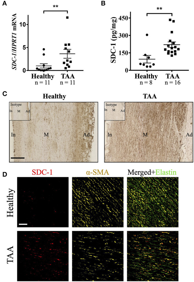
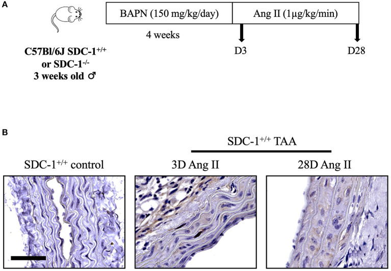
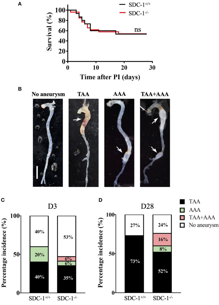
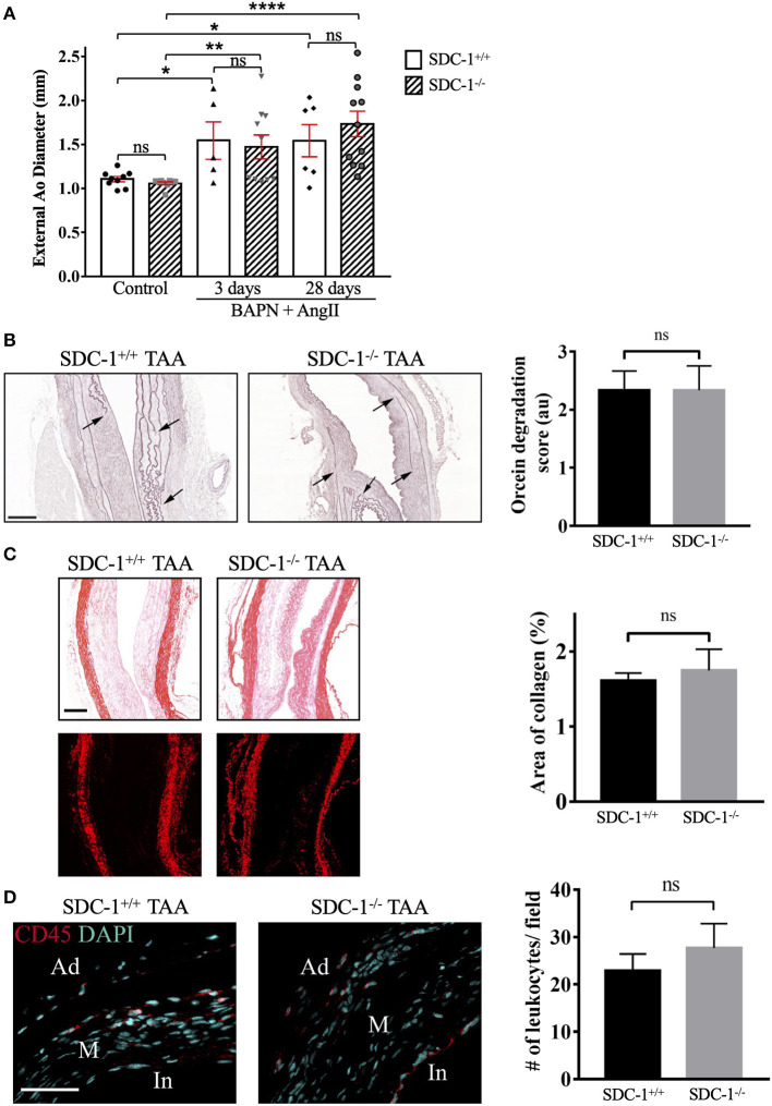
Glycosaminoglycans (GAGs) pooling has long been considered as one of the histopathological characteristics defining thoracic aortic aneurysm (TAA) together with smooth muscle cells (SMCs) apoptosis and elastin fibers degradation. However, little information is known about GAGs composition or their potential implication in TAA pathology. Syndecan-1 (SDC-1) is a heparan sulfate proteoglycan that is implicated in extracellular matrix (ECM) interaction and assembly, regulation of SMCs phenotype, and various aspects of inflammation in the vascular wall. Therefore, the aim of this study was to determine whether SDC-1 expression was regulated in human TAA and to analyze its role in a mouse model of this disease. In the current work, the regulation of SDC-1 was examined in human biopsies by RT-qPCR, ELISA, and immunohistochemistry. In addition, the role of SDC-1 was evaluated in descending TAA in vivo using a mouse model combining both aortic wall weakening and hypertension. Our results showed that both SDC-1 mRNA and protein are overexpressed in the media layer of human TAA specimens. RT-qPCR experiments revealed a 3.6-fold overexpression of SDC-1 mRNA (p = 0.0024) and ELISA assays showed that SDC-1 protein was increased 2.3 times in TAA samples compared with healthy counterparts (221 ± 24 vs. 96 ± 33 pg/mg of tissue, respectively, p = 0.0012). Immunofluorescence imaging provided evidence that SMCs are the major cell type expressing SDC-1 in TAA media. Similarly, in the mouse model used, SDC-1 expression was increased in TAA specimens compared to healthy samples. Although its protective role against abdominal aneurysm has been reported, we observed that SDC-1 was dispensable for TAA prevalence or rupture. In addition, SDC-1 deficiency did not alter the extent of aortic wall dilatation, elastin degradation, collagen deposition, or leukocyte recruitment in our TAA model. These findings suggest that SDC-1 could be a biomarker revealing TAA pathology. Future investigations could uncover the underlying mechanisms leading to regulation of SDC-1 expression in TAA.

Keywords: Syndecan-1; aneurysm; extracellular matrix; proteoglycan; smooth muscle cell (SMC).


Conflict of interest statement

The authors declare that the research was conducted in the absence of any commercial or financial relationships that could be construed as a potential conflict of interest.

Figures

Figure 1
Figure 1
SDC-1 mRNA and protein levels are increased in the media layer of human TAA compared to healthy counterparts and is expressed by SMCs. (A) mRNA expression level of SDC-1 in the media layer of healthy aorta and TAA was measured by RT-qPCR and normalized to HPRT1. (B) The abundance of SDC-1 protein in human aortic media was assessed by ELISA from healthy or TAA donors. (C) Representative images of SDC-1 staining by immunohistochemistry in healthy human and TAA media. Staining with an isotype control was performed as a negative control and shown on the top left of images. Scale bar corresponds to 200 μm. In, intima; M, media; Ad, adventitia. (D) Representative immunofluorescence images of SDC-1 (red) and α-SMA (yellow) co-staining in the media of healthy human or TAA media. Green color corresponds to elastin autofluorescence in the merged images. Scale bar corresponds to 50 μm. (A,B) Data are presented as mean ± SEM and p-values were calculated using non-parametric two-tailed Mann-Whitney test; **p < 0.01.
Figure 2
Figure 2
SDC-1 is overexpressed in TAA compared to control aortas in the BAPN/AngII aneurysm mouse model. (A) Experimental model used. SDC-1+/+ or SDC-1−/− male C57Bl/6J mice at 3 weeks old received intraperitoneal injection of β-amino propionitrile (BAPN) for 4 weeks followed by subcutaneous infusion of angiotensin II (Ang II) by pump implantation for 3 (D3) or 28 (D28) days, then sacrificed. (B) Representative images of SDC-1 immunostaining in control (n = 3) and TAA specimens after 3 (n = 2) or 28 days (n = 3) of Ang II infusion. Scale bar corresponds to 50 μm.
Figure 3
Figure 3
SDC-1 is dispensable for TAA incidence or rupture but tends to protect from AAA development in mice. (A) Survival rate of mice after 28 days following Ang II infusion, compared with the Gehan-Breslow-Wilcoxon test. PI, pump implantation; SDC-1+/+: n = 15, SDC-1−/−: n = 26; ns, non-significant. (B) Aorta macroscopic images with/without aneurysms 28 days after Ang II infusion. Scale bar corresponds to 5 mm. (C,D) Percentage of TAA or AAA incidence in SDC-1+/+ or SDC-1−/− mice for 3 (C) or 28 days (D) of Ang II infusion. (C) SDC-1+/+: n = 5, SDC-1−/−: n = 17. (D) SDC-1+/+: n = 15, SDC-1−/−: n = 26.
Figure 4
Figure 4
SDC-1 deficiency does not alter the extent of aortic dilatation, ECM remodeling, or leukocytes recruitment in descending TAA in mice. (A) Measurement of external descending thoracic diameter in SDC-1+/+ and SDC-1−/− mice that received BAPN and Ang II for 3 or 28 days or control (sham) mice. Aorta with a diameter ≥ 1.5 mm was considered as an aorta that developed TAA. SDC-1+/+ or SDC-1−/− control: n = 9 for both; SDC-1+/+ and SDC-1−/− after 3 days: n = 5 and n = 10, respectively; SDC-1+/+ and SDC-1−/− after 28 days: n = 6 and n = 11, respectively. (B) Elastin degradation. Representative images of orcein staining for SDC-1+/+ or SDC-1−/− TAA specimens after 28 days following Ang II infusion and quantification of elastin degradation (to the right). Arrows show sites of elastic laminae degradation. SDC-1+/+: n = 3, SDC-1−/−: n = 6. Scale bar corresponds to 100 μm. (C) Collagen deposition. Representative images of Sirius red staining for SDC-1+/+ or SDC-1−/− TAA specimens after 28 days following Ang II infusion visualized by bright-field microscopy (upper images) or polarization microscopy (lower images) and its quantification (to the right) by image J software. Scale bar corresponds to 100 μm. (D) CD45 expression. Representative immunofluorescence images of TAA specimens after 28 days of Ang II infusion stained with Ab against CD45 and counterstained with DAPI and its quantification (to the right) by Image J software. In, intima; M, media; Ad, adventitia. Scale bar corresponds to 50 μm. (C,D) SDC-1+/+: n = 3, SDC-1−/−: n = 7. (A–D) Data are presented as mean ± SEM and p-values were calculated using non-parametric two-tailed Mann-Whitney test; *p < 0.05, **p < 0.01, ****p < 0.0001, ns: not significant.

References

    1. Olsson C, Thelin S, Ståhle E, Ekbom A, Granath F. Thoracic Aortic Aneurysm and Dissection. Circulation. (2006) 114:2611–8. 10.1161/CIRCULATIONAHA.106.630400 - DOI - PubMed
    1. Kuzmik GA, Sang AX, Elefteriades JA. Natural history of thoracic aortic aneurysms. J Vasc Surg. (2012) 56:565–71. 10.1016/j.jvs.2012.04.053 - DOI - PubMed
    1. Elefteriades JA, Botta DM. Indications for the Treatment of Thoracic Aortic Aneurysms. Surg Clin North Am. (2009) 89:845–67. 10.1016/j.suc.2009.06.005 - DOI - PubMed
    1. Shen Ying H, Lu Hong S, LeMaire Scott A, Daugherty A. Unfolding the story of proteoglycan accumulation in thoracic aortic aneurysm and dissection. Arterioscler Thromb Vasc Biol. (2019) 39:1899–901. 10.1161/ATVBAHA.119.313279 - DOI - PMC - PubMed
    1. Halushka MK, Angelini A, Bartoloni G, Basso C, Batoroeva L, Bruneval P, et al. . Consensus statement on surgical pathology of the aorta from the Society for Cardiovascular Pathology and the Association For European Cardiovascular Pathology: II Noninflammatory degenerative diseases — nomenclature and diagnostic criteria Cardiovascular Pathology. (2016) 25:247–57. 10.1016/j.carpath.2016.03.002 - DOI - PubMed