. 2022 Apr 25:12:811474.
doi: 10.3389/fcimb.2022.811474. eCollection 2022.

Pseudomonas aeruginosa Infection Modulates the Immune Response and Increases Mice Resistance to Cryptococcus gattii

Affiliations

Pseudomonas aeruginosa Infection Modulates the Immune Response and Increases Mice Resistance to Cryptococcus gattii

Eluzia C Peres-Emidio et al. Front Cell Infect Microbiol. .

Abstract

Cryptococcosis is an invasive mycosis caused by Cryptococcus spp. that affects the lungs and the central nervous system (CNS). Due to the severity of the disease, it may occur concomitantly with other pathogens, as a coinfection. Pseudomonas aeruginosa (Pa), an opportunistic pathogen, can also cause pneumonia. In this work, we studied the interaction of C. gattii (Cg) and Pa, both in vitro and in vivo. Pa reduced growth of Cg by the secretion of inhibitory molecules in vitro. Macrophages previously stimulated with Pa presented increased fungicidal activity. In vivo, previous Pa infection reduced morbidity and delayed the lethality due to cryptococcosis. This phenotype was correlated with the decreased fungal burden in the lungs and brain, showing a delay of Cg translocation to the CNS. Also, there was increased production of IL-1β, CXCL-1, and IL-10, together with the influx of iNOS-positive macrophages and neutrophils to the lungs. Altogether, Pa turned the lung into a hostile environment to the growth of a secondary pathogen, making it difficult for the fungus to translocate to the CNS. Further, iNOS inhibition reverted the Pa protective phenotype, suggesting its important role in the coinfection. Altogether, the primary Pa infection leads to balanced pro-inflammatory and anti-inflammatory responses during Cg infection. This response provided better control of cryptococcosis and was decisive for the mild evolution of the disease and prolonged survival of coinfected mice in a mechanism dependent on iNOS.

Keywords: Cryptococcosis; Cryptococcus gattii; Pseudomonas aeruginosa; coinfection; iNOS.


Conflict of interest statement

The authors declare that the research was conducted in the absence of any commercial or financial relationships that could be construed as a potential conflict of interest.

Figures

Figure 1
Figure 1
Inhibition of Cryptococcus spp. growth by P. aeruginosa. (A–C) Colony-forming units (CFU) of Cryptococcus spp. strains (L27/01, R265, and H99) when co-cultured with different P. aeruginosa strains (Pa ATCC27853, PAK, PA103, and PAO1). *p<0.05 when compared with control (fungal culture only). (D–F) Growth curves of L27/01, R265, and H99 in the presence of 6.4 µg/mL of pyocyanin (PCN). *p <0.05 compared with control without pyocyanin. MIC: PCN minimum inhibitory concentration. MFC: PCN minimum fungicidal concentration. (G) Inhibition of Cryptococcus spp. growth obtained from the spot-on-the-lawn technique with Pa (ATCC27853), PAK, PA103, PAO1, PA14 and mutants PAKΔexoS, PA103ΔexoU, PA14ΔpvdA, PA14ΔpqsE, PA14ΔphZA1. Photos show the inhibition zone for each pair of strains tested. Data are representative of three independent experiments.
Figure 2
Figure 2
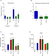
Phagocytic index (PI), intracellular proliferation rate (IPR), and production of reactive oxygen species (ROS) and peroxynitrite (PRN) by bone marrow-derived macrophages (BMDM). Results represent two different protocols: BMDM were stimulated with P. aeruginosa for 30 min (Pa30min), then washed and infected with C. gattii (Cg) (Pa30min+Cg), or Pa was maintained during all the protocol. (A) Phagocytic index after 3h and 6h of the protocol. (B) IPR, ratio of 6h/3h CFU obtained in each time point. (C) ROS and (D) PRN production after 6 hours of infection by C. gattii. Cg: BMDM infected only with C. gattii; Pa30min: BMDM infected with Pa and washed after 30 min; Pa: Pa was maintained during all the protocol; Cg: C. gattii; M0: non-infected BMDM. *p <0.05, different from Cg group. #p <0.05 different from M0 group (one-way ANOVA). Data are representative of three independent experiments consisting of six replicates each.
Figure 3
Figure 3
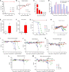
Survival and behavior of mice. (A) Six mice per group were inoculated with different inocula of P. aeruginosa (Pa) by intranasal infection (B) Weight variation after Pa infection with 105 colony forming units (CFU). (C) Bacterial burden recovered from the lungs after 1, 3, 6, 10, and 15 days of infection with Pa. *p<0.05. (D) Bronchoalveolar lavage fluid (BALF) differential cell counting at different time-points of infection with Pa. *p<0.05. (E) Reactive oxygen species (ROS) and (F) Peroxynitrite (PRN) levels in (BALF) after three days of infection with Pa. *p<0.05. (G) Six mice per group were inoculated with 104 cells of L27/01 strain by intratracheal line inoculation (Cg+Pa - three days before Pa; Pa+Cg - three days after Pa). #p<0.0001 Pa+Cg compared with Cg group (Log-Rank test). (H) Weight variation of mice expressed in %. (I–M) Five animals per group were submitted to the SHIRPA Protocol. # p <0.05 compared with Cg group (One-Way ANOVA). *p <0.05 compared with the NI group. NI, non-infected; ND, non-detectable. All the experiments were performed at least three times to confirm the data, and the results were always reproducible.
Figure 4
Figure 4
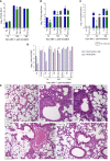
Fungal burden, cell recruitment to bronchoalveolar lavage fluid (BALF), and histopathology. Six mice per group were inoculated with 104 cells of L27/01 strain by intratracheal line inoculation (Cg+Pa - three days before Pa; Pa+Cg - three days after Pa) analyzed at 1, 10 and 18 dpi: Fungal burden in the BALF (A), lungs (B), and brain (C) of mice infected with C. gattii (Cg) or coinfected with P. aeruginosa. #p<0.05 compared to Cg group at the same time-point (ANOVA/Tukey’s multiple comparisons test). (D) Cell recruitment in the BALF. *p<0.05 compared to the NI group. #p<0.05 compared to the Cg group. (E) Histopathological HE staining of the lungs. Representative pictures of the histopathology after ten days of Cg infection; except for the Pa group, which was analyzed 3 dpi of the bacteria infection. Amplification of 200 X. Scale bars of 50 µm. The arrows in Cg and Cg+Pa point to yeasts inside the alveoli, seen as spherical or oval structures, forming clusters, which induce anatomical deformation of the alveoli. The yeasts can also be seen, to a lesser extent, in the group Pa+Cg. NI, non-infected; Cg, C. gattii; Pa, P. aeruginosa; Cg+Pa, Pa 3 days after infection to Cg; Pa+Cg, Pa 3 days before infection to Cg.
Figure 5
Figure 5
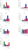
N-acetylglucosaminidase (NAG) and Myeloperoxidase (MPO) activities and cytokines and chemokine levels in the lungs of mice. NAG (A) and MPO (B) activities. Concentrations of CXCL-1 (C), IL-1β (D), IFN-γ (E), IL-17 (F), and IL-10 (G). *p<0.05 compared with the NI group; #p<0.05 compared with the Cg group at the same time-point (ANOVA/Newman-Keuls multiple comparisons test). Pa: group infected with P. aeruginosa and euthanized 3 days after inoculation; Cg: group infected with C. gattii; Pa+Cg: group infected with P. aeruginosa 3 days before infection with C. gattii. Tests were carried out after 1 and 10 days of fungal infection. Data are representative of three independent experiments.
Figure 6
Figure 6
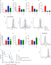
Cellular recruitment to the bronchoalveolar lavage fluid (BALF) of mice and effects of an iNOS inhibitor on the survival curve. (A) The number of total macrophages (CD45+ CD11b+ F4/80+); (B) total macrophage MFI (CD11b+); (C) MFI of activated macrophages (MHCII+) and (D) MFI of iNOS+ macrophages; (E) Representative histograms of CD11b+ (left), MHC-II (center) and iNOS (right) staining in macrophages (CD45+F4/80+CD11b+ cells) from lung homogenates of mono and co-infected mice; (F) Number of total neutrophils (CD11b+ LY6G+); (G) MFI of activated neutrophils (MHCII+) and (H) MFI of iNOS+ neutrophils; (I) Representative histograms of MHC-II (top) and iNOS (bottom) staining in neutrophils CD45+Ly6G+CD11b+ cells) from BALF of mono and coinfected mice. *p<0.05 compared with the NI group. #p<0.05 compared with the Cg group. (ANOVA/Newman-Keuls multiple comparisons test). NI: non-infected group; Pa3d: group infected with P. aeruginosa and euthanized 3 days after; Cg: group infected with C. gattii; Pa+Cg: group infected with P. aeruginosa 3 days before C. gattii infection. Cg and Pa+Cg groups were euthanized 10 days after Cg infection. MFI (Median Fluorescence Intensity). (J) Survival curve of mice treated and non-treated (NT) with Aminoguanidine Hydrochloride. *** p <0.001 compared with all the other groups. ** p <0.01 compared with the CgNT group (Log-rank test). Flow cytometry data are representative of three independent experiments.
Figure 7
Figure 7
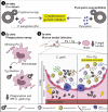
Summary of results. 1) Pa and pyocyanin reduced growth of Cg in vitro. 2) Macrophages previously stimulated with Pa presented increased fungicidal activity. 3) In vivo, previous Pa infection reduced morbidity and delayed the lethality due to cryptococcosis. This phenotype showed decreased fungal burden in the lungs associated with an increase in IL-1β, CXCL-1 and IL-10 and influx of iNOS positive neutrophils and MHCII neutrophils. Also, we observed a lower fungal burden in the brain at 10 and 18 dpi when mice were previously infected with Pa.

References

    1. Aguirre K. M., Gibson G. W. (2000). Differing Requirement for Inducible Nitric Oxide Synthase Activity in Clearance of Primary and Secondary Cryptococcus Neoform Ans Infection. Med. Mycol. 38 (5), 343–353. doi: 10.1080/mmy.38.5.343.353 - DOI - PubMed
    1. Bora L. (2016). Anticandidal Activity of Medicinal Plants and Pseudomonas Aeruginosa Strains of Clinical Specimens. J. Mcrobiol. Immunol. Infect. 49, 276–280. doi: 10.1016/j.jmii.2014.10.002 - DOI - PubMed
    1. Byrnes E. J., III, Bartlett K. H., Perfect J. R., Heitman J.. (2011). Cryptococcus Gattii: An Emerging Fungal Pathogen Infecting Humans and Animals. Microbes Infect. 13, 895–907. doi: 10.1016/j.micinf.2011.05.009 - DOI - PMC - PubMed
    1. Caldwell C. C., Chen Y., Goetzmann H. S., Hao Y., Borchers M. T., Hassett D.J. (2009). Pseudomonas Aeruginosa Exotoxin Pyocyanin Causes Cystic Fibrosis Airway Pathogenesis. Am. J. Pathology. Elsevier Inc. 175 (6), 2473–2488. doi: 10.2353/ajpath.2009.090166 - DOI - PMC - PubMed
    1. Chen S. C., Meyer W., Sorrell C. (2014). Cryptococcus Gattii Infections. Clin. Microbiol. Rev. 27 (4), 980–1024. doi: 10.1128/CMR.00126-13 - DOI - PMC - PubMed

Publication types