. 2018 Oct 9;8(60):34656-34669.
doi: 10.1039/c8ra06740f. eCollection 2018 Oct 4.

Accessing local structural disruption of Bid protein during thermal denaturation by absorption-mode ESR spectroscopy

Affiliations

Accessing local structural disruption of Bid protein during thermal denaturation by absorption-mode ESR spectroscopy

Chien-Lun Hung et al. RSC Adv. .

Abstract

Bid is a requisite protein that connects death receptors to the initiation of mitochondrial-dependent apoptosis. Its structure was determined more than a decade ago, but its structure-function relationship remains largely unexplored. Here we investigate the thermostability of Bid protein and explore how the death-promoting function of Bid is affected by thermally-induced unfolding. First, we show by circular dichroism (CD) spectroscopy that Bid remains partially folded at high temperatures (350-368 K), and that the thermal unfolding of Bid is irreversible and accompanied with intermolecular associations that lead to protein aggregation. In 3 M GdnHCl, the onset of unfolding can, however, be conveniently observed at much lower temperatures around 320 K. We employ pulsed ESR dipolar spectroscopy to show that the structure of Bid remains almost unchanged between 0 and 3 M GdnHCl before thermal denaturation. More than 30 single-labeled Bid mutants are studied using the peak-height analysis method based on ESR absorption spectroscopy, in the temperature range of 300-345 K. The ESR results provide site-specific information about the temperature dependence of the local environment of Bid, thus enabling the discrimination between the onsets of unfolding and aggregation for respective sites. Consequently, we map out the local thermostability over the Bid structure and identify a new interface between helices 3, 6, and 8 as the beginning of structural unfolding. This study also investigates the apoptotic activity of the thermally-induced Bid aggregates and shows that Bid retains the death-promoting function even when unfolded and aggregated. The applicability of the new ESR absorption peak-height method is demonstrated for protein thermostability.


Conflict of interest statement

There are no conflicts of interest to declare.

Figures

Fig. 1
Fig. 1. Thermal denaturation study of Bid by CD spectroscopy. (A) The sequence of mouse wt-Bid protein. The sites, which were mutated to cysteines to form disulfide bonds with the MTSSL probe (designated as R1), are highlighted with red boxes. D59 in mouse Bid is the cleavage site for caspase-8. (B) Thermal denaturation (298–368 K, or 25–95 °C) study of wt-Bid by CD in the far-UV region. Bid is not completely unfolded at high temperatures. (C) MRE at 222 nm for wt-Bid in the heating and then cooling cycle (298–368 K, with intervals of 5 K) in 0 M GdnHCl. (D) Wt-Bid samples (10 μM) were incubated at the indicated temperatures for 1 h. The extent of aggregation was determined by turbidity (absorbance at 395 nm). Buffer without proteins serves as control. Values represent means ± S.D. (n = 5). (E) MRE at 222 nm in the heating process when wt-Bid was incubated in different concentrations of GdnHCl. (F) MRE at 222 nm in the heating and cooling cycle for wt-Bid in 3 M GdnHCl. Measurements were repeated to verify the irreversibility of the thermal unfolding.
Fig. 2
Fig. 2. Exploring structural differences between 0 and 3 M GdnHCl with DEER spectroscopy. (A) Cartoon model of Bid protein (PDB: 1DDB) highlighting helices 1, 2, 3, and 5. Three pairs of cysteine variants of Bid were engineered to obtain three double-labeled mutants, 30/126R1, 30/82R1, and 82/126R1. As displayed, the global structure of Bid is well represented by the three sites. (B) Raw experimental data (blue) of the DEER measurements for the indicated mutants in 0 M versus 3 M GdnHCl. Fits to DEER background signals are shown in red. (C) Interspin distance distributions and average distances obtained from analyzing the DEER data in (B). Results of 0 M and 3 M GdnHCl are shown in black and gray lines, respectively. The distance distributions are highly similar between the two GdnHCl conditions.
Fig. 3
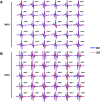
Fig. 3. Studying local structural changes in Bid using cw-ESR. Cw-ESR spectra of various single-labeled Bid mutants in 0 M and 3 M GdnHCl conditions, reordered at temperatures (A) 300 K and (B) 345 K. All spectra are scaled to have the same height in the central peak for a clear presentation. Spectra of individual sites in the two [GdnHCl] conditions are reasonably similar at the lower temperature 300 K, supporting a view that Bid structure remains undisturbed. Whereas, many spectra of individual sites show clear dissimilarity between the two [GdnHCl] conditions at high temperature 345 K. At 345 K, the spectra of 0 M GdnHCl are generally characterized by a greater linewidth and spectral anisotropy as compared to the spectra of 3 M GdnHCl. This indicates that some local structures in Bid are not completely disrupted in 0 M GdnHCl at 345 K.
Fig. 4
Fig. 4. Cw-ESR spectra of 39R1 and 108R1 in 0 M versus 3 M GdnHCl, recorded in the temperature range of 300–345 K (in increments of 5 K). (A) Spectra displayed in the first-derivative mode. Blues lines are guides to the eye indicating the shift in position of lower-field peak, important spectroscopic evidence, with temperature. (B) Spectra converted to the absorption mode. Blue boxes highlight that when displayed in the absorption mode, the lower-field peak is more sensitive to present the temperature-dependent effect (which includes sharp and broadened spectral components) on spin label mobility. Totally, 30 single-labeled Bid mutants were studied. See Fig. S2 for the temperature-dependent ESR spectra of the all Bid mutants.
Fig. 5
Fig. 5. Monitoring the onset of local unfolding in Bid during thermal denaturation with the peak height of the spectra in the absorption mode. The peak-height data were analyzed in a linear regression fit with criteria given in Experimental section. Basically, the peak height increases with increasing temperature, consistent with the general expectation for temperature dependence of ESR lineshape, whereas at high temperatures it drops largely with increasing temperature due to the increased intermolecular associations in the thermally unfolded proteins. For the results of 3 M GdnHCl, we can easily identify a changeover in the slope of the peak-height data. The changeover temperature (Tx), denoted in each of the subplots, indicates the onset of local unfolding in Bid during thermal denaturation.
Fig. 6
Fig. 6. Mapping thermal unfolding events in Bid protein. (A) The onset temperatures (Tx) of Bid unfolding (for 3 M GdnHCl) determined from the peak-height data, with estimated errors of ±2 K. The average of the Tx temperatures is 321 K, denoted by red dashed line, and is close to the onset temperature (ca. 321 ± 2 K) in the sigmoidal curve of CD spectra shown in Fig. 1E. This observation provides some support to the suitability of the ESR method for protein stability. (B) A cartoon model illustrating the positions of the studied sites in Bid protein. They are colored according to Tx values. (C) A cartoon model exhibiting the 8 helices of Bid protein, for a convenient comparison with the presentation in B. (D) A cartoon model illustrating that the disruption of the interface between helices 3, 6, and 8, which leads to the exposure of the BH3 domain, is the initial event during thermal denaturation.
Fig. 7
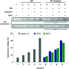
Fig. 7. Assay for cytochrome c release from mitochondria. Lanes 1–5 show that the incubated Bid is most potent for promoting the BAX-induced cytochrome c release from mitochondria when caspase-8 is present that causes the cleavage of Bid, consistent with the understanding of Bid. Lanes 6–9 show that after the heat treatments (70 or 95 °C incubation for 1 h) to Bid, the activity of Bid to promote the BAX-induced cytochrome c release is barely affected. It suggests that the heat-treated Bid proteins retain the death-promoting function. Error bar represents the mean ± SD (n = 5). See also Fig. S8.

Similar articles

Cited by

References

    1. Delbridge A. R. D. Strasser A. The BCL-2 protein family, BH3-mimetics and cancer therapy. Cell Death Differ. 2015;22:1071–1080. doi: 10.1038/cdd.2015.50. - DOI - PMC - PubMed
    1. Czabotar P. E. Lessene G. Strasser A. Adams J. M. Control of apoptosis by the BCL-2 protein family: implications for physiology and therapy. Nat. Rev. Mol. Cell Biol. 2014;15:49–63. doi: 10.1038/nrm3722. - DOI - PubMed
    1. Shamas-Din A. Brahmbhatt H. Leber B. Andrews D. W. Biochim. Biophys. Acta, Mol. Cell Res. 2011;1813:508–520. doi: 10.1016/j.bbamcr.2010.11.024. - DOI - PubMed
    1. Kantari C. Walczak H. Biochim. Biophys. Acta, Mol. Cell Res. 2011;1813:558–563. doi: 10.1016/j.bbamcr.2011.01.026. - DOI - PubMed
    1. Billen L. P. Shamas-Din A. Andrews D. W. Bid: a Bax-like BH3 protein. Oncogene. 2008;27:S93–S104. doi: 10.1038/onc.2009.47. - DOI - PubMed