. 2018 May 30;8(35):19835-19845.
doi: 10.1039/c8ra03368d. eCollection 2018 May 25.

Elucidation of the structural stability and dynamics of heterogeneous intermediate ensembles in unfolding pathway of the N-terminal domain of TDP-43

Affiliations

Elucidation of the structural stability and dynamics of heterogeneous intermediate ensembles in unfolding pathway of the N-terminal domain of TDP-43

Amresh Prakash et al. RSC Adv. .

Abstract

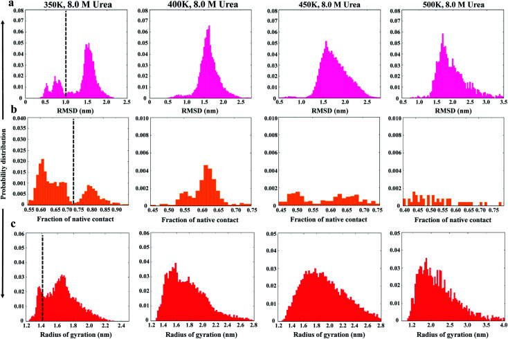
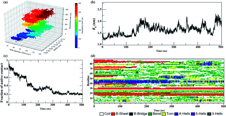
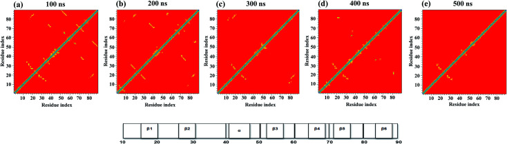
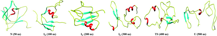
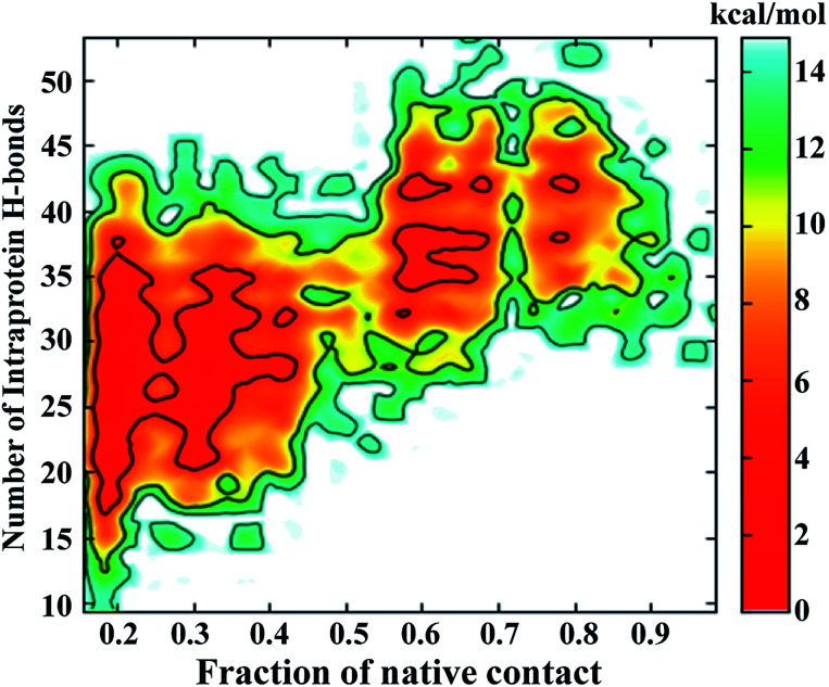
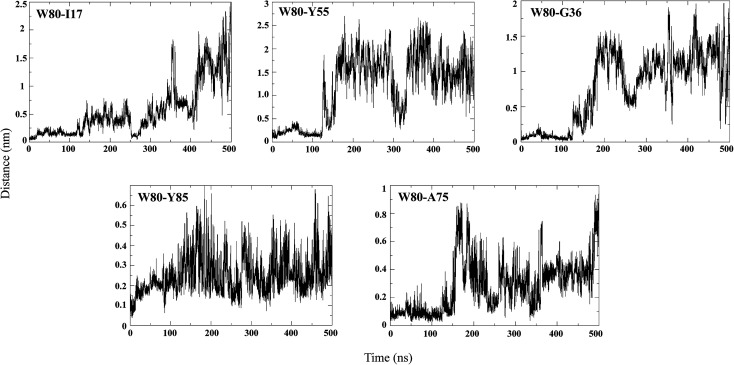
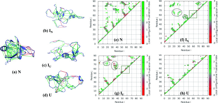
The N-terminal domain of the RNA binding protein TDP-43 (NTD) is essential to both physiology and proteinopathy; however, elucidation of its folding/unfolding still remains a major quest. In this study, we have investigated the biophysical behavior of intermediate ensembles employing all-atom molecular dynamics simulations in 8 M urea accelerated with high temperatures to achieve unfolded states in a confined computation time. The cumulative results of the 2.75 μs simulations show that unfolding of the NTD at 350 K evolves through different stable and meta-stable intermediate states. The free-energy landscape reveals two meta-stable intermediates (IN and IU) stabilized by non-native interactions, which are largely hydrophilic and highly energetically frustrated. A single buried tryptophan residue, W80, undergoes solvent exposure to different extents during unfolding; this suggests a structurally heterogeneous population of intermediate ensembles. Furthermore, the structure properties of the IN state show a resemblance to the molten globule (MG) state with most of the secondary structures intact. The unfolding of the NTD is initiated by the loss of β-strands, and the unfolded (U) states exhibit a population of non-native α-helices. These non-native unfolded intermediate ensembles may mediate protein oligomerization, leading to the formation of pathological, irreversible aggregates, characteristics of disease pathogenesis.


Conflict of interest statement

The authors declare no conflict of interest.

Figures

Fig. 1
Fig. 1. Probability distributions of structural parameters: (a) Cα-RMSD, (b) number of native contacts (Nc), and (c) radius of gyration (Rg) in 8 M urea at 350 K (left panel) to 500 K (right panel). The dashed line defines the two different conformational states as native and non-native states.
Fig. 2
Fig. 2. Unfolding pathways of the NTD in 8 M urea at 350 K. (a) Evolution of the solvent-exposed surface area, SASA, of the W80 side chain and Cα-RMSD during unfolding. Each point on this plot is colored according to its time of occurrence according to the color scale shown. Time evolution of (b) Rg, (c) Nc, and (d) secondary structures.
Fig. 3
Fig. 3. Tertiary contact map of the NTD unfolding pathway in 8 M urea at 350 K as seen at different time points. (a) 50 ns represents the N state, (b) 100 ns represents the IN state, (c) 200 ns represents the IU state, (d) 400 ns represents the transition state, and (e) 500 ns represents the U state. The horizontal bar on the right denotes the secondary structures present in the original NMR structure.
Fig. 4
Fig. 4. Snapshots corresponding to the N, IN, IU, TS and U conformational states observed during the unfolding pathway of the NTD at 350 K.
Fig. 5
Fig. 5. Free energy contour maps constructed from (a) Cα-RMSD versus native contact (left panel) and (b) Rgversus native contact (right panel) in 8 M urea at 350 K. The color is scaled according to kcal mol−1. Key minima (basins) in the maps are marked.
Fig. 6
Fig. 6. FEL of the number of intraprotein hydrogen bonds versus the number of native contacts at 350 K.
Fig. 7
Fig. 7. Pairwise distance distribution of representative hydrophobic contacts during the simulation at 350 K.
Fig. 8
Fig. 8. Residual frustration analysis in the selected states of the NTD. (a) Local frustration is depicted on the native NTD structure (PDB : 2N4P). The large cluster of minimally frustrated interactions (green) defines the core of the protein, whereas some highly frustrated interactions (red) occur on the surface of the protein. Local frustration in the (b) IN, (c) IU, and (d) U states depicting the large changes in frustration profiles. (f–h) Contact maps of the corresponding transitions of the NTD compared with the native state, along with their frustration profiles. The distinct non-native clusters in the intermediate states are circled in black. Within the β-hairpin region (residues 55–62), many frustrated contacts remain unformed in the intermediate states (orange box). (e) Energetically favorable residue pair contacts of NTD.

Similar articles

Cited by

References

    1. Ling S. C. Polymenidou M. Cleveland D. W. Neuron. 2013;79:416–438. doi: 10.1016/j.neuron.2013.07.033. - DOI - PMC - PubMed
    1. Kumar V. Islam A. Hassan M. I. Ahmad F. Eur. J. Med. Chem. 2016;121:903–917. doi: 10.1016/j.ejmech.2016.06.017. - DOI - PubMed
    1. Kumar V. Islam A. Hassan M. I. Ahmad F. Biochim. Biophys. Acta. 2016;1862:1742–1754. doi: 10.1016/j.bbadis.2016.06.011. - DOI - PubMed
    1. Neumann M. Sampathu D. M. Kwong L. K. Truax A. C. Micsenyi M. C. Chou T. T. Bruce J. Schuck T. Grossman M. Clark C. M. McCluskey L. F. Miller B. L. Masliah E. Mackenzie I. R. Feldman H. Feiden W. Kretzschmar H. A. Trojanowski J. Q. Lee V. M. Science. 2006;314:130–133. doi: 10.1126/science.1134108. - DOI - PubMed
    1. Lagier-Tourenne C. Cleveland D. W. Cell. 2009;136:1001–1004. doi: 10.1016/j.cell.2009.03.006. - DOI - PMC - PubMed