

Nociception in the Glycine Receptor Deficient Mutant Mouse Spastic
- PMID: 35548669
- PMCID: PMC9082815
- DOI: 10.3389/fnmol.2022.832490
Nociception in the Glycine Receptor Deficient Mutant Mouse Spastic
Abstract
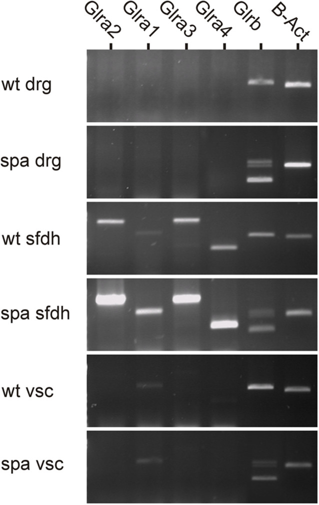
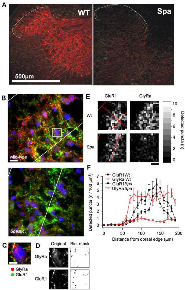
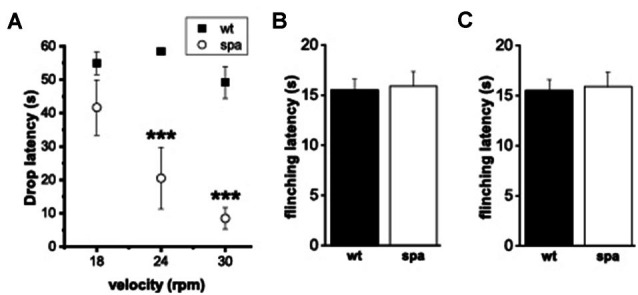
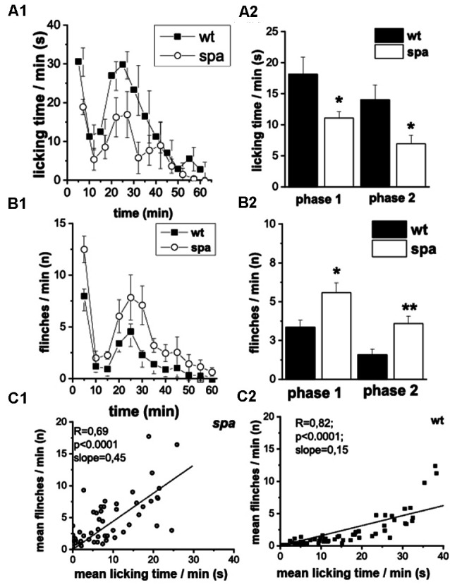
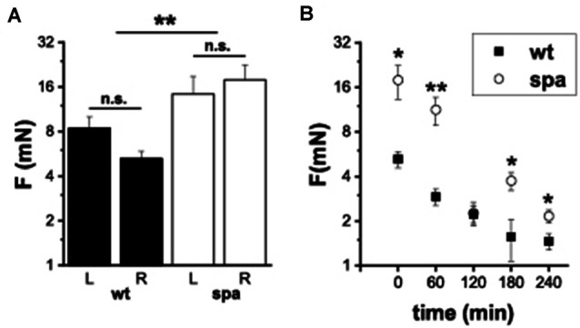
Glycine receptors (GlyRs) are the primary mediators of fast inhibitory transmission in the mammalian spinal cord, where they modulate sensory and motor signaling. Mutations in GlyR genes as well as some other genes underlie the hereditary disorder hyperekplexia, characterized by episodic muscle stiffness and exaggerated startle responses. Here, we have investigated pain-related behavior and GlyR expression in the spinal cord of the GlyR deficient mutant mouse spastic (spa). In spastic mice, the GlyR number is reduced due to a β subunit gene (Glrb) mutation resulting in aberrant splicing of GlyRβ transcripts. Via direct physical interaction with the GlyR anchoring protein gephyrin, this subunit is crucially involved in the postsynaptic clustering of heteromeric GlyRs. We show that the mutation differentially affects aspects of the pain-related behavior of homozygous Glrbspa/Glrbspa mice. While response latencies to noxious heat were unchanged, chemically induced pain-related behavior revealed a reduction of the licking time and an increase in flinching in spastic homozygotes during both phases of the formalin test. Mechanically induced nocifensive behavior was reduced in spastic mice, although hind paw inflammation (by zymosan) resulted in allodynia comparable to wild-type mice. Immunohistochemical staining of the spinal cord revealed a massive reduction of dotted GlyRα subunit immunoreactivity in both ventral and dorsal horns, suggesting a reduction of clustered receptors at synaptic sites. Transcripts for all GlyRα subunit variants, however, were not reduced throughout the dorsal horn of spastic mice. These findings suggest that the loss of functional GlyRβ subunits and hence synaptically localized GlyRs compromises sensory processing differentially, depending on stimulus modality.
Keywords: glycine receptor; glycinergic inhibition; nociception; pain; spastic; synaptic clustering.
Copyright © 2022 Groemer, Triller, Zeilhofer, Becker, Eulenburg and Becker.
Conflict of interest statement
VE works as a consultant for Disc medicine, this was, however, completely independent of the work presented here. The remaining authors declare that the research was conducted in the absence of any commercial or financial relationships that could be construed as a potential conflict of interest.
Figures
Similar articles
-
A Missense Mutation A384P Associated with Human Hyperekplexia Reveals a Desensitization Site of Glycine Receptors.J Neurosci. 2018 Mar 14;38(11):2818-2831. doi: 10.1523/JNEUROSCI.0674-16.2018. Epub 2018 Feb 13. J Neurosci. 2018. PMID: 29440552 Free PMC article.
-
Novel Functional Properties of Missense Mutations in the Glycine Receptor β Subunit in Startle Disease.Front Mol Neurosci. 2021 Sep 24;14:745275. doi: 10.3389/fnmol.2021.745275. eCollection 2021. Front Mol Neurosci. 2021. PMID: 34630038 Free PMC article.
-
Role of the Glycine Receptor β Subunit in Synaptic Localization and Pathogenicity in Severe Startle Disease.J Neurosci. 2024 Jan 10;44(2):e0837232023. doi: 10.1523/JNEUROSCI.0837-23.2023. J Neurosci. 2024. PMID: 37963764 Free PMC article.
-
Impaired Glycine Receptor Trafficking in Neurological Diseases.Front Mol Neurosci. 2018 Aug 21;11:291. doi: 10.3389/fnmol.2018.00291. eCollection 2018. Front Mol Neurosci. 2018. PMID: 30186111 Free PMC article. Review.
-
Glycine Receptor Drug Discovery.Adv Pharmacol. 2017;79:225-253. doi: 10.1016/bs.apha.2017.01.003. Epub 2017 Mar 21. Adv Pharmacol. 2017. PMID: 28528670 Review.
Cited by
-
Unraveling the parameters and biological mechanisms of CO2 laser therapy for acute pain relief.Front Neurol. 2023 Oct 20;14:1271655. doi: 10.3389/fneur.2023.1271655. eCollection 2023. Front Neurol. 2023. PMID: 37928139 Free PMC article.
-
Metabolic Biomarkers Differentiate a Surgical Intervertebral Disc from a Nonsurgical Intervertebral Disc.Int J Mol Sci. 2023 Jun 24;24(13):10572. doi: 10.3390/ijms241310572. Int J Mol Sci. 2023. PMID: 37445750 Free PMC article.
-
The glycine receptor alpha 3 subunit mRNA expression shows sex-dependent differences in the adult mouse brain.BMC Neurosci. 2023 Jun 1;24(1):32. doi: 10.1186/s12868-023-00800-9. BMC Neurosci. 2023. PMID: 37264306 Free PMC article.
-
Spinal Glycine Receptor Alpha 3 Cells Communicate Sensations of Chemical Itch in Hairy Skin.J Neurosci. 2024 May 8;44(19):e1585232024. doi: 10.1523/JNEUROSCI.1585-23.2024. J Neurosci. 2024. PMID: 38553047 Free PMC article.
References
-
- Becker K., Braune M., Benderska N., Buratti E., Baralle F., Villmann C., et al. . (2012). A retroelement modifies pre-mRNA splicing: the murine Glrbspa allele is a splicing signal polymorphism amplified by long interspersed nuclear element insertion. J. Biol. Chem. 287, 31185–31194. 10.1074/jbc.M112.375691 - DOI - PMC - PubMed
-
- Becker L., Hartenstein B., Schenkel J., Kuhse J., Betz H., Weiher H. (2000). Transient neuromotor phenotype in transgenic spastic mice expressing low levels of glycine receptor beta-subunit: an animal model of startle disease. Eur. J. Neurosci. 12, 27–32. 10.1046/j.1460-9568.2000.00877.x - DOI - PMC - PubMed
LinkOut - more resources
Full Text Sources