Case Reports
. 2022 Apr 29;26(1):2387.
doi: 10.4102/sajr.v26i1.2387. eCollection 2022.

Diagnosing rare intraductal biliary neoplasms - Intraductal papillary neoplasm of the bile duct: A case report with typical imaging findings

Affiliations
Case Reports

Diagnosing rare intraductal biliary neoplasms - Intraductal papillary neoplasm of the bile duct: A case report with typical imaging findings

Saumya Pandey et al. SA J Radiol. .

Abstract

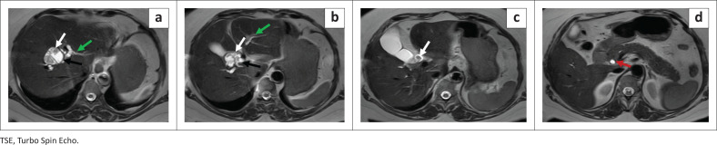
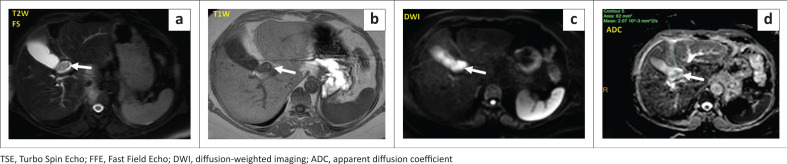
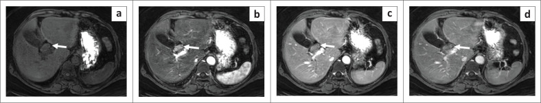
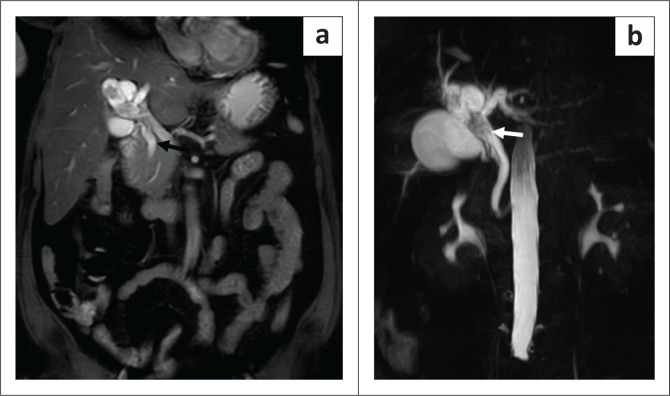
Intraductal papillary neoplasm of the bile duct (IPN-B) is a rare preinvasive intraductal pathology of the biliary tract. It should be differentiated from other more common benign or malignant causes of biliary obstruction and dilatation such as calculi or cholangiocarcinoma because the management and prognosis of this condition differs significantly. This case report describes a case of IPN-B in a 45-year-old female patient who presented with non-specific complaints of chronic abdominal pain without jaundice for three months.

Keywords: biliary dilatation; communicating; hyperenhancing; intraductal neoplasms; mucin production; papillary growth; solid-cystic.


Conflict of interest statement

The authors declare that they have no financial or personal relationships that may have inappropriately influenced them in writing this case report.

Figures

FIGURE 1
FIGURE 1
T2W–TSE axial sections showing cystic dilatation (black arrow) at the primary confluence with internal, mural-based, frond-like hypointense contents (white arrow), extending to the common hepatic duct/CBD. Upstream dilated biliary radicles (green arrow) are observed with a mildly dilated CBD distally (red arrow).
FIGURE 2
FIGURE 2
Axial T2W–TSE (a) and T1-FFE (b) sections demonstrating an iso to hypointense mural-based growth (white arrow) projecting into the lumen of the common hepatic duct or CBD. High signal is observed within the lesion on the axial b-1000 DWI image (c) with low signal on the corresponding ADC map (d).
FIGURE 3
FIGURE 3
Dynamic post-contrast axial sequences revealing iso-intensity of the tumour (white arrow) on noncontrast (a), hyperenhancement on the late arterial phase (b), reduced hyperintensity on the porto-venous (c) and delayed (d) phases.
FIGURE 4
FIGURE 4
(a) Balanced Turbo Field Echo (BTFE) coronal section showing the dilated downstream common hepatic duct and CBD (black arrow), separate from the lesion at the primary confluence. The site and extent of the intraductal tumour is best seen on the volumetric MIP of the magnetic resonance pancreatico-cholangiography (b), indicating extension of the tumour into the common hepatic duct (white arrow) with upstream and downstream ductal dilatation.
FIGURE 5
FIGURE 5
(a) The patient underwent open hilar resection with standard lymphadenectomy and right and left Roux-en-Y-cholangiojejunostomy. Papillary growth was seen at the primary confluence, extending into the right hepatic duct. (b) Low–power photomicrograph (Hematoxylin & eosin [H & E] stain; magnification 10×) of the resected specimen showing papillary projections (black arrow) with a central fibrovascular core (green arrow). (c) High-power photomicrograph (H & E stain; magnification- 20×) showing irregular papillae with features of high-grade dysplasia and hyperchromatic nuclei (black arrow); goblet cells are also seen (white arrow). (d) Positive immunohistochemistry staining (brown) for MUC2 and MUC5 using recombinant antibodies.

Similar articles

References

    1. Ainechi S, Lee H. Updates on precancerous lesions of the biliary tract: Biliary precancerous lesion. Arch Pathol Lab Med. 2016;140(11):1285–1289. 10.5858/arpa.2015-0396-RS - DOI - PubMed
    1. Chatterjee A, Vendrami CL, Nikolaidis P, et al. . Uncommon intraluminal tumors of the gallbladder and biliary tract: Spectrum of imaging appearances. RadioGraphics. 2019;39(2):388–412. 10.1148/rg.2019180164 - DOI - PubMed
    1. Wan X-S, Xu Y-Y, Qian J-Y, et al. . Intraductal papillary neoplasm of the bile duct. World J Gastroenterol. 2013;19(46):8595–8604. 10.3748/wjg.v19.i46.8595 - DOI - PMC - PubMed
    1. Lingegowda D, Gehani A, Mukhopadhyay S, Midha D, Banerjee S, Gupta B. Intraductal papillary neoplasm of the bile ducts: Case reports with review of the literature. J Gastrointest Abdominal Radiol. 2020;4(1):58–61.
    1. Bosman FT, Carneiro F, Hruban RH, Theise ND. WHO classification of tumours of the digestive system. Geneva: World Health Organization; 2010.

Publication types

LinkOut - more resources