. 2018 Sep 5;8(54):31201-31212.
doi: 10.1039/c8ra06691d. eCollection 2018 Aug 30.

Synthesis of novel genistein amino acid derivatives and investigation on their interactions with bovine serum albumin by spectroscopy and molecular docking

Affiliations

Synthesis of novel genistein amino acid derivatives and investigation on their interactions with bovine serum albumin by spectroscopy and molecular docking

Xiaokang Long et al. RSC Adv. .

Abstract

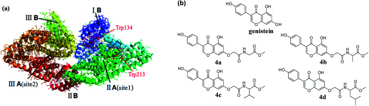
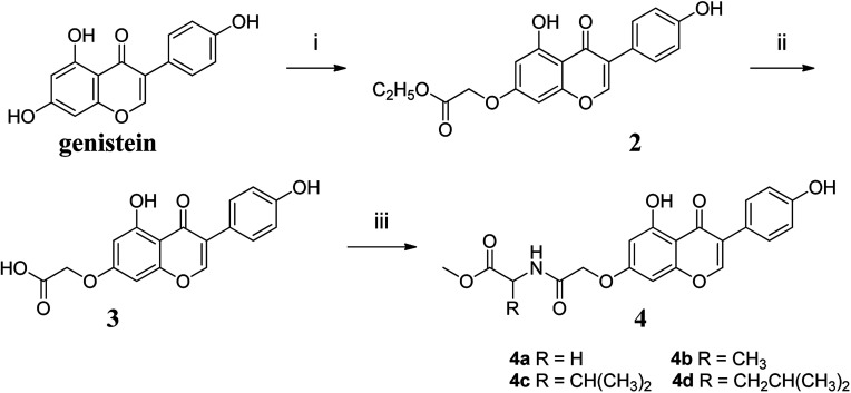
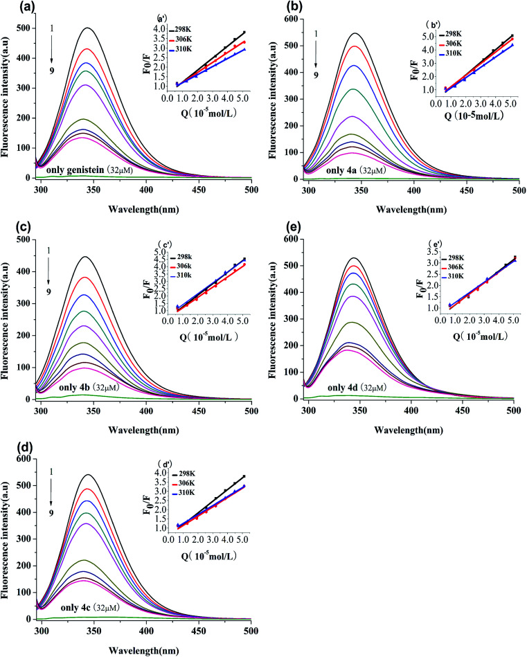
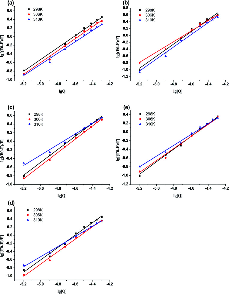
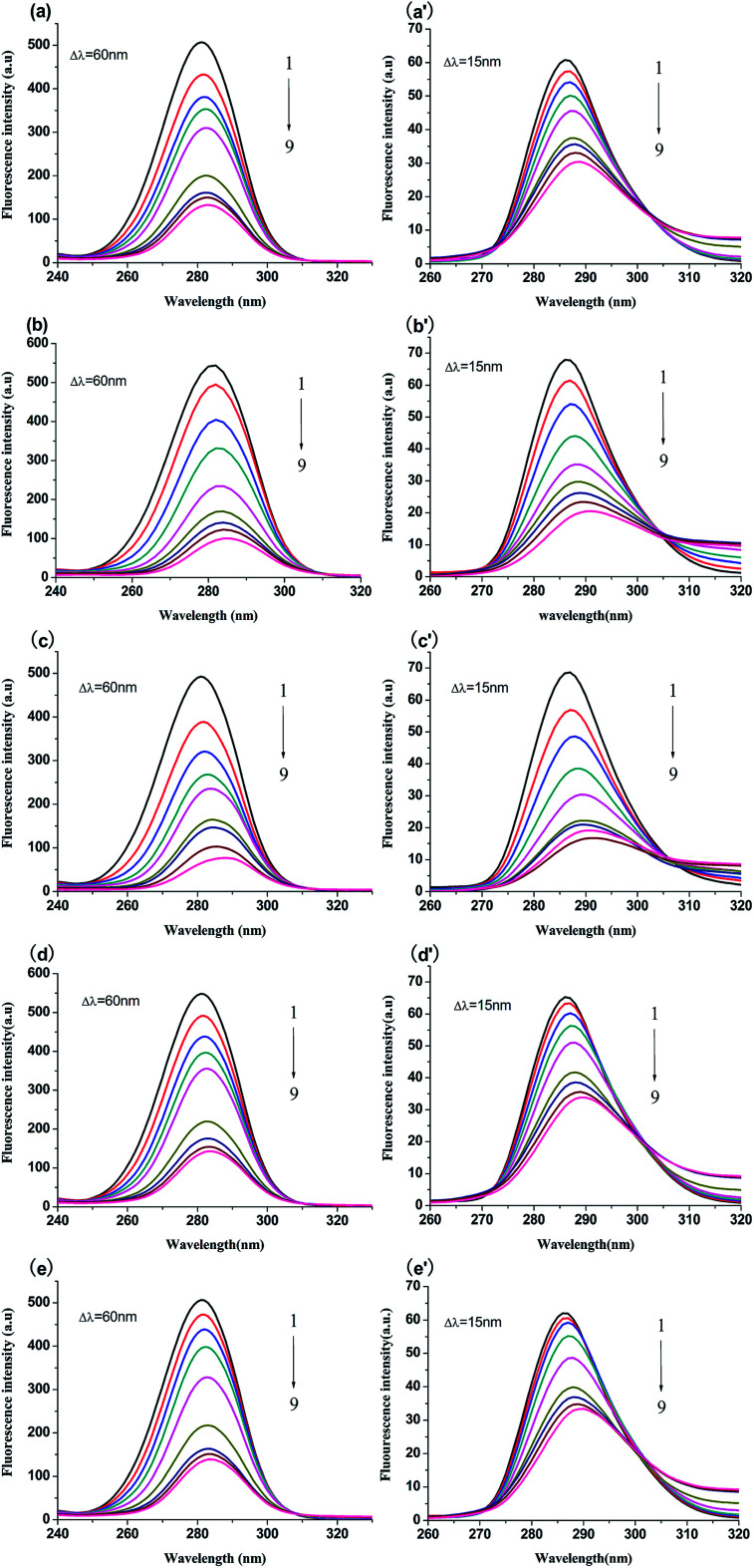
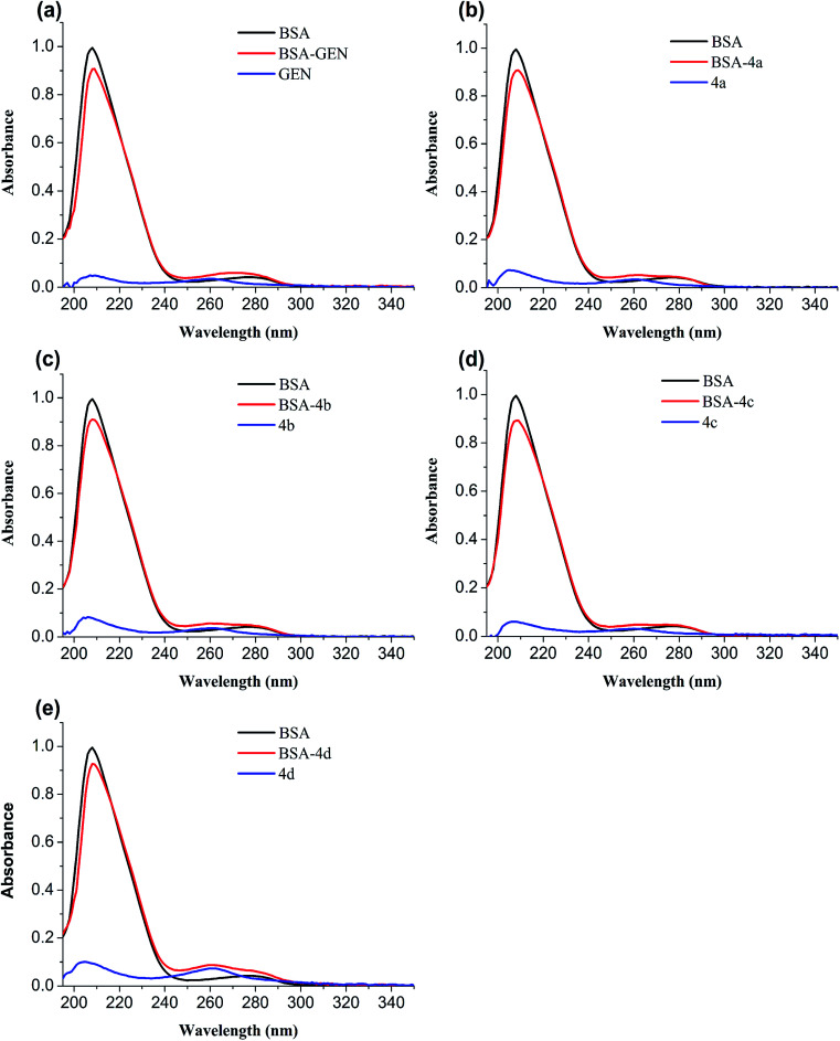
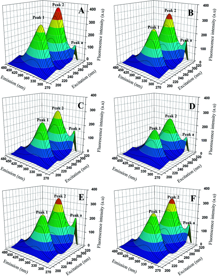
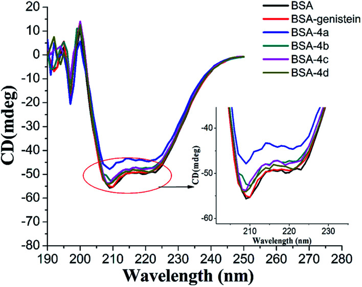
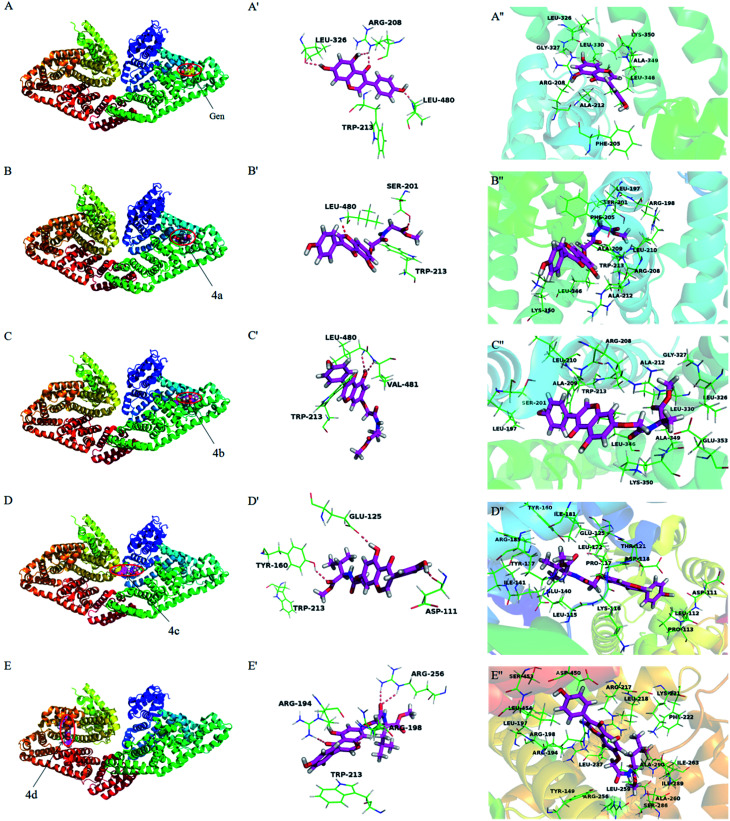
Genistein amino acid derivatives 4a-4d were synthesized and evaluated for their cytotoxic activities against MCF-7, Hela, MGC-803 and HCT-116 cell lines by MTT assays in vitro. The results revealed that compounds 4a-4d showed better activity than the parent compound genistein. Particularly, compound 4b displayed the most significant anticancer activity against MGC-803 with an IC50 value of 12.08 μM. In addition, the mechanisms of interaction between genistein, compounds 4a-4d and BSA were investigated via multi-spectroscopic techniques such as ultraviolet (UV) spectroscopy, fluorescence, circular dichroism (CD), and molecular docking under physiological conditions. The results suggested that endogenous fluorescence of BSA could be quenched by genistein and compounds 4a-4dvia forming BSA-compound complex, which meant a static quenching mechanism was involved. The negative values of enthalpy (ΔH) and entropy (ΔS) indicated that interactions between BSA and the ligands were spontaneous, and hydrogen bonding and van der Waals interactions were involved in the BSA-compound complexion formation. The UV, synchronous and 3D fluorescence results revealed that the micro-environment of tryptophan and conformation of BSA were changed after binding to ligands. CD analysis demonstrated the variation in the secondary structure and that the α-helix content of BSA decreased. Eventually, molecular docking was executed to forecast the binding forces and binding sites between BSA and compounds 4a-4d.


Conflict of interest statement

There are no conflicts to declare.

Figures

Fig. 1
Fig. 1. (a) The crystal structure of BSA; (b) the chemical structures of genistein and 4a–4d.
Fig. 2
Fig. 2. Reagents and conditions: (i) KOH, DMF, 60 °C, 1 h, then BrCH2COOC2H5, 60 °C, 5 h; (ii) CH3OH, H2O, KOH (1 mol L−1), 60 °C, 0.5 h, then H2SO4 (0.5 mol L−1), at room temperature, 0.5 h; (iii) EDCI, HOBT, DMF, 0 °C, 4 h, then DIPEA, DMAP, amino acid methyl ester, r.t., 24 h.
Fig. 3
Fig. 3. Fluorescence spectra of BSA (1 μM) solution in the presence of genistein (a), 4a (b), 4b (c), 4c (d), 4d (e), at λex = 280 nm at 298 K. The concentrations of all compounds were from 0 to 51.2 μM at intervals of 6.4 μM. Inset: Stern–Volmer plots of genistein (a′), 4a (b′), 4b (c′), 4c (d′), 4d (e′).
Fig. 4
Fig. 4. Double-log plots of genistein (a), 4a (b), 4b (c), 4c (d) or 4d (e) binding to BSA at different temperatures.
Fig. 5
Fig. 5. Synchronous fluorescence spectra of BSA (1 μM) with and without genistein, 4a, 4b, 4c or 4d at 298 K when Δλ = 60 nm (a–e) and Δλ = 15 nm (a′–e′). The concentrations of all compounds from 0 to 51.2 μM at intervals of 6.4 μM.
Fig. 6
Fig. 6. UV absorption spectra of BSA in the absence and presence of genistein (a), 4a (b), 4b (c), 4c (d), or 4d (e). The concentrations of BSA and compounds 4a–4d were 1 μM.
Fig. 7
Fig. 7. 3D fluorescence spectra of BSA (A) and BSA-genistein (B), BSA-4a (C), BSA-4b (D), BSA-4c (E), BSA-4d (F). C(compounds) = 3.0 μM, C(BSA) = 1.0 μM.
Fig. 8
Fig. 8. CD spectra of BSA with and without genistein, 4a, 4b, 4c, and 4d.The concentrations of BSA and all compounds were 4 μM.
Fig. 9
Fig. 9. The lowest energy conformation of compounds-BSA complexes obtained from molecular docking (A–E). The hydrogen bonding interactions of genistein and compounds 4a–4d with amino acid residues of BSA (A′–E′). Amino acid residues surrounding genistein and compounds 4a–4d within 3 Å (A′′–E′′). Notes: labels of A–E correspond to genistein and compounds 4a–4d, respectively.

Similar articles

Cited by

References

    1. FrostellKarlsson Å. Remaeus A. Roos H. Andersson K. Borg P. Markku Hämäläinen A. Karlsson R. J. Med. Chem. 2000;43:1986–1992. doi: 10.1021/jm991174y. - DOI - PubMed
    1. Pal S. Saha C. J. Biomol. Struct. Dyn. 2014;32:1132–1147. doi: 10.1080/07391102.2013.811700. - DOI - PubMed
    1. Tian Z. Zang F. Luo W. Zhao Z. Wang Y. Xu X. Wang C. J. Photochem. Photobiol., B. 2015;142:103–109. doi: 10.1016/j.jphotobiol.2014.10.013. - DOI - PubMed
    1. Carter D. C. Chang B. Ho J. X. Keeling K. Krishnasami Z. FEBS J. 1994;226:1049–1052. - PubMed
    1. Lou Y. Y. Zhou K. L. Shi J. H. Pan D. Q. J. Photochem. Photobiol., B. 2017;173:589–597. doi: 10.1016/j.jphotobiol.2017.06.037. - DOI - PubMed