. 2022 Jul 29;7(73):eabq3511.
doi: 10.1126/sciimmunol.abq3511. Epub 2022 Jul 29.

Recall of preexisting cross-reactive B cell memory after Omicron BA.1 breakthrough infection

Affiliations

Recall of preexisting cross-reactive B cell memory after Omicron BA.1 breakthrough infection

Chengzi I Kaku et al. Sci Immunol. .

Abstract

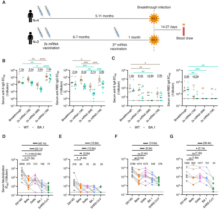
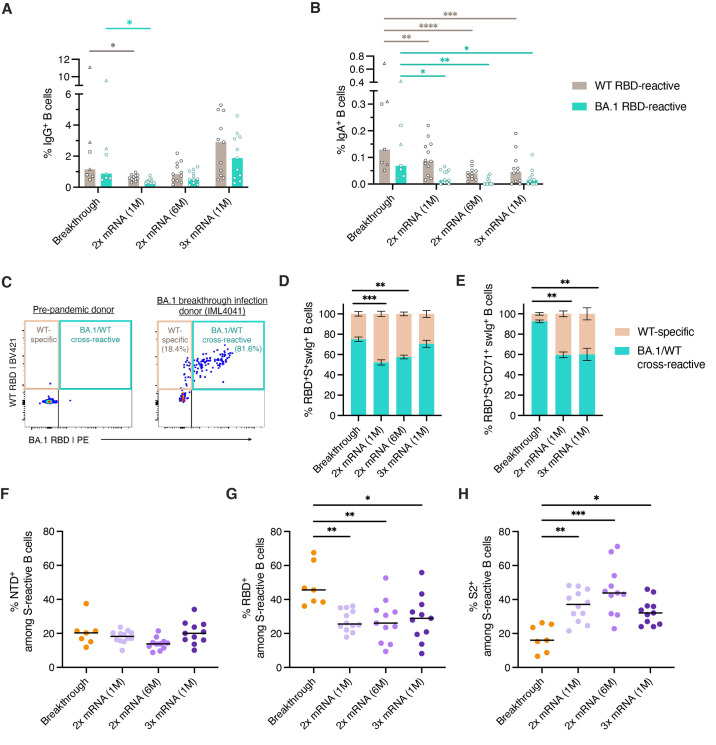
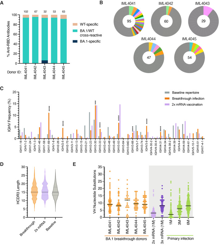
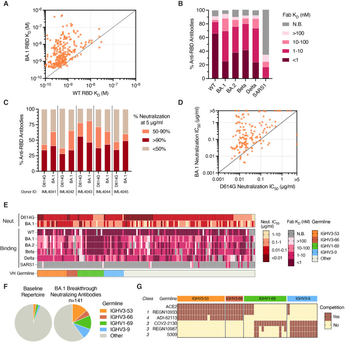
Understanding immune responses after severe acute respiratory syndrome coronavirus 2 (SARS-CoV-2) breakthrough infection will facilitate the development of next-generation vaccines. Here, we profiled spike (S)-specific B cell responses after Omicron/BA.1 infection in messenger RNA-vaccinated donors. The acute antibody response was characterized by high levels of somatic hypermutation and a bias toward recognition of ancestral SARS-CoV-2 strains, suggesting the early activation of vaccine-induced memory B cells. BA.1 breakthrough infection induced a shift in B cell immunodominance hierarchy from the S2 subunit, which is highly conserved across SARS-CoV-2 variants of concern (VOCs), and toward the antigenically variable receptor binding domain (RBD). A large proportion of RBD-directed neutralizing antibodies isolated from BA.1 breakthrough infection donors displayed convergent sequence features and broadly recognized SARS-CoV-2 VOCs. Together, these findings provide insights into the role of preexisting immunity in shaping the B cell response to heterologous SARS-CoV-2 variant exposure.


Figures

Fig. 1.
Fig. 1.
Serum binding and neutralizing activity following BA.1 breakthrough infection. (A) Vaccination, infection, and blood draw timelines. (B-C) Serum (B) IgG and (C) IgA reactivity with recombinant WT and BA.1 (left) Hexapro-stabilized S proteins and (right) RBDs following BA.1 breakthrough infection. Serum samples from uninfected/vaccinated donors at one month or six months following primary vaccination (2x mRNA) or one month following booster mRNA vaccination (3x mRNA) are shown for comparison. The fold change in median EC50 against BA.1 relative to D614G is shown above each paired set of measurements. Black bars represent median binding EC50 titers. Dotted lines represent the lower limit of detection. (D-G) Serum neutralizing activity against SARS-CoV-2 D614G, Beta, Delta, and BA.1 and SARS-CoV (D) one month after primary mRNA vaccination (n=12), (E) six months after primary mRNA vaccination (n=10), (F) one month after mRNA booster vaccination (n=11), and (G) 14 to 27 days after BA.1 breakthrough infection (n=7), as measured using an MLV-based pseudovirus neutralization assay. Plotted values represent serum neutralizing IC50 titers and values shown above the data points indicate the median IC50 titer. The fold change in IC50 titer for each virus relative to D614G is shown in parentheses. Breakthrough infection donors infected after primary mRNA vaccination (n=4) are shown as squares and those infected after mRNA booster vaccination (n=3) are shown as triangles. Statistical comparisons were determined by (B-C) two-sided Kruskal-Wallis test with Dunn's multiple comparisons or (D) Friedman's test with multiple comparisons. 1M, one month; 6M, six months; EC50, 50% effective concentration; IC50, 50% inhibitory concentration; WT, wild type. *P < 0.05, **P < 0.01, ***P < 0.001, ****P < 0.0001.
Fig. 2.
Fig. 2.
SARS-CoV-2 S-specific B cell responses induced by BA.1 breakthrough infection. (A-B) Frequency of circulating B cells that recognize recombinant WT and BA.1 RBDs among (A) IgG+ and (B) IgA+ B cells in BA.1 breakthrough infection donors (n=7) and uninfected/vaccinated donors at one month (n=12) or six months (n=11) following primary vaccination (2x mRNA) or one month following booster mRNA vaccination (3x mRNA, n=11), as measured by flow cytometry. Bars indicate median frequencies. Donors with breakthrough infections occurring after primary mRNA vaccination are shown as squares and those infected after booster mRNA vaccination are shown as triangles. (C) Representative fluorescence-activated cell sorting (FACS) gating strategy used to identify RBD-directed B cells that are WT-specific or WT/BA.1 cross-reactive, shown for a pre-pandemic donor and a breakthrough infection donor. Percentages of WT-specific and WT/BA.1 cross-reactive B cells out of total RBD-reactive cells are shown in parentheses. (D-E) Mean proportions of RBD-reactive B cells that bind WT and/or BA.1 RBDs among (D) total S+swIg+ B cells or (E) S+swIg+ CD7+ B cells. Error bars represent standard errors of the mean. A median of 65 RBD-specific B cells (ranging 12 to 310 cells) were collected from each donor for this analysis. Samples collected six months following mRNA vaccination were excluded from this analysis due to low numbers of RBD-specific CD71+ cells at this time point. (F-H) Percentage of S-reactive swIg+ B cells that target the (F) NTD, (G) RBD, and (H) Hexapro-stabilized S2 subunits. Black bars represent median percentages. For breakthrough infection donors, this analysis was restricted to S+swIg+CD71+ B cells to capture the activated response. 87 to 1721 S-reactive B cells were collected from each donor for this analysis. Statistical comparisons were determined by (A-B) two-way ANOVA with subsequent Dunnett's multiple comparisons test or (D-H) two-sided Kruskal-Wallis test with Dunn's multiple comparisons. 1M, one month; 6M, six months; swIg, class-switched (IgG+ or IgA+) immunoglobulin; WT, wild type. *P < 0.05, **P < 0.01, ***P < 0.001, ****P < 0.0001.
Fig. 3.
Fig. 3.
Sequence features of RBD-directed monoclonal antibodies isolated from BA.1 breakthrough infection donors. (A) Proportion of antibodies that bind recombinant WT and/or BA.1 RBD antigens from each donor, as determined by IgG binding via biolayer interferometry (BLI). Antibodies were isolated from breakthrough infection donors after two-dose vaccination (IML4041 through IML4044) or three-dose vaccination (IML4045). The number of antibodies isolated from each donor is indicated at the top of each bar. (B) Clonal lineage analysis of BA.1-reactive B cells. Clonally expanded lineages are represented as colored slices, with each differentially colored slice representing a separate lineage and the size of the slice proportional to the lineage size. Unique clones are combined and shown as a single grey segment. The total number of antibodies is shown in the center of each pie. (C) Germline IGHV gene usage frequencies among anti-RBD antibodies derived from breakthrough infection donors. Anti-RBD antibodies isolated from mRNA-vaccinated donors (purple bars) were obtained from the CoV-AbDab database ( 45 ). Unselected (baseline) memory B cell repertoires (grey bars) are included for reference ( 22 ). (D) Distribution of HCDR3 amino acid lengths in BA.1-reactive antibodies. Antibodies isolated from two-dose mRNA vaccinated individuals (from CoV-AbDab) and baseline human repertoire antibodies are shown for reference ( 22 , 45 ). The dotted black line represents the median, and the lower and upper lines represent the first and third quartile, respectively. (E) Somatic hypermutation levels are shown as the number of VH nucleotide substitutions in antibodies isolated from each breakthrough infection donor (orange), with medians shown by black bars. Antibodies isolated one month after two-dose mRNA vaccination (light green); one month after three-dose mRNA vaccination (dark green); and one, three, or six months after primary D614G infection (purple) are shown for comparison ( 21 ). Statistical significance was determined by Fisher's exact test compared to the baseline repertoire. 1M, one month; 3M, three months; 6M, six months; CDR, complementarity determining region; IGHV, immunoglobulin heavy variable domain; VH, variable heavy chain; WT, wild type. ***P < 0.001, ****P < 0.0001.
Fig. 4.
Fig. 4.
Binding and neutralization properties of anti-RBD antibodies isolated following BA.1 breakthrough infection. (A) Fab binding affinities for recombinant WT and BA.1 RBD antigens, as measured by BLI. Fabs with no detectable binding activity or with binding kinetics that could not be fit to a 1:1 binding model were excluded. (B) Proportion of BA.1-reactive antibodies with the indicated binding affinities for SARS-CoV-2 VOC RBDs and SARS-CoV RBD, as measured by BLI. Antibodies with weak binding affinities that could not be fit to a 1:1 binding model are shown as >100 nM, and antibodies with no detectable Fab binding, including those that bind only avidly, are indicated as non-binding (N.B.). (C) Proportion of antibodies from each donor with the indicated levels of neutralizing activity against MLV-SARS-CoV-2 D614G and BA.1 at a concentration of 5 μg/ml. (D) MLV-SARS-CoV-2 D614G and BA.1 neutralization IC50s for antibodies that displayed >90% neutralization against D614G and/or BA.1 at a concentration of 5 μg/ml. (E) Heatmap showing neutralization potency and binding breadth of BA.1 neutralizing antibodies. The bottom bar shows convergent IGHV germline gene families. Antibodies with weak binding affinities that could not be fit to a 1:1 binding model are shown as >100 nM, and antibodies with no detectable Fab binding are indicated as non-binding (N.B.). (F) Pie charts showing convergent germline gene usage among (right) BA.1-neutralizing antibodies compared to the (left) baseline human antibody repertoires ( 22 ). (G) Competitive binding profiles of BA.1 neutralizing antibodies utilizing convergent IGHV germline genes, as determined by BLI sandwich competition assay using ACE2 and the indicated comparator antibodies. Fab, antigen binding fragment; IC50, 50% inhibitory concentration; IGHV, immunoglobulin gene heavy variable; KD, equilibrium dissociation constant; N.B., non-binding; WT, wild type.

Comment in

References

    1. Pilishvili T., Gierke R., Fleming-Dutra K. E., Farrar J. L., Mohr N. M., Talan D. A., Krishnadasan A., Harland K. K., Smithline H. A., Hou P. C., Lee L. C., Lim S. C., Moran G. J., Krebs E., Steele M. T., Beiser D. G., Faine B., Haran J. P., Nandi U., Schrading W. A., Chinnock B., Henning D. J., Lovecchio F., Lee J., Barter D., Brackney M., Fridkin S. K., Marceaux-Galli K., Lim S., Phipps E. C., Dumyati G., Pierce R., Markus T. M., Anderson D. J., Debes A. K., Lin M. Y., Mayer J., Kwon J. H., Safdar N., Fischer M., Singleton R., Chea N., Magill S. S., Verani J. R., Schrag S. J.; Vaccine Effectiveness among Healthcare Personnel Study Team , Effectiveness of mRNA Covid-19 Vaccine among U.S. Health Care Personnel. N. Engl. J. Med. 385, e90 (2021). 10.1056/NEJMoa2106599 - DOI - PMC - PubMed
    1. Feikin D. R., Higdon M. M., Abu-Raddad L. J., Andrews N., Araos R., Goldberg Y., Groome M. J., Huppert A., O’Brien K. L., Smith P. G., Wilder-Smith A., Zeger S., Deloria Knoll M., Patel M. K., Duration of effectiveness of vaccines against SARS-CoV-2 infection and COVID-19 disease: Results of a systematic review and meta-regression. Lancet 399, 924–944 (2022). 10.1016/S0140-6736(22)00152-0 - DOI - PMC - PubMed
    1. Goldberg Y., Mandel M., Bar-On Y. M., Bodenheimer O., Freedman L., Haas E. J., Milo R., Alroy-Preis S., Ash N., Huppert A., Waning Immunity after the BNT162b2 Vaccine in Israel. N. Engl. J. Med. 385, e85 (2021). 10.1056/NEJMoa2114228 - DOI - PMC - PubMed
    1. Pouwels K. B., Pritchard E., Matthews P. C., Stoesser N., Eyre D. W., Vihta K. D., House T., Hay J., Bell J. I., Newton J. N., Farrar J., Crook D., Cook D., Rourke E., Studley R., Peto T. E. A., Diamond I., Walker A. S., Effect of Delta variant on viral burden and vaccine effectiveness against new SARS-CoV-2 infections in the UK. Nat. Med. 27, 2127–2135 (2021). 10.1038/s41591-021-01548-7 - DOI - PMC - PubMed
    1. Abu-Raddad L. J., Chemaitelly H., Butt A. A.; National Study Group for COVID-19 Vaccination , Effectiveness of the BNT162b2 Covid-19 Vaccine against the B.1.1.7 and B.1.351 Variants. N. Engl. J. Med. 385, 187–189 (2021). 10.1056/NEJMc2104974 - DOI - PMC - PubMed

Publication types

Supplementary concepts

LinkOut - more resources