. 2022 May 12;20(1):102.
doi: 10.1186/s12915-022-01288-1.

Decreasing dorsal cochlear nucleus activity ameliorates noise-induced tinnitus perception in mice

Affiliations

Decreasing dorsal cochlear nucleus activity ameliorates noise-induced tinnitus perception in mice

Thawann Malfatti et al. BMC Biol. .

Abstract

Background: The dorsal cochlear nucleus (DCN) is a region known to integrate somatosensory and auditory inputs and is identified as a potential key structure in the generation of phantom sound perception, especially noise-induced tinnitus. Yet, how altered homeostatic plasticity of the DCN induces and maintains the sensation of tinnitus is not clear. Here, we chemogenetically decrease activity of a subgroup of DCN neurons, Ca2+/Calmodulin kinase 2 α (CaMKII α)-positive DCN neurons, using Gi-coupled human M4 Designer Receptors Exclusively Activated by Designer Drugs (hM4Di DREADDs), to investigate their role in noise-induced tinnitus.

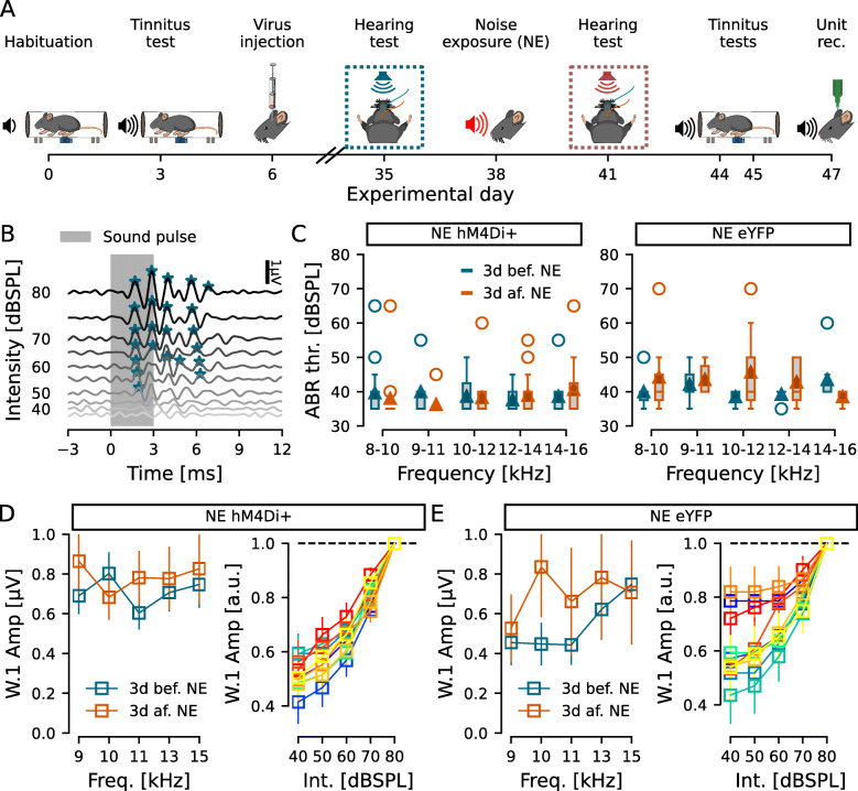
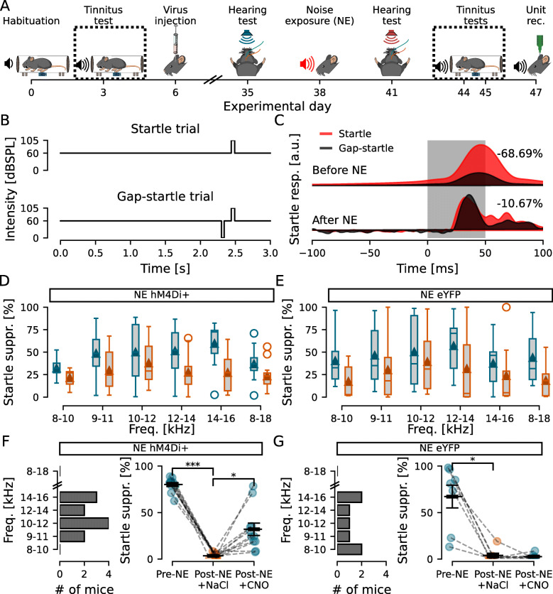
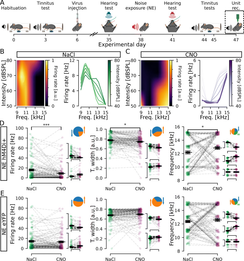
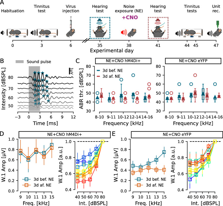
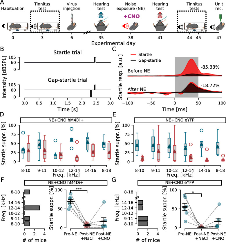
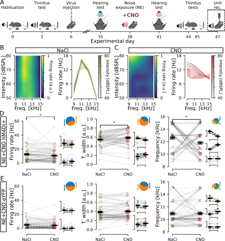
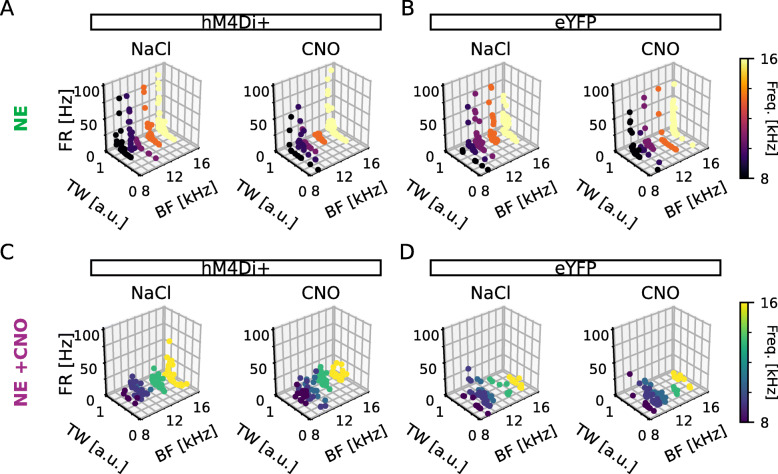
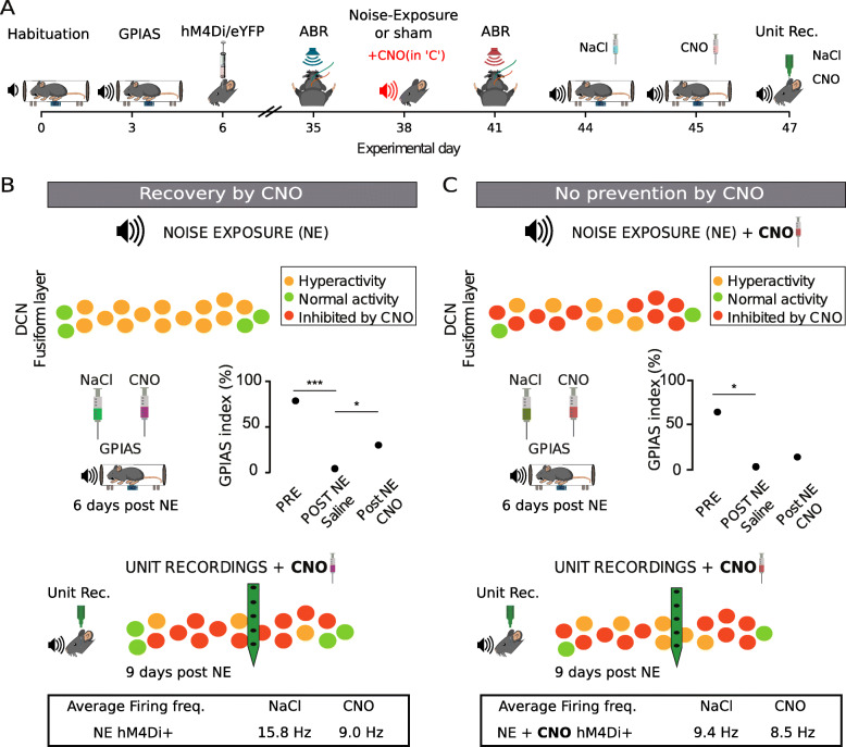
Results: Mice were exposed to loud noise (9-11kHz, 90dBSPL, 1h, followed by 2h of silence), and auditory brainstem responses (ABRs) and gap prepulse inhibition of acoustic startle (GPIAS) were recorded 2 days before and 2 weeks after noise exposure to identify animals with a significantly decreased inhibition of startle, indicating tinnitus but without permanent hearing loss. Neuronal activity of CaMKII α+ neurons expressing hM4Di in the DCN was lowered by administration of clozapine-N-oxide (CNO). We found that acutely decreasing firing rate of CaMKII α+ DCN units decrease tinnitus-like responses (p = 3e -3, n = 11 mice), compared to the control group that showed no improvement in GPIAS (control virus; CaMKII α-YFP + CNO, p = 0.696, n = 7 mice). Extracellular recordings confirmed CNO to decrease unit firing frequency of CaMKII α-hM4Di+ mice and alter best frequency and tuning width of response to sound. However, these effects were not seen if CNO had been previously administered during the noise exposure (n = 6 experimental and 6 control mice).

Conclusion: We found that lowering DCN activity in mice displaying tinnitus-related behavior reduces tinnitus, but lowering DCN activity during noise exposure does not prevent noise-induced tinnitus. Our results suggest that CaMKII α-positive cells in the DCN are not crucial for tinnitus induction but play a significant role in maintaining tinnitus perception in mice.

Keywords: Chemogenetics; Dorsal cochlear nucleus; GPIAS; Tinnitus; Unit recording.


Conflict of interest statement

The authors declare that they have no competing interests.

Figures

Fig. 1.
Fig. 1.
Noise exposure did not induce hearing threshold shifts. A Experimental timeline highlighting the ABR recordings before and after noise exposure. B ABR representative example for 8–10-kHz frequency presented from 80 to 35dBSPL. Algorith-detected response peaks are marked with asterisks. Hearing threshold was defined at the last intensity with an identified peak, in this example, 50dBSPL. C Group hearing thresholds for each frequency tested before (blue) and after (orange) noise exposure (NE) for hM4Di (left) and eYFP (right) injected animals. D, E ABR Wave 1 (W.1) amplitudes at 80dBSPL (left) and growth functions (right) for the hM4Di (D) and eYFP (E) injected animals. Left, amplitude of wave 1 for each frequency tested 3d before (blue) and 3d after (orange) nose exposure. Right, wave 1 amplitude at each tested intensity, normalized by amplitude at 80dBSPL, at each tested frequency (cold blue-green colors: 3d before; warm red-yellow colors: 3d after noise exposure). n = 11 and 7 for NE hM4Di+ and NE eYFP, respectively
Fig. 2.
Fig. 2.
Inhibition of DCN CaMKII α-hM4Di-positive cells after noise exposure decreases tinnitus-like behavior. A Experimental timeline highlighting the GPIAS recordings before and after noise exposure. B Schematic drawing of the Startle and Gap-startle protocols. C Representative GPIAS recording of a mouse showing 68.69% suppression of acoustic startle before and 10.67% suppression after noise exposure when comparing Startle (red) and Gap-startle (black) responses, indicating tinnitus-like behavior for the tested frequency (9–11kHz). D, E GPIAS group performance before (blue) and after (orange) noise exposure for hM4Di (D) and eYFP (E) injected mice. F, G Left, histogram showing the number of hM4Di (F) and eYFP (G) injected animals in function of the frequency with the greatest decrease in GPIAS index. Right, GPIAS group index showed that hM4Di+ mice (F) decreased startle suppression after noise exposure and regained startle suppression under the effect of CNO, while eYFP-injected mice shows that, although presenting tinnitus-like responses after noise exposure, no difference was observed between NaCl and CNO treatments (p = 0.696). n = 11 and 7 for NE hM4Di+ and NE eYFP, respectively; *p >0.05; **p = 1.3e −10
Fig. 3.
Fig. 3.
Decreasing CaMKII α-hM4Di-positive cell activity in the DCN changes firing properties of the circuitry. A Timeline of experiments highlighting unit recordings. B, C Left, normalized firing rate (colormap) of a representative unit after NaCl (B) and CNO (C) administration for each intensity (lines) and each frequency (columns) tested. Right, a different representation of the same representative examples in the left, showing firing rate per frequency for each intensity. Data was upsampled 3 times in the intensity and frequency dimensions. D, E Units firing rate (left), tuning width (middle), and best frequency (right) for stimulation at 80dBSPL, at each unit best frequency. Animals expressing hM4Di (D) showed a significant decrease in firing rate (left), decrease in tuning width (middle), and increase in best frequency (right). Control animals expressing eYFP (E) showed no significant change in any of those parameters. Individual unit values are shown in green (NaCl) or purple (CNO) condition. Black line indicates the mean ± SEM. Insets D and E (pie) shows the portion of units decreasing (blue), increasing (orange), or not changing (green) values upon CNO administration. Insets D and E (scatters) show distribution of unit values divided in groups for decrease, increase, or no change (for larger representation see Additional file 1: Fig. S1). n = 11 and 7 mice, 122 and 102 units, for NE hM4Di+ and NE eYFP, respectively; *p <0.05; ***p = 1.3e −4
Fig. 4.
Fig. 4.
Inhibiting DCN CaMKII α-hM4Di-positive cell activity during noise exposure does not interfere with hearing at the tested frequencies. A Timeline of experiments highlighting ABR recordings before and after noise exposure. B, C Representative ABR traces (B) and group responses for hM4Di (C left) and eYFP (C right) injected mice that received i.p. CNO injection 30 min before noise exposure. D, E ABR Wave 1 (W.1) amplitudes at 80dBSPL (left) and growth functions (right) for the hM4Di (D) and eYFP (E) injected animals that received CNO before noise exposure. Left, amplitude of wave 1 for each frequency tested 3d before (blue) and 3d after (orange) noise exposure. Right, wave 1 amplitude at each tested intensity, normalized by amplitude at 80dBSPL, at each tested frequency (cold blue-green colors: 3d before; warm red-yellow colors: 3d after noise exposure). n = 6 and 6 for NE+CNO hM4Di+ and NE+CNO eYFP
Fig. 5.
Fig. 5.
Decreasing CaMKII α-hM4Di-positive DCN cell activity during noise exposure does not prevent tinnitus-like behavior and abolishes hM4Di-dependent recovery. A Schematic timeline highlighting GPIAS recordings before and after noise exposure. B Schematic outline of Startle and Startle-gap protocols. C Representative GPIAS response. D, E Group results for startle suppression of all frequencies tested before (blue) and after (dark red) noise exposure in the presence of CNO for hM4Di (D) and eYFP (E) injected mice. F, G Left, quantification of most affected frequency of each animal. Right, Inhibition of CaMKII α-hM4Di-positive DCN cells during noise exposure did not prevent a decrease in the startle suppression, indicating tinnitus, and also the second CNO injection before GPIAS recording (in mice receiving CNO 30 min before the noise exposure) did not recover startle suppression (F; p = 0.404). The eYFP injected group showed tinnitus-like behavior after noise exposure with CNO and did not recover the startle suppression after CNO injection upon testing GPIAS (G; p = 0.176). n = 6 and 6 for NE+CNO hM4Di+ and NE+CNO eYFP; *p <0.05
Fig. 6.
Fig. 6.
Decreasing activity of CaMKII α-hM4Di-positive DCN cells that were also inhibited during noise exposure changes firing properties of the circuitry. A Timeline of experiments highlighting the unit recordings. BC Left, normalized firing rate (colormap) of a representative unit after NaCl (B) and CNO (C) injection for each intensity (lines) and each frequency (columns) tested. Right, a different representation of the same representative examples in the left, showing firing rate per frequency for each intensity. D, E Units firing rate (left), tuning width (middle), and best frequency (right) for stimulation at 80dBSPL, at each unit best frequency in the 8–16kHz interval. D Units from mice expressing hM4Di, showing significant difference after CNO application for firing rate, tuning width, and best frequency. Individual unit values are shown in olive (NaCl) or red (CNO) condition. Black line indicates the mean ± SEM. E Units from mice expressing eYFP showing no significant difference for any of the parameters. Insets show the proportion of units decreasing (blue), increasing (orange) or not changing (green) parameters of each graph (see Additional file 2: Fig. S2 for greater detail). n = 6 and 6 mice, 85 and 91 units, for NE+CNO hM4Di+ and NE+CNO eYFP; *p <0.05
Fig. 7.
Fig. 7.
Three-dimensional scatter plots of firing rate, tuning width, and best frequency of DCN units of noise-exposed hM4Di+ or eYFP+ animals in the presence of NaCl or CNO. A, B 3D scatters representing each unit by firing rate x tuning width x best frequency for hM4Di (experimental; A and eYFP (control; B animals under NaCl (left) or CNO (right) treatment. C, D Same as A and B for experiments where animals were administered CNO (0.5mg/kg) 30 min prior to noise exposure. Colors represent the best frequency response between 8 and 16kHz. FR, firing rate; TW, tuning width; BF, best frequency; n = 11, 7, 6, and 6 mice; 122, 102, 85, and 91 units; for NE hM4Di+, NE eYFP, NE+CNO hM4Di+ and NE+CNO eYFP, respectively
Fig. 8.
Fig. 8.
DCN unit depth profile. A Schematic representation of the probe location within the DCN according to coordinates used highlighting the dorsoventral depth of unit recordings. B Distribution of recorded DCN units along the dorsoventral axis for noise-exposed animals expressing CaMKII α-hM4Di or CaMKII α-eYFP. C The same as B but for experimental and control animals that were pre-treated with CNO 30 min before noise exposure. Black bars indicate mean ± SEM. n = 11, 7, 6, and 6 mice; 122, 102, 85, and 91 units; for NE hM4Di+, NE eYFP, NE+CNO hM4Di+, and NE+CNO eYFP, respectively
Fig. 9.
Fig. 9.
Recovery from tinnitus-behavior when decreasing activity of CaMKII α+ DCN neurons acutely but not if activity was chemogenetically reduced during the noise exposure. A Schematic outline highlighting the hypothetical difference between experiments of recovery in tinnitus-like animals (B) and animals where the aim was to prevent tinnitus (CNO administered before the noise exposure, second set of experiments as shown in C, red font). B Top, schematic drawing of the DCN fusiform layer showing hypothetical hyperactive neurons following noise exposure. Middle, CNO improved gap detection. Bottom, CNO administration causes a reduction in average firing frequency that may be due to inhibiting hyperactive DCN neurons. C Top, schematic drawing showing the DCN fusiform layer chemogenetically inhibited during the noise exposure; however, some neurons are probably not affected by CNO and may still render some neurons hyperactive. Middle, acute CNO exposure does not improve gap detection in mice administered CNO during the noise exposure. Bottom, unit recordings in animals administered CNO during the noise exposure shows that acute additional CNO administration reduces the overall firing frequency less dramatically compared to “B,” suggesting units expressing hM4Di receptors were not hyperactivated during the noise trauma

References

    1. Heller AJ. Classification and epidemiology of tinnitus. Otolaryngol Clin N Am. 2003;36(2):239–48. doi: 10.1016/S0030-6665(02)00160-3. - DOI - PubMed
    1. Gallus S, Lugo A, Garavello W, Bosetti C, Santoro E, Colombo P, Perin P, La Vecchia C, Langguth B. Prevalence and determinants of tinnitus in the Italian adult population. Neuroepidemiology. 2015;45(1):12–19. doi: 10.1159/000431376. - DOI - PubMed
    1. Møller AR. Tinnitus: presence and future. Tinnitus Pathophysiol Treat. 2007;3–16. 10.1016/s0079-6123(07)66001-4.
    1. Langguth B, Landgrebe M, Kleinjung T, Sand GP, Hajak G. Tinnitus and depression. World J Biol Psychiatry. 2011;12(7):489–500. doi: 10.3109/15622975.2011.575178. - DOI - PubMed
    1. Shore SE, Roberts LE, Langguth B. Maladaptive plasticity in tinnitus — triggers, mechanisms and treatment. Nat Rev Neurol. 2016;12(3):150–60. doi: 10.1038/nrneurol.2016.12. - DOI - PMC - PubMed

Publication types

Substances