. 2022 May 13;20(1):109.
doi: 10.1186/s12915-022-01318-y.

Asynchronous embryonic germ cell development leads to a heterogeneity of postnatal ovarian follicle activation and may influence the timing of puberty onset in mice

Affiliations

Asynchronous embryonic germ cell development leads to a heterogeneity of postnatal ovarian follicle activation and may influence the timing of puberty onset in mice

Yanli Dai et al. BMC Biol. .

Abstract

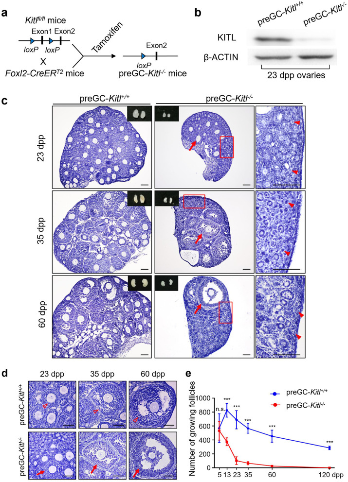
Background: Ovarian follicles, which are the basic units of female reproduction, are composed of oocytes and surrounding somatic (pre) granulosa cells (GCs). A recent study revealed that signaling in somatic preGCs controlled the activation (initial recruitment) of follicles in the adult ovaries, but it is also known that there are two waves of follicle with age-related heterogeneity in their developmental dynamics in mammals. Although this heterogeneity was proposed to be crucial for female reproduction, our understanding of how it arises and its significance is still elusive.

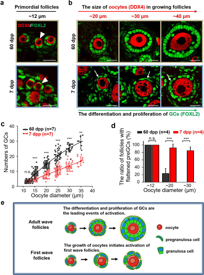
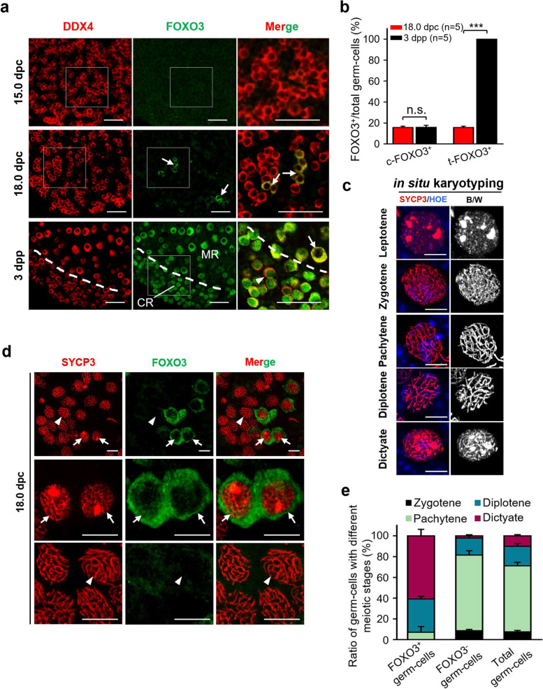
Results: In the current study, by deleting the key secreted factor KIT ligand from preGCs and analyzing the follicle cell developmental dynamics, we revealed distinct patterns of activation and growth associated with the two waves of follicles in mouse ovary. Our results confirmed that activation of adult wave follicles is initiated by somatic preGCs and dependent on the KIT ligand. By contrast, activation of first wave follicles, which are awakened from germ cells before follicle formation, can occur in the absence of preGC-secreted KIT ligand in postnatal ovaries and appears to be oocyte-initiated. We also found that the asynchronous activity of phosphatidylinositol 3 kinases (PI3K) signaling and meiotic process in embryonic germ cells lead to the follicle heterogeneity in postnatal ovaries. In addition, we supplied evidence that the time sequence of embryonic germ cell development and its related first wave follicle growth are correlated to the time of puberty onset in females.

Conclusion: Taken together, our study provides evidence that asynchronous development of embryonic oocytes leads to the heterogeneity of postnatal ovarian follicle activation and development, and affects the timing of onset of puberty in females.

Keywords: Embryonic germ cells; Meiosis; Ovarian follicle heterogeneity; Primordial follicle activation; Puberty onset.


Conflict of interest statement

The authors declare that they have no competing interests.

Figures

Fig. 1
Fig. 1
Primordial follicle activation in early life is independent of preGC-KITL. a Illustration of the deleting strategy of Kitl in preGC-Kitl−/− mice. By neonatal tamoxifen administration before the primordial follicle formation, exon 1 of Kitl was deleted from preGCs in the ovary. b Western blot showing the deleting efficiency of KITL in the ovaries of 23 dpp preGC-Kitl−/− females. β-ACTIN was used as the internal control. c Representative images of ovarian morphological changes in preGC-Kitl+/+ (left panel) and preGC-Kitl−/− (right panel) females from 23 to 60 dpp, showing a dramatic suppression (arrowheads) of primordial follicle activation in the cortical region of preGC-Kitl−/− ovaries, and a small portion of follicles (arrows) were activated and developed normally in the medulla region of mutant ovaries. d A comparable developmental dynamics of activated follicles in preGC-Kitl+/+ (hollow arrowheads) and preGC-Kitl−/− (arrows) mice, showing the activated follicles were developed to the secondary (23 dpp), pre-antal (35 dpp), and antral stages (60 dpp) in the preGC-Kitl−/− ovaries. e Quantification of growing follicles in preGC-Kitl−/− ovaries at different ages. Showing the number of growing follicles decreased continuously and exhausted at around 120 dpp (red line) but not in the preGC-Kitl+/+ ovaries (blue line) (n ≥ 3). All experiments were repeated more than three times, and representative images are shown. Data are presented as the mean ± SD and analyzed by a two-tailed unpaired Student’s t-test, n.s. P ≥ 0.05 and ***P < 0.001. Scale bars, 100 μm
Fig. 2
Fig. 2
Distinct developmental patterns of growing follicles exist in postnatal and adult ovaries. a Showing a comparable morphology of dormant primordial follicles (arrowheads) in the ovaries at 60 dpp and 7 dpp. DDX4, red; FOXL2, green. b Distinct developmental patterns of growing follicles exist in the postnatal and adult ovaries. With the increase of oocyte size (DDX4, red), major GCs (FOXL2, green) differentiated to a cuboidal state with the oocyte enlargement in the 60 dpp ovaries, whereas a dramatic retardation of GC development (arrows) was found in follicles at 7 dpp. c Counting the increase of GC number with oocyte enlargement showing different growth kinetics of the follicle growth in the ovaries at 60 dpp and 7 dpp. Showing the average number of GCs increased from 3 to 30 in the 60 dpp ovaries (n = 7), while just increased from 3 to 18 in 7 dpp (n = 7). Each dot showed the index of a single follicle. d The ratio of growing follicles with flattened preGCs, showing a large proportion of growing follicles at 7 dpp (red column) contained flattened preGCs (n = 4). e The model of distinct developmental patterns of follicle growth in the ovaries. Showing the differentiation and proliferation of GCs are the leading events of adult wave follicles, whereas the growth of oocytes initiates activation of first wave follicles. All experiments were repeated more than three times, and representative images are shown. Data are presented as the mean ± SD and analyzed by a two-tailed unpaired Student’s t-test, n.s. P ≥ 0.05, **P < 0.01 and *** P < 0.001. Scale bars, 20 μm
Fig. 3
Fig. 3
Asynchronous activity of PI3K in germ cells of embryonic ovaries leads to the formation of first wave follicles. a Detecting the expression of FOXO3 showed an asynchronous activity of PI3K signaling in embryonic germ cells. No FOXO3 expressions were observed in germ cells at 15.0 dpc, whereas part of germ cells in cysts expressed FOXO3 (arrows) in the cytoplasm (c-FOXO3+), indicating an activity of PI3K signaling in them at 18.0 dpc. At 3 dpp, all oocytes expressed FOXO3 whereas only the oocytes in the medulla region were with c-FOXO3+ (arrow), and the oocytes in cortical region were with nuclear-FOXO3 localization (arrowhead). DDX4, red; FOXO3, green. CR, cortical region; MR, medulla region. b The germ cell counting result showing the ratio of total FOXO3+ (t-FOXO3+) germ cells significantly increased from 18.0 dpc to 3 dpp, but the c-FOXO3+ germ cells kept a stable proportion in the ovaries (n = 5). c Representative in situ karyotyping images of the meiotic phases in embryonic germ cells. SYCP3, red; HOE, blue. B/W, the red fluorescence of SYCP3 was inverted to black/white (B/W) to highlight the meiotic stages in the right panel. d Co-localization of FOXO3 and SYCP3 in 18.0-dpc mouse ovaries, showing the majority of FOXO3+ germ cells were at diplotene or dictyate stage (arrows), while most of FOXO3 germ cells were at pachytene stage (arrowheads). SYCP3, red; FOXO3, green. e Counting the ratio of meiotic stages in FOXO3+, FOXO3 and total germ cells in 18.0 dpc, confirming that the FOXO3+ germ cells present a faster meiotic process (n = 3). All experiments were repeated at least three times, and representative images are shown. Data are presented as the mean ± SD and analyzed by a two-tailed unpaired Student’s t-test, n.s. P ≥ 0.05 and ***P < 0.001. Scale bars, 50 μm (a), 5 μm (c), 10 μm (d)
Fig. 4
Fig. 4
The pattern of embryonic germ cell development is correlated to the process of first wave follicle growth and the time of puberty onset. a The vaginal opening ages of C57 and C3H strains, showing a significantly earlier vaginal opening in C3H compared to C57 (n = 19). b The age of first vaginal cornification in C57 and C3H, showing a significantly earlier onset of first vaginal cornification in the C3H compared to C57 (n = 19). c The age-related FOXO3 expressing profiles in embryonic germ cells of C57 and C3H females. The FOXO3+ germ cells (arrows) were observed in the C3H ovaries at 16.0 dpc, whereas the FOXO3+ germ cells started to appear in C57 ovaries at 18.0 dpc. FOXO3, green; DDX4, gray. d The ratio of activated germ cells at 16.0 dpc and 18.0 dpc showed a significant difference in C3H ovaries compared to that in C57 (n = 6). e Histological analysis of the ovarian development in C57 and C3H at 5 dpp, showing the secondary follicles with multi-layer of GCs (arrows) in C3H ovaries. f Follicle counting results showing a high proportion of secondary follicles in C3H ovaries compared to C57 at 5 dpp (n = 7). g Histological analysis of the ovarian development in C57 and C3H at 13 dpp, showing the antral follicles (arrow) existed at 13 dpp C3H ovaries. h, i Calculating results of the size and the GC layer number in 5 largest follicles per ovaries in C57 and C3H at 13 dpp. Showing a significantly larger size (h) and GC layer number (i) of pioneering follicles in C3H ovaries than that in C57 ovaries (n = 8). All experiments were repeated more than three times, and representative images are shown. Data are presented as the mean ± SD and analyzed by a two-tailed unpaired Student’s t-test, ***P < 0.001. Scale bars, 50 μm (c), 100 μm (e, g)

References

    1. Zhang H, Liu L, Li X, Busayavalasa K, Shen Y, Hovatta O, et al. Life-long in vivo cell-lineage tracing shows that no oogenesis originates from putative germline stem cells in adult mice. Proc Natl Acad Sci U S A. 2014;111(50):17983–17988. doi: 10.1073/pnas.1421047111. - DOI - PMC - PubMed
    1. Pepling ME. Follicular assembly: mechanisms of action. Reproduction. 2012;143(2):139–149. doi: 10.1530/REP-11-0299. - DOI - PubMed
    1. Wang Y, Teng Z, Li G, Mu X, Wang Z, Feng L, et al. Cyclic AMP in oocytes controls meiotic prophase I and primordial folliculogenesis in the perinatal mouse ovary. Development. 2015;142(2):343–351. - PubMed
    1. McGee EA, Hsueh AJ. Initial and cyclic recruitment of ovarian follicles. Endocr Rev. 2000;21(2):200–214. - PubMed
    1. Nelson LM. Clinical practice. Primary ovarian insufficiency. N Engl J Med. 2009;360(6):606–614. doi: 10.1056/NEJMcp0808697. - DOI - PMC - PubMed

Publication types

Substances

LinkOut - more resources