. 2022 Jul:61:101512.
doi: 10.1016/j.molmet.2022.101512. Epub 2022 May 9.

Beta-hydroxybutyrate dampens adipose progenitors' profibrotic activation through canonical Tgfβ signaling and non-canonical ZFP36-dependent mechanisms

Affiliations

Beta-hydroxybutyrate dampens adipose progenitors' profibrotic activation through canonical Tgfβ signaling and non-canonical ZFP36-dependent mechanisms

Simon Lecoutre et al. Mol Metab. 2022 Jul.

Abstract

Background/purpose: Adipose tissue contains progenitor cells that contribute to beneficial tissue expansion when needed by de novo adipocyte formation (classical white or beige fat cells with thermogenic potential). However, in chronic obesity, they can exhibit an activated pro-fibrotic, extracellular matrix (ECM)-depositing phenotype that highly aggravates obesity-related adipose tissue dysfunction.

Methods: Given that progenitors' fibrotic activation and fat cell browning appear to be antagonistic cell fates, we have examined the anti-fibrotic potential of pro-browning agents in an obesogenic condition.

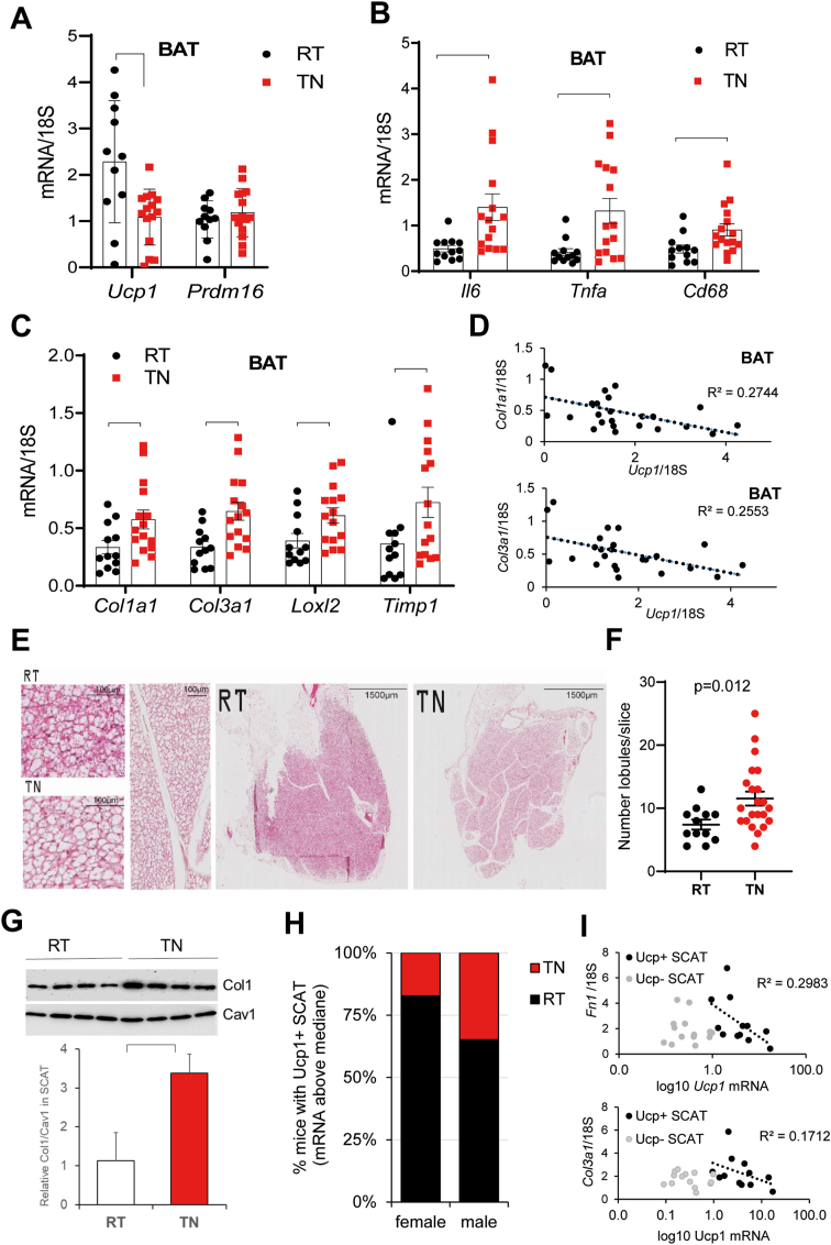
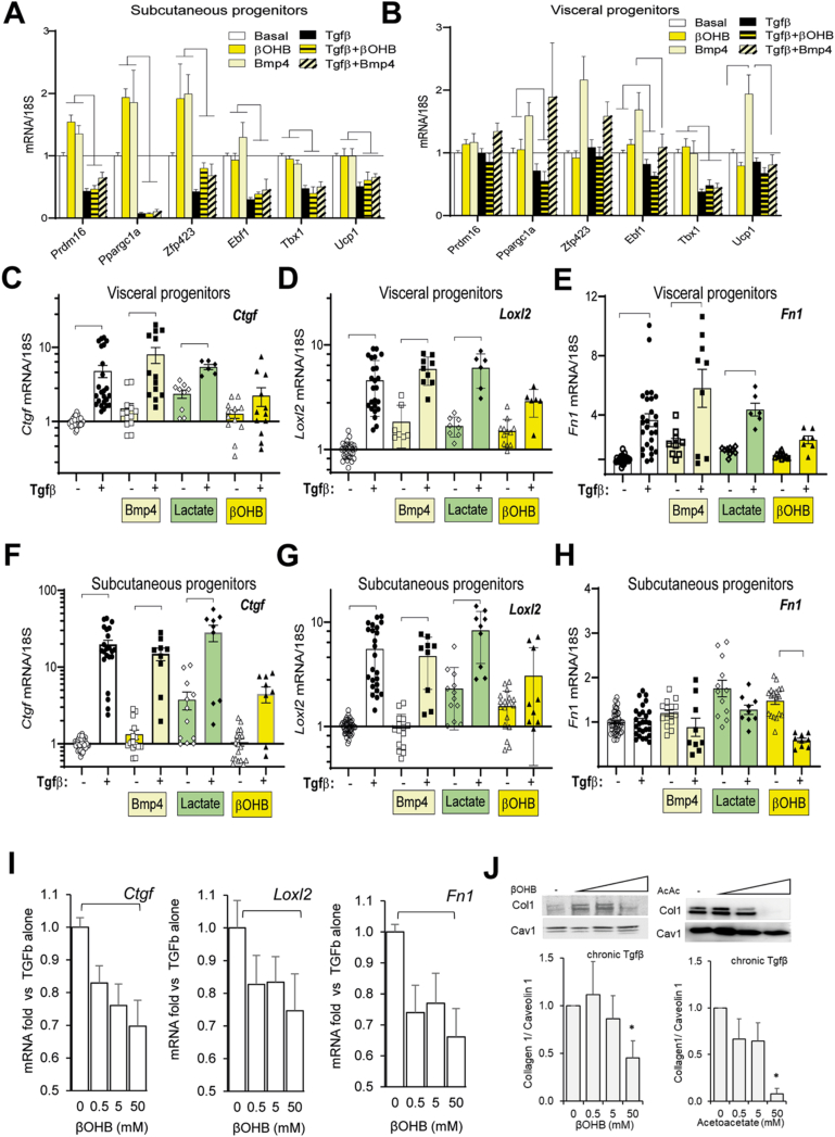
Results: In obese mice fed a high fat diet, thermoneutral housing, which induces brown fat cell dormancy, increases the expression of ECM gene programs compared to conventionally raised animals, indicating aggravation of obesity-related tissue fibrosis at thermoneutrality. In a model of primary cultured murine adipose progenitors, we found that exposure to β-hydroxybutyrate selectively reduced Tgfβ-dependent profibrotic responses of ECM genes like Ctgf, Loxl2 and Fn1. This effect is observed in both subcutaneous and visceral-derived adipose progenitors, as well as in 3T3-L1 fibroblasts. In 30 patients with obesity eligible for bariatric surgery, those with higher circulating β-hydroxybutyrate levels have lower subcutaneous adipose tissue fibrotic scores. Mechanistically, β-hydroxybutyrate limits Tgfβ-dependent collagen accumulation and reduces Smad2-3 protein expression and phosphorylation in visceral progenitors. Moreover, β-hydroxybutyrate induces the expression of the ZFP36 gene, encoding a post-transcriptional regulator that promotes the degradation of mRNA by binding to AU-rich sites within 3'UTRs. Importantly, complete ZFP36 deficiency in a mouse embryonic fibroblast line from null mice, or siRNA knock-down in primary progenitors, indicate that ZFP36 is required for β-hydroxybutyrate anti-fibrotic effects.

Conclusion: These data unravel the potential of β-hydroxybutyrate to limit adipose tissue matrix deposition, a finding that might exploited in an obesogenic context.

Keywords: Adipocyte; Extracellular matrix; Fibrosis; Progenitors.


Figures

Image 1
Graphical abstract
Figure 1
Figure 1
Thermoneutral housing aggravates HFD-induced adipose tissue ECM gene program in mice. Mice were housed for 9 weeks at room temperature (RT, 22 °C) or, thermoneutrality (TN, 30 °C) and received ad libitum HFD feeding. A-D: Brown adipose tissue (BAT) gene expression after 9 weeks on HFD was evaluated by Real time Q-PCR and normalized to 18 S. Values from individual mice are plotted, parentheses indicate significant differences between groups assessed by Student's t test. E: Hematoxilin/eosin BAT slices from RT and TN mice at different focus level (see scale bar in each image). Left panels show increased lipid droplet size in TN compared to RT (left panels). White spans consisting of acellular material (vertical image) were identified in whole tissue scans, delimiting lobules. F: Semi quantitative analysis of lobule number. Each point is from individual mice. G: Western blot analysis of SCAT Collagen1 content in RT and TN mice. Four tissues from individual mice are shown in each group; signals are normalized to Caveolin 1 for quantification. Parenthesis indicates significant difference (p < 0.05) by t test. H: Proportion of mice with Ucp1 positive SCAT (defined as above median Ucp1 mRNA value) in RT and TN groups. P value = 0.035 by Chi2 Test I: Correlation between Ucp1 expression in SCAT and ECM genes (Fn1: upper panel, Col3a1: lower panel). Significant correlation (p < 0.05) is found in Ucp1+ SCAT (black) but not Ucp1- SCAT (grey). (For interpretation of the references to colour in this figure legend, the reader is referred to the Web version of this article.)
Figure 2
Figure 2
βOHB counteracts Tgfβ-mediated stimulation of ECM genes. Combinatorial treatment with browning agents and Tgfb of visceral (A) or subcutaneous progenitors (B) on the expression of browning genes. βOHB (50 mM) or Bmp4 (10 ng/ml) was added 24 h after plating and Tgfβ (10 ng/ml) was added the following day, until day 5. Bars are mean+/- sem of three independent cell preparations. The same experimental scheme was used in (C–H), to assess Ctgf, Loxl2 and Fn1 mRNA responses. Each point is an individual well from triplicates in at least 4 independent cell preparations. I: Dose-dependent response of Ctgf, Loxl2 and Fn1 gene expression to βOHB in the presence of Tgfβ in primary subcutaneous progenitors. Parentheses indicate significant differences between conditions, by Student's t test. J: Western blot analysis of Collagen1 protein expression in Tgfβ-stimulated visceral progenitors in the presence of βOHB or Acetoacetate. βOHB or Acetoacetate was added 1 day post plating and was maintained until cell harvest (Day 5). Chronic Tgfβ stimulation started from Day 2 to Day 5. Collagen1 antibody was from Proteintech, ref14695-1. Representative blot is shown, with quantification from 2 to 4 independent cell preparations.
Figure 3
Figure 3
Serum βOHB levels negatively associate with subcutaneous adipose tissue fibrosis in patients with obesity. A: Serum βOHB levels were assessed in a group of 29 obese patients eligible for bariatric surgery, in which the intensity of subcutaneous fat fibrosis had been scored as previously described. Men are represented as open symbols. B: Patient stratification according to serum βOHB (relative to median value) indicates preferential clustering of patients with low Fat score in the high βOHB group. Numbers represent the total number of patients in stratification groups, with the number of men in parenthesis C: Negative association (Spearman correlation) of serum βOHB levels and intensity of ECM labelling by Picrosirius-red in histological biopsies of subcutaneous adipose tissue in a subgroup of 16 patients. Open symbols are men. D: Association of clinical parameters to serum βOHB levels in the patients studied. E: Patients distribution according to Fat score and biopsy-proven liver status. Numbers represent the total number of patients in stratification groups, with the number of men in parenthesis. (For interpretation of the references to colour in this figure legend, the reader is referred to the Web version of this article.)
Figure 4
Figure 4
Mechanisms of fibrotic attenuation by βOHB. A: Smads mRNA expression in the presence of βOHB or chronic Tgfβ. Bars are mean values ± sem from 4 independent cell preparations. B: A representative western blot probed with antibodies against Smad2-3 (Cell signalling, ref 8685), Phospho Smad2 (Ser465/467)/Smad3 (Ser423/425) (Cell signalling, ref 8828 and Caveolin 1 (BD Transduction Laboratory, ref 610,060). βOHB concentration range is as in Figure 1, in the presence or absence of a chronic Tgfβ stimulation. C-D: Quantitative analysis of protein signal intensity after normalization with Caveolin-1 as a loading control. Bars are mean values from 3 independent cell preparations. Parentheses indicate significant differences between groups by t test. E:ZFP36 mRNA upregulation by βOHB. F: Expression of ZFP36 and related transcripts by βOHB and Tgfβ in subcutaneous and visceral progenitors. Bars are mean values ± sem from 3 to 5 independent cell preparations. ∗ indicate significant differences compared to basal by Student t test. G–H: Gene expression in fibroblast cell lines from Wild Type (WT) or ZFP36 KO mice. Note that in KO mice, ZFP36 mRNA is transcribed but contains an insertion in exon 2 which prevents protein production. I-J: Knock-down of ZFP36 with siRNA in primary progenitors maintained with Tgfβ with or without βOHB. Bars are mean values from 3 independent experiments. Parentheses indicate significant differences between groups by t test.
figs1
figs1
figs2
figs2
figs3
figs3
figs4
figs4

References

    1. Olefsky J.M., Glass C.K. Macrophages, inflammation, and insulin resistance. Annual Review of Physiology. 2010;72(1):219–246. - PubMed
    1. Sun K., Tordjman J., Clément K., Scherer P.E. Fibrosis and adipose tissue dysfunction. Cell Metabolism. 2013;18(4):470–477. - PMC - PubMed
    1. Divoux A., Tordjman J., Lacasa D., Veyrie N., Hugol D., Aissat A., et al. Fibrosis in human adipose tissue : composition , distribution , and link with lipid metabolism and fat. Diabetes. 2010;59:2817–2825. - PMC - PubMed
    1. Marcelin G., Ferreira A., Liu Y., Atlan M., Aron-Wisnewsky J., Pelloux V., et al. A pdgfrα-mediated switch toward CD9highAdipocyte progenitors controls obesity-induced adipose tissue fibrosis. Cell Metabolism. 2017;25(3):673–685. - PubMed
    1. Hasegawa Y., Ikeda K., Chen Y., Alba D.L., Stifler D., Shinoda K., et al. Repression of adipose tissue fibrosis through a PRDM16-GTF2IRD1 complex improves systemic glucose homeostasis. Cell Metabolism. 2018 Jan;27(1):180–194.e6. - PMC - PubMed

Publication types