Review
. 2022 May 11;30(5):684-695.
doi: 10.1016/j.chom.2022.02.016.

Early life host-microbe interactions in skin

Affiliations
Review

Early life host-microbe interactions in skin

Laura R Dwyer et al. Cell Host Microbe. .

Abstract

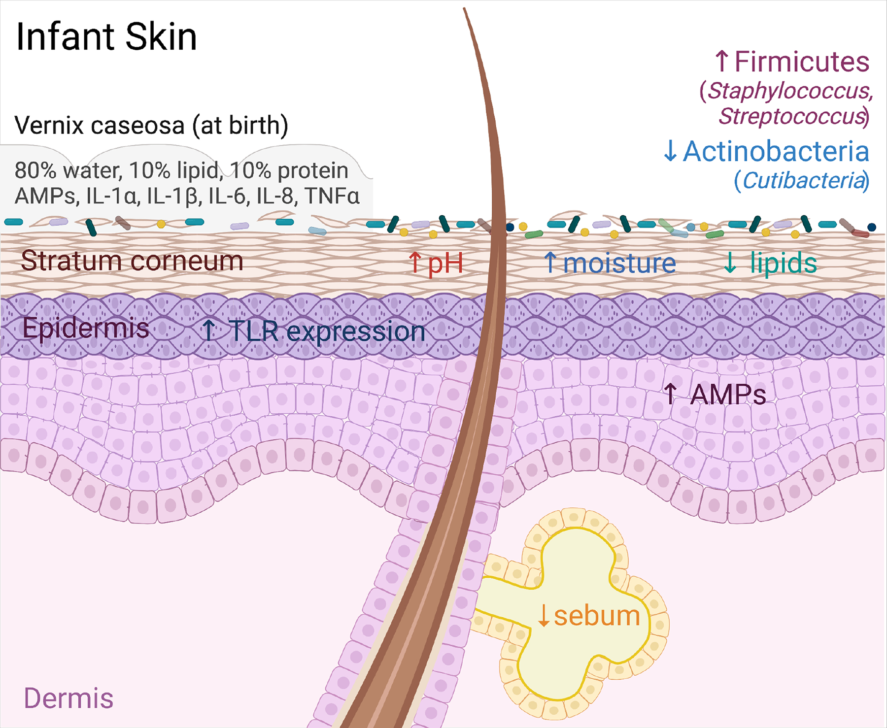
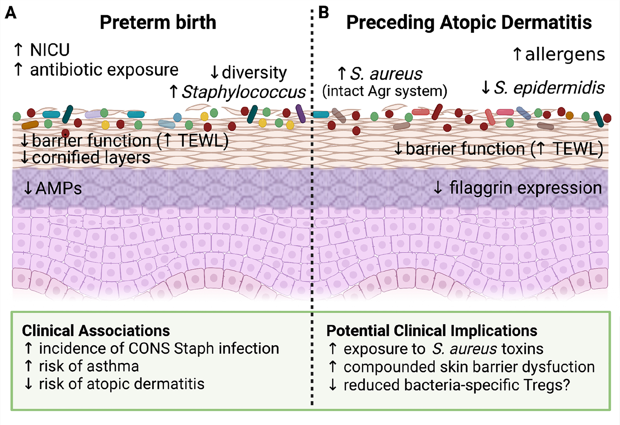
Our skin is the interface through which we mediate lifelong interactions with our surrounding environment. Initial development of the skin's epidermis, adnexal structures, and barrier function is necessary for normal cutaneous microbial colonization, immune development, and prevention of disease. Early life microbial exposures can have unique and long-lasting impacts on skin health. The identity of neonatal skin microbes and the context in which they are first encountered, i.e., through a compromised skin barrier or in conjunction with cutaneous inflammation, can have additional short- and long-term health consequences. Here, we discuss key attributes of infant skin and endogenous and exogenous factors that shape its relationship to the early life cutaneous microbiome, with a focus on their clinical implications.


Conflict of interest statement

Declaration of interests T.C.S. serves as a member of the scientific advisory board of Concerto Biosciences.

Figures

Figure 1:
Figure 1:. Physiologic Features of Infant skin.
Infant skin at birth is covered in the vernix caseosa (VC). The VC is made up of 80% water, 10% lipid, 10% protein, and contains various antimicrobial peptides (AMPs) and cytokines. The VC likely influences initial microbial colonization of the skin via its role as both a physical and chemical barrier as well as a nutrient substrate for microbes. Compared to adults, infant skin has increased expression of AMPs and toll like receptors (TLRs), an elevated pH, increased moisture content, and decreased lipid and sebum production. Collectively these features shape early life skin microbial ecology, which is distinguished by relatively increased colonization by Firmicutes, including members of Staphylococcus and Streptococcus, and decreased colonization by Actinobacteria, including Cutibacteria.
Figure 2:
Figure 2:. Early life microbial-immune interactions.
(A) 5-OP-RU, a microbially-produced riboflavin-derived antigen stimulates production of MAIT cells specifically in the neonatal versus adult thymus. As shown in mice, inadequate microbial exposure during this early window results in a lost opportunity to expand this lymphocyte population. MAIT cells travel from the thymus to skin where subsequent local production of 5-OP-RU by S. epidermidis and other skin bacteria promotes proliferation of MAITs and stimulates their IL-17 production. These cutaneous MAIT cells contribute to skin homeostasis for example by augmenting local would healing. (B) A polyclonal wave of regulatory T cells (Tregs) migrates from the thymus and secondary lymphoid organs into murine skin between the first and second week of postnatal life. Microbial colonization of developing hair follicles during this postnatal window in mice augments production of the ligand Ccl20 in the hair follicle infundibulum, which helps draw this polyclonal Ccr6+ Treg population into the tissue. Cutaneous exposure to commensal antigens in this early window, for example via neonatal S. epidermidis colonization, leads to a persistent enrichment of Tregs among commensal-specific CD4+ T cells in skin. They help to limit skin inflammation upon subsequent re-exposure to S. epidermidis under an inflammatory context. Expansion of these commensal-specific Tregs is dependent on dendritic-cell mediated uptake of bacterial antigens and their trafficking to the skin-draining lymph nodes (SDLN) for presentation to naïve CD4+ T cells. When initial skin exposure to S. epidermidis is delayed until adulthood, the repertoire of CD4+ T cells specific for that bacteria is shifted instead towards effector CD4+ T cells (Teffs). The mechanistic basis for this neonatal capacity to establish commensal-specific tolerance is likely multifactorial, but the enriched presence of Tregs in neonatal skin has been shown to be one contributing factor.
Figure 3:
Figure 3:. Early life pathologies associated with changes in skin physiology and colonization.
(A) Preterm birth (PTB) has a significant impact on neonatal skin. These infants are often exposed to the NICU environment as well as more frequent topical and systemic antibiotics. Because full maturation of the epidermis occurs during the final weeks of normal in utero gestation, the stratum corneum in PTB infants has fewer cornified layers, reduced antimicrobial peptide (AMP) expression and decreased barrier function as evidenced by increased transepidermal water loss (TEWL). These changes in skin physiology are accompanied by reduced diversity of the PTB skin microbiome, with a notable increase in Staphylococcus prevalence. If and how these altered host and microbial features in preterm infants contribute to the clinical associations seen in this population, such as increased opportunistic infections and decreased later risk of atopic dermatitis, remains an open area of investigation. (B) Blooms of S. aureus and S. epidermidis are well documented during flares of established atopic dermatitis (AD). However, there is also evidence for skin microbial changes that precede disease onset. Specifically, increased colonization by S. aureus, especially stains with a preserved Agr quorum sensing system, and a relative decrease in the prevalence of S. epidermidis have both been observed among infants that go on to develop AD. These ecological differences are accompanied by distinct skin physiological features, namely decreased filaggrin expression and skin barrier function (increased TEWL) that could alter the host response to skin microbes. Collectively, this suggests that an altered relationship with commensal microbes may be established prior to AD onset and contribute to its early pathogenesis alongside other key environmental or allergic exposures.

References

    1. Adachi T, Kobayashi T, Sugihara E, Yamada T, Ikuta K, Pittaluga S, Saya H, Amagai M, and Nagao K (2015). Hair follicle–derived IL-7 and IL-15 mediate skin-resident memory T cell homeostasis and lymphoma. Nature Medicine 21, 1272–1279. - PMC - PubMed
    1. Arrieta M-C, Stiemsma LT, Dimitriu PA, Thorson L, Russell S, Yurist-Doutsch S, Kuzeljevic B, Gold MJ, Britton HM, Lefebvre DL, et al. (2015). Early infancy microbial and metabolic alterations affect risk of childhood asthma. Science Translational Medicine 7. - PubMed
    1. Aversa Z, Atkinson EJ, Schafer MJ, Theiler RN, Rocca WA, Blaser MJ, and LeBrasseur NK (2021). Association of Infant Antibiotic Exposure With Childhood Health Outcomes. Mayo Clinic Proceedings 96, 66–77. - PMC - PubMed
    1. Ayhan M, Sancak B, Karaduman A, Arikan S, and Sahin S (2007). Colonization of neonate skin by Malassezia species: relationship with neonatal cephalic pustulosis. Journal of the American Academy of Dermatology 57, 1012–1018. - PubMed
    1. Bager P, Wohlfahrt J, and Westergaard T (2008). Caesarean delivery and risk of atopy and allergic disesase: Meta-analyses. Clinical and Experimental Allergy 38, 634–642. - PubMed