. 2022;107(7-8):406-416.
doi: 10.1159/000524386. Epub 2022 May 12.

Fenretinide Reduces Intestinal Mucin-2-Positive Goblet Cells in Chronic Alcohol Abuse

Affiliations

Fenretinide Reduces Intestinal Mucin-2-Positive Goblet Cells in Chronic Alcohol Abuse

Marta Melis et al. Pharmacology. 2022.

Abstract

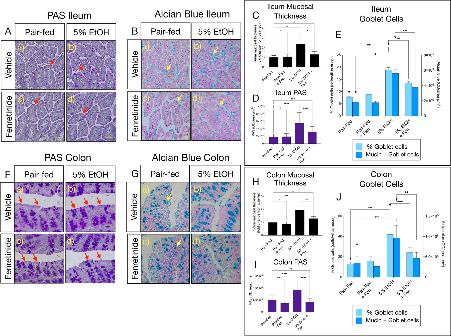
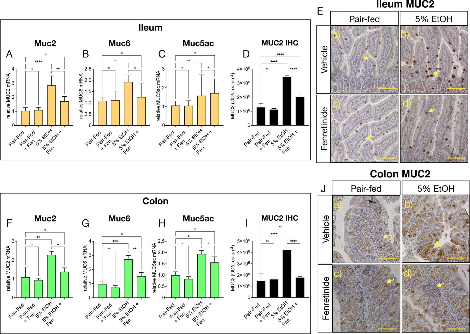
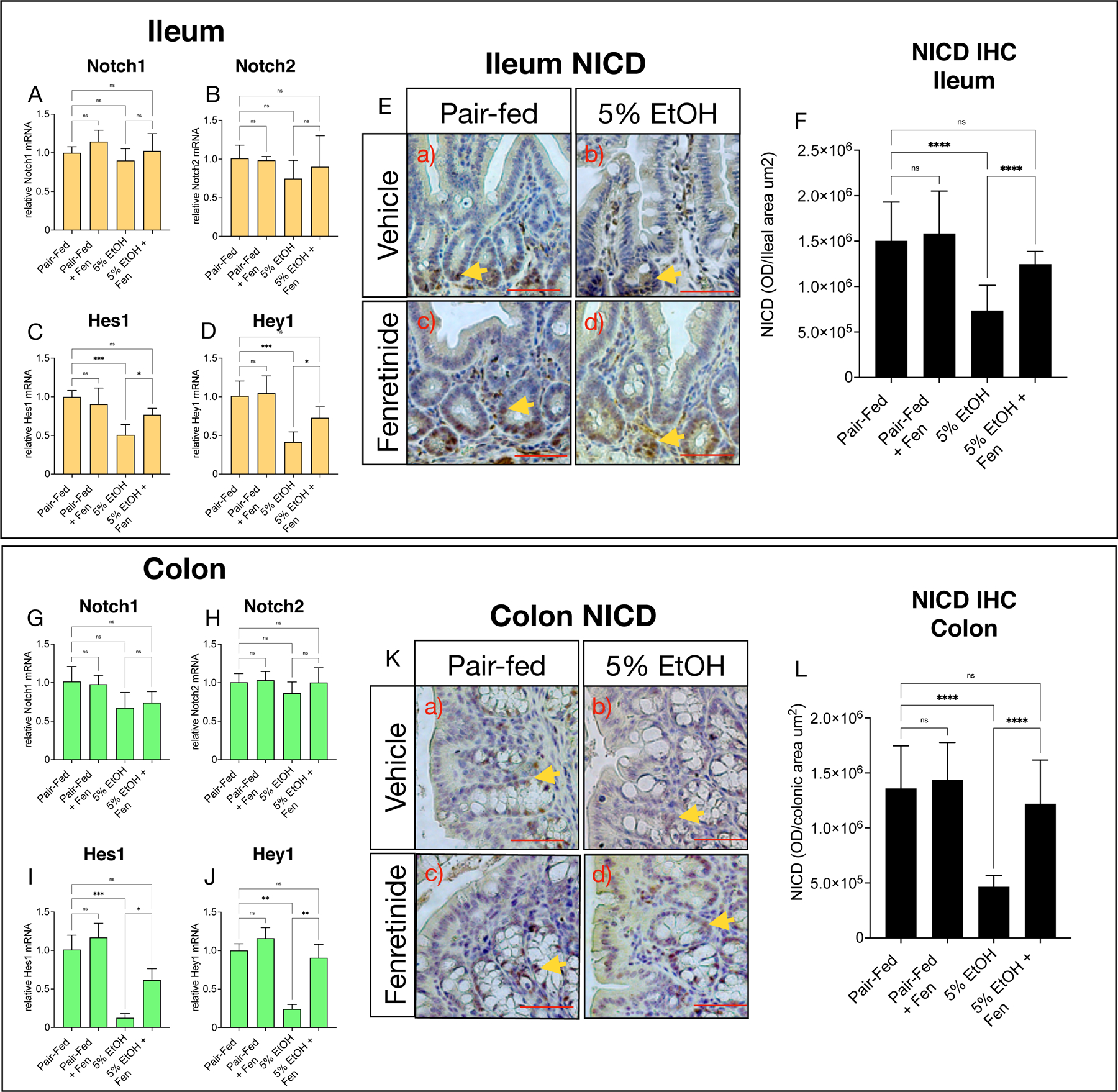
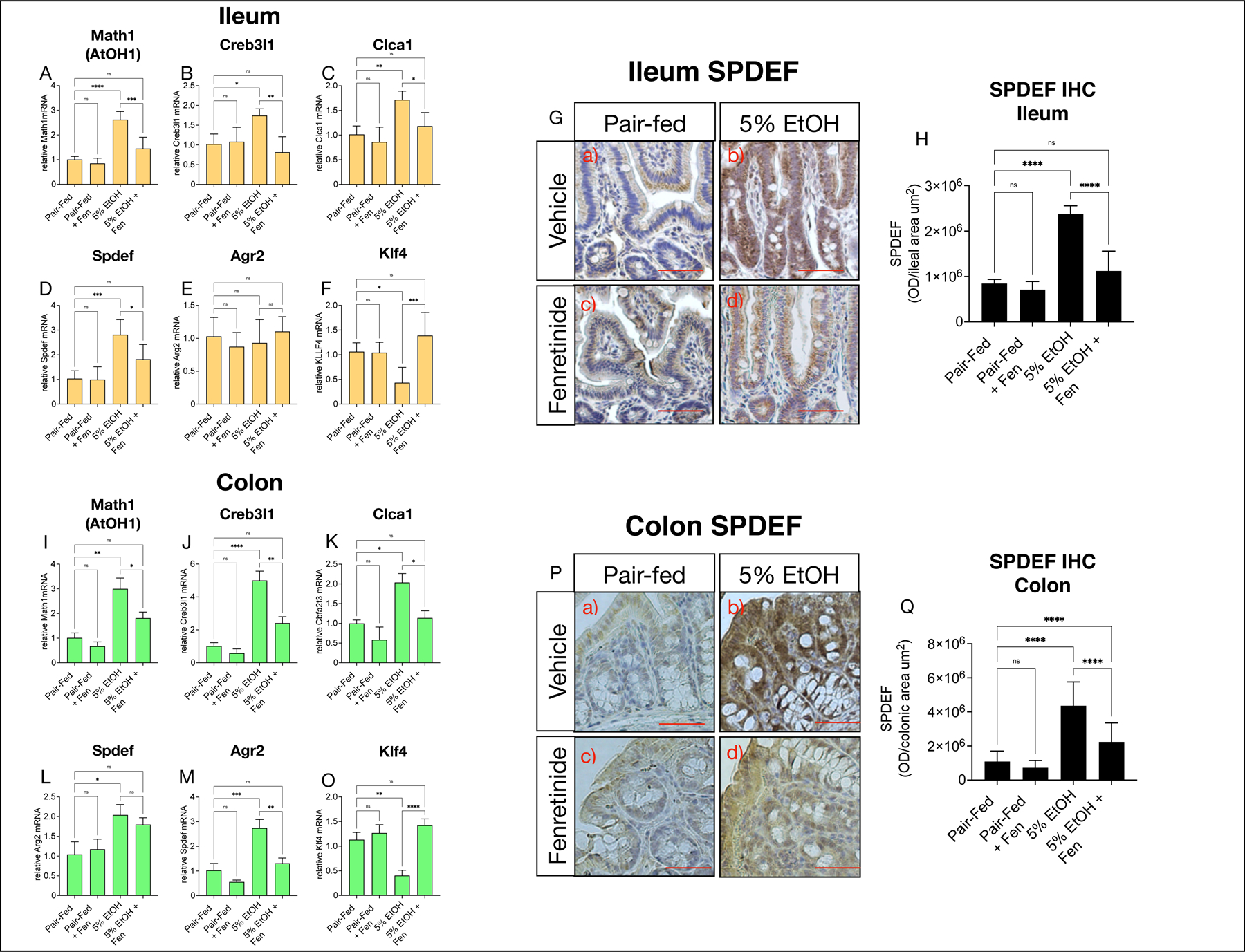
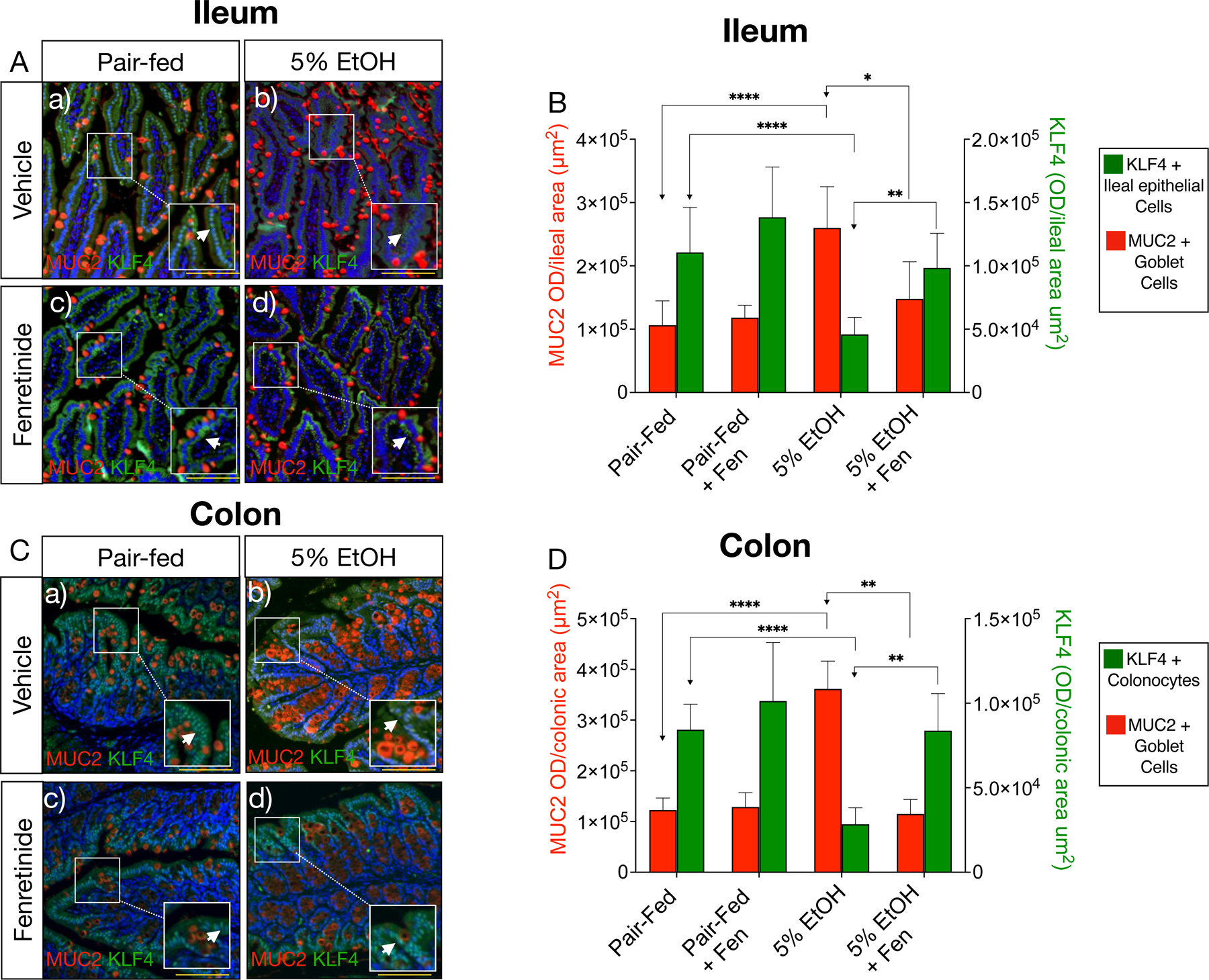
Introduction: Alcohol-induced thickening of the gut mucosal layer and increased expression of goblet cell gel-forming mucins, such as mucin-2 (MUC2) are associated with disruptions to the gut barrier in alcoholic liver disease (ALD). Interest in drugs that can target gut mucins in ALD has grown; however to date, no studies have examined the properties of drugs on expression of gut mucins in models of ALD. We previously demonstrated that at 10 mg/kg/day, the drug fenretinide (N-[4-hydroxyphenyl] retinamide [Fen]), a synthetic retinoid, mitigates alcohol-associated damage to the gut barrier and liver injury in a murine model of ALD.

Methods: In this study, we specifically sought to examine the effects of Fen on gut goblet cells, and expression of mucins, including MUC2 using a 25-day Lieber-DeCarli model of chronic alcohol intake.

Results: Our results show that chronic alcohol intake increased gut-mucosal thickening, goblet cell numbers, and mRNA and protein expression of MUC2 in both the ileum and colon. Alcohol intake was associated with marked decreases in ileal and colonic Notch signaling, levels of Notch ligands Dll1 and Dll4, and increases in the expression of Notch-associated genes indispensable for goblet cell specification, including Math1 and Spdef. Interestingly, ileal and colonic expression of KLF4, which is involved in terminal differentiation of goblet cells, was reduced in mice chronically fed alcohol. Coadministration of alcohol with Fen at 10 mg/kg/day significantly reduced alcohol-associated increases in ileal and colonic mucosal thickening, ileal Muc2, colonic Muc2, Muc5ac and Muc6 mRNAs, and goblet cell numbers. We also found that Fen strongly prevented alcohol-mediated suppression of the Notch ligand Dll1, Notch signaling, and alcohol-induced increases in expression of Notch-associated goblet cell specification genes in both the ileum and colon. In the absence of alcohol, Fen treatments alone at 10 mg/kg/day had no effects on any of the goblet cell-related endpoints.

Conclusion: These data show for the first time that the drug Fen possesses mucosal layer-modulating properties in response to chronic alcohol abuse. These data warrant further preclinical examination of Fen given the need for anti-ALD drugs and emerging evidence of a role for intestinal goblet cell mucins in the progression of ALD.

Keywords: Alcohol; Fenretinide; Goblet cells; Gut; Mucin-2.


Conflict of interest statement

Conflict of Interest Statement: The authors have no conflicts of interest to declare.

Figures

Fig. 1.
Fig. 1.. Fenretinide reduces ileal and colonic mucosal thickness and goblet cell numbers.
A) and B) Representative images of Periodic Acid Schiff (PAS), and Alcian blue stained goblet cells (yellow arrows) in the ileum, and F) and G) the colon. C) and D) Quantification of mucosal thickness (red arrows), and PAS staining in the ileum, and H) and I) the colon. E) and J) Quantification of goblet cell numbers (light blue bars), and Alcian blue optical intensity (dark blue bars) in the ileum and colon. All image magnification is ×100; scale bar = 50 μm. All data errors bars represent ±SD, with **p < 0.01, ***p < 0.001, ****p < 0.0001, ns = not significant.
Fig. 2.
Fig. 2.. Fenretinide reduces ileal and colonic mucin mRNA and protein levels.
Quantitative PCR (qPCR) histograms of A-C) ileal, and F-H) colonic relative mRNA levels of the genes Muc2, Muc5ac, and Muc6. E) and J) Representative images, and D) and I), quantification of ileal, and) colonic MUC2 immunohistochemistry (IHC). Magnification: ×100; scale bar = 100 μm. All histogram errors bars represent ±SD, with *p< 0.05, **p < 0.01, ***p < 0.001, ****p < 0.0001, ns = not significant. qPCR results represent gene fold changes relative to pair-fed mice normalized against the housekeeping gene 36B4.
Fig. 3.
Fig. 3.. Fenretinide prevents alcohol suppression of gut canonical Notch signaling.
Quantitative PCR (qPCR) histograms of A-D) ileal, and G-J) colonic relative mRNA levels of genes involved in canonical Notch signaling, Notch1, Notch2, Hes1 and Hey1. E) and K) Representative images, and F) and L) quantification of ileal, and colonic Notch intra-cellular domain (NICD) immunohistochemistry (IHC) (yellow arrows). Magnification: ×200; scale bar = 100 μm. All histogram errors bars represent ±SD, with *p< 0.05, **p < 0.01, ***p < 0.001, ****p < 0.0001, ns = not significant. qPCR results represent gene fold changes relative to pair-fed mice normalized against the housekeeping gene 36B4.
Fig. 4.
Fig. 4.. Fenretinide opposes alcohol-mediated increases in gut goblet specification genes.
Quantitative PCR (qPCR) histograms of A-F) ileal, and I-O) colonic relative mRNA levels of the goblet specification genes Creb3l1, Clca1, Spdef, Agr2, Klf4. G) and P) Representative images, and H) and Q) quantification of ileal, and colonic SPDEF (SAM Pointed Domain Containing ETS Transcription Factor). Magnification: ×200; scale bar = 100 μm. All histogram errors bars represent ±SD, with *p< 0.05, **p < 0.01, ***p < 0.001, ****p < 0.0001, ns = not significant. qPCR results represent gene fold changes relative to pair-fed mice normalized against the housekeeping gene 36B4.
Fig. 5.
Fig. 5.. Fenretinide increases ileal and colonic KLF4 positive epithelial cells.
A) and C) Representative double immunofluorescence microscopy (IFM) images of ileal, and colonic tissue double stained with antibodies against MUC2 (red channel) and KLF4 (green channel, white arrows). Magnification: ×100 (inset 200X), scale bar = 100 μm; B) and D) Quantification of ileal and colonic IFM optical intensity for MUC2 (red bars) and KLF4 (green bars). All data error bars represent ±SD, with **p < 0.01, ****p < 0.0001 and ns = not significant.

Similar articles

Cited by

References

    1. Bode C, Bode JC. Effect of alcohol consumption on the gut. Best Pract Res Clin Gastroenterol 2003;17(4):575–92. - PubMed
    1. Osna NA, Donohue TM, Kharbanda KK. Alcoholic Liver Disease: Pathogenesis and Current Management. Alcohol Res 2017;38(2):147–61. - PMC - PubMed
    1. Rao R Endotoxemia and gut barrier dysfunction in alcoholic liver disease. Hepatology 2009;50(2):638–44. - PMC - PubMed
    1. Cornick S, Tawiah A, Chadee K. Roles and regulation of the mucus barrier in the gut. Tissue Barriers 2015;3(1–2):e982426. - PMC - PubMed
    1. Hartmann P, Chen P, Wang HJ, Wang L, McCole DF, Brandl K, et al. Deficiency of intestinal mucin-2 ameliorates experimental alcoholic liver disease in mice. Hepatology 2013;58(1):108–19. - PMC - PubMed