. 2022 May 12;13(5):451.
doi: 10.1038/s41419-022-04914-6.

UBR5 targets tumor suppressor CDC73 proteolytically to promote aggressive breast cancer

Affiliations

UBR5 targets tumor suppressor CDC73 proteolytically to promote aggressive breast cancer

Gang Xiang et al. Cell Death Dis. .

Abstract

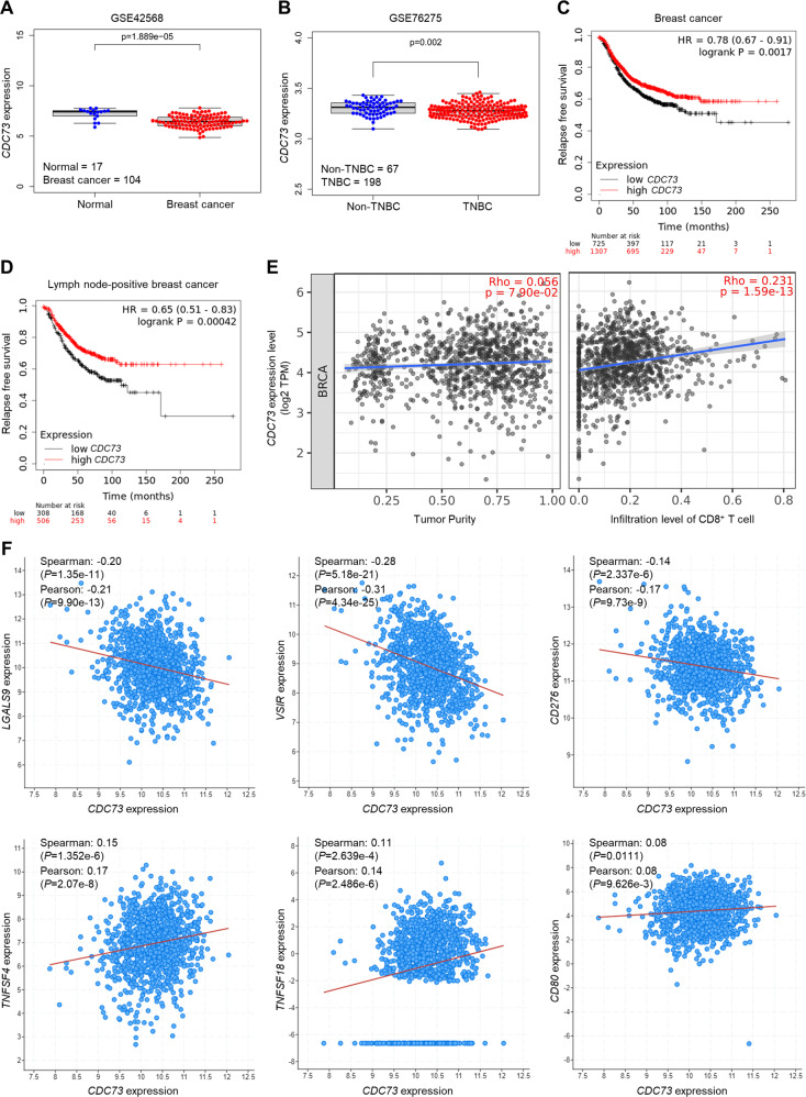
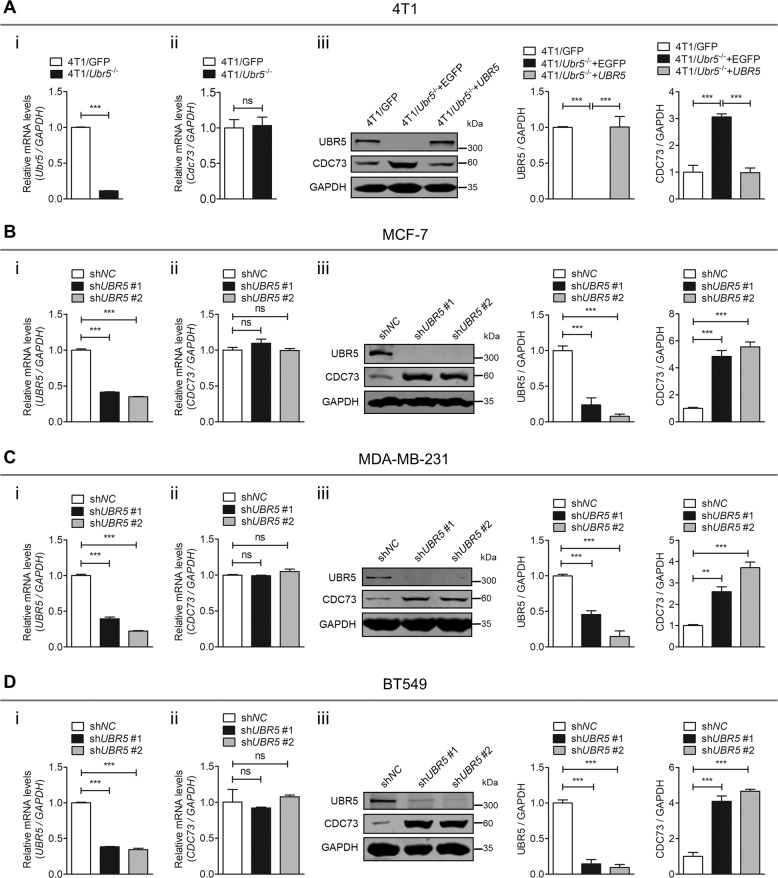
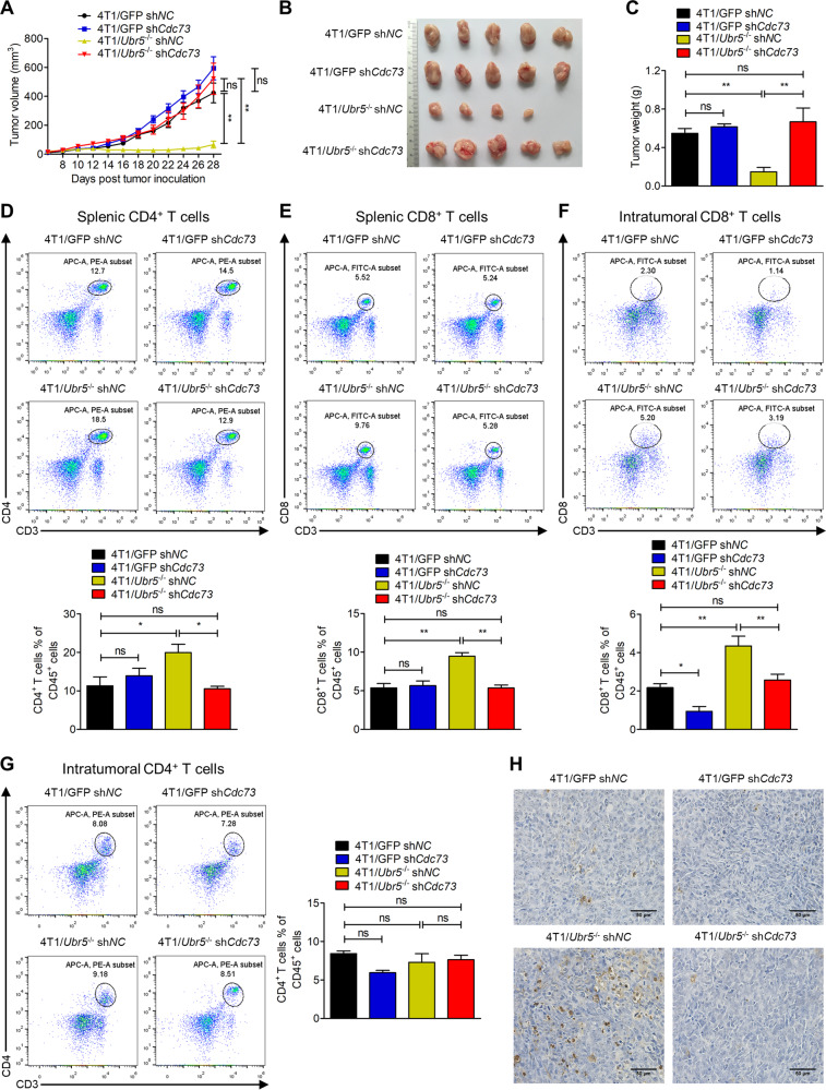
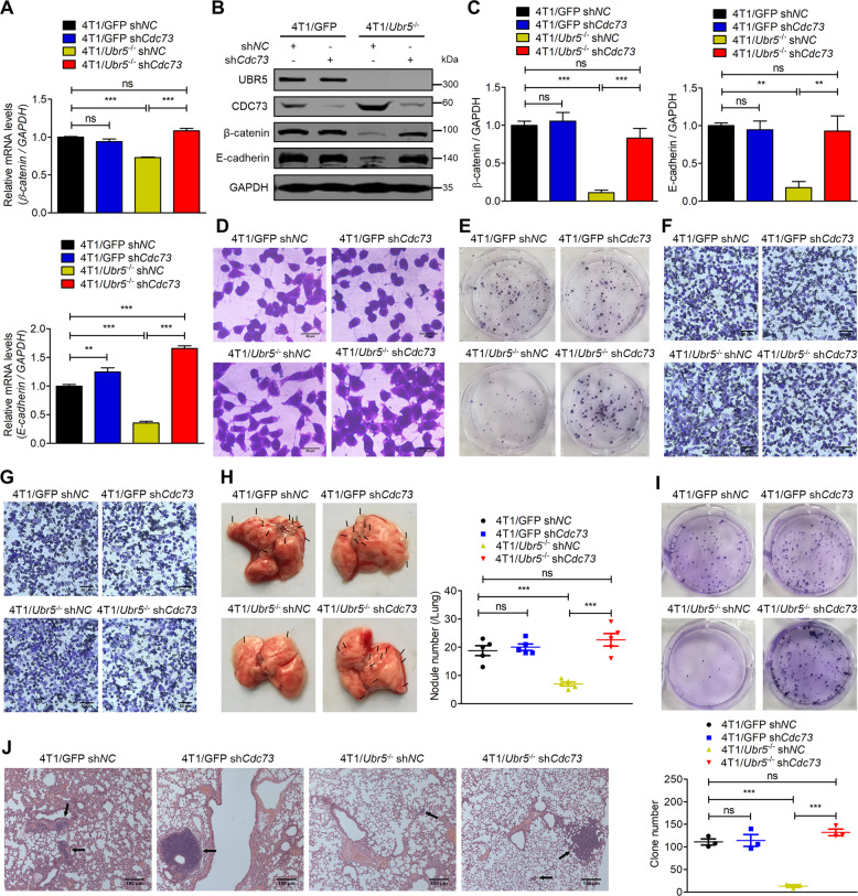
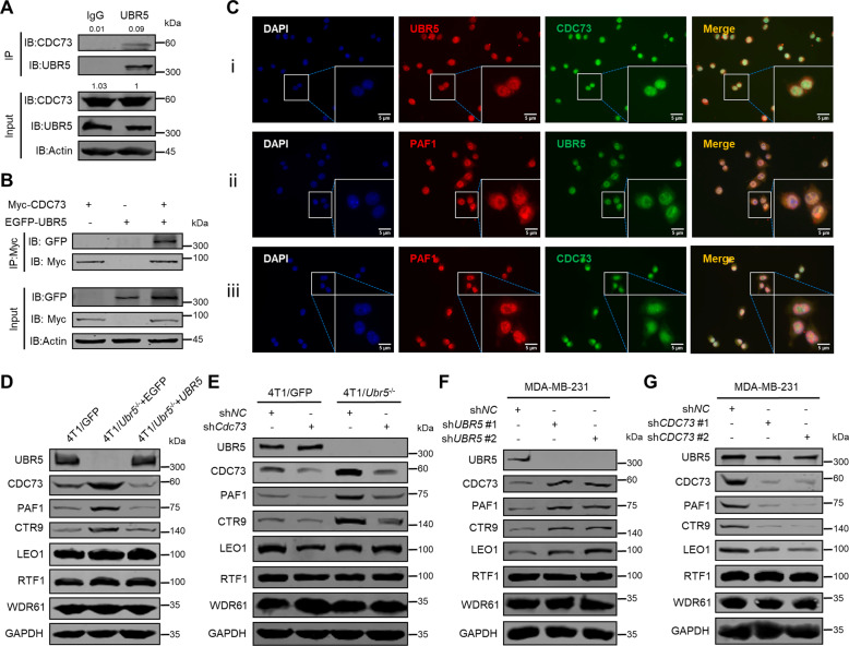
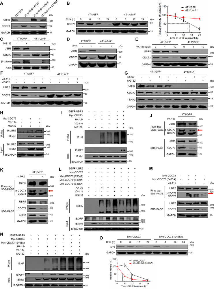
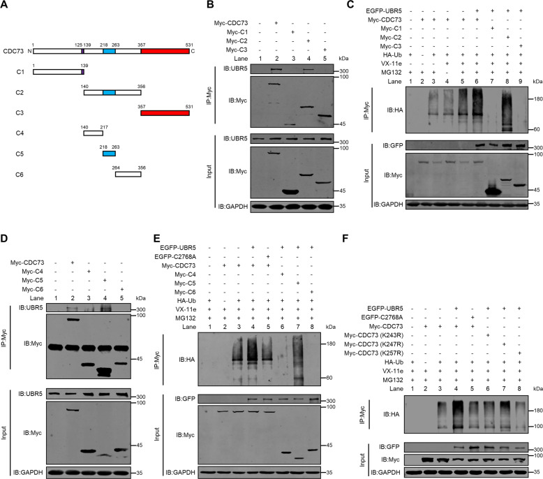
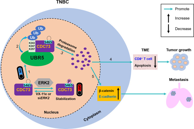
UBR5, a HECT-domain E3 ubiquitin ligase, is an attractive therapeutic target for aggressive breast cancers. Defining the substrates of UBR5 is crucial for scientific understanding and clinical intervention. Here, we demonstrate that CDC73, a component of the RNA polymerase II-associated factor 1 complex, is a key substrate that impedes UBR5's profound tumorigenic and metastatic activities in triple-negative breast cancer (TNBC) via mechanisms of regulating the expression of β-catenin and E-cadherin, tumor cell apoptosis and CD8+ T cell infiltration. Expression of CDC73 is also negatively associated with the progression of breast cancer patients. Moreover, we show that UBR5 destabilizes CDC73 by polyubiquitination at Lys243, Lys247, and Lys257 in a non-canonical manner that is dependent on the non-phosphorylation state of CDC73 at Ser465. CDC73 could serve as a molecular switch to modulate UBR5's pro-tumor activities and may provide a potential approach to developing breast cancer therapeutic interventions.


Conflict of interest statement

The authors declare no competing interests.

Figures

Fig. 1
Fig. 1. Expression of CDC73 negatively correlates with human breast cancer progression.
A The mRNA level of CDC73 in patient normal breast tissue (n = 17) and breast cancer tissue (n = 104) from the subset GSE42568. B The mRNA level of CDC73 in patient Non-TNBC tissue (n = 67) and TNBC tissue (n = 198) from the subset GSE76275. Correlations of CDC73 expression with relapse free survival of breast cancer patients (n = 2032) (C) and lymph node-positive breast cancer patients (n = 814) (D) were analyzed by using the Kaplan–Meier plotter database. E Correlations of CDC73 expression with the infiltration level of CD8+ T cells (right) in human breast cancer from TCGA BRCA cohort were analyzed by the TIMER2.0 database (n = 1100). F Correlations of the mRNA expression of CDC73 with the expression of immune response regulators LGALS9, VSIR, CD276, TNFSF4, TNFSF18, and CD80 in human breast cancer from TCGA were analyzed by using the cBioPortal database (n = 1084).
Fig. 2
Fig. 2. UBR5 inhibits the protein level of CDC73 in breast cancer cells.
A The mRNA expression of Ubr5 (i) and Cdc73 (ii) was measured by qPCR, and their protein level (iii) was analyzed by western blotting in 4T1/GFP and 4T1/Ubr5−/− cells. shNC and UBR5-knocked down MCF-7 cells (B), MDA-MB-231 cells (C), and BT549 cells (D) were analyzed for the mRNA expression of UBR5 (i) and CDC73 (ii), and their protein level (iii) by qPCR and western blotting, respectively. Data were shown as mean ± SEM for triplicates; ns not significant; **p < 0.01; ***p < 0.001.
Fig. 3
Fig. 3. CDC73 antagonizes UBR5’s tumorigenic and immunoregulatory activities.
A Tumor size was measured every 2 days in BALB/c mice injected with 5 × 105 4T1/GFP shNC, 4T1/GFP shCdc73, 4T1/Ubr5−/− shNC, and 4T1/Ubr5−/− shCdc73 cells into the mammary fat pad, respectively, and the tumor-bearing mice were sacrificed on day 28 after injection (n = 5). Representative tumor images (B) were documented and tumor weight (C) was measured on day 28. Representative FACS images and quantification of splenic CD4+ T cells (D) and splenic CD8+ T cells (E), as well as intratumoral CD8+ T cells (F) and intratumoral CD4+ T cells (G) were analyzed on day 28 (n = 3). H Representative images of TUNEL staining of the tumor sections analyzed on day 28. Data were shown as mean ± SEM; ns not significant; *p < 0.05; **p < 0.01.
Fig. 4
Fig. 4. CDC73 inhibits UBR5’s metastatic activities.
The mRNA expression (A), protein expression (B) and quantification (C) of β-catenin and E-cadherin were measured by qPCR and western blotting, respectively, in 4T1/GFP shNC, 4T1/GFP shCdc73, 4T1/Ubr5−/− shNC, and 4T1/Ubr5−/− shCdc73 cells. D Representative images of the cell morphology analyzed for 4T1/GFP shNC, 4T1/GFP shCdc73, 4T1/Ubr5−/− shNC, and 4T1/Ubr5−/− shCdc73 cells stained with 0.5% crystal violet. E Representative images of colonies of 4T1/GFP shNC, 4T1/GFP shCdc73, 4T1/Ubr5−/− shNC, and 4T1/Ubr5−/− shCdc73 cells in colony formation assay in six-well plates (100 cells/well). Representative images of transwell migration (F) and invasion (G) were analyzed for 4T1/GFP shNC, 4T1/GFP shCdc73, 4T1/Ubr5−/− shNC, and 4T1/Ubr5−/− shCdc73 cells, respectively. H Representative images of lungs in the tumor-bearing mice and the visible metastatic nodules on lung surfaces were counted. Female BALB/c mice were intravenously injected with 2 × 105 4T1/GFP shNC, 4T1/GFP shCdc73, 4T1/Ubr5−/− shNC, and 4T1/Ubr5−/− shCdc73 cells, respectively. The tumor-bearing mice were sacrificed and the lungs were harvested on day 21 (n = 5). I Metastatic tumor cells of the lungs as described in (H) were visualized and quantitated by the 6-thioguanine clonogenicity assay (n = 3). J Representative images of H&E staining of lung sections. Data were presented as mean ± SEM; ns not significant; **p < 0.01; ***p < 0.001.
Fig. 5
Fig. 5. Interaction of UBR5 with CDC73/PAF1C.
A Physical interaction of endogenous UBR5 with endogenous CDC73 was detected by co-IP with anti-UBR5 antibody in 4T1 cells which were treated with MG132 (10 μM) for 8 h before being collected for co-IP. B Physical interaction of exogenous UBR5 with exogenous CDC73 was monitored by co-IP with anti-Myc antibody in HEK293T cells transfected with empty vector, Myc-CDC73 or/and EGFP-UBR5 expressing plasmids for 40 h and then treated with MG132 (10 μM) for 8 h before being collected for co-IP. C The co-localization of endogenous UBR5 and CDC73 (i), UBR5 and PAF1 (ii), and CDC73 and PAF1 (iii) in 4T1 cells was monitored by immunofluorescence with a fluorescent microscope. The protein levels of UBR5 and the subunits of PAF1C including CDC73, PAF1, CTR9, LEO1, RTF1, and WDR61 were detected by western blotting in 4T1/GFP and 4T1/Ubr5−/− cells reconstituted with EGFP or UBR5 (D), 4T1/GFP shNC, 4T1/GFP shCdc73, 4T1/Ubr5−/− shNC, and 4T1/Ubr5−/− shCdc73 cells (E), shNC and UBR5 knockdown MDA-MB-231 cells (F), and shNC and CDC73 knockdown MDA-MB-231 cells (G).
Fig. 6
Fig. 6. Degradation of non-phosphorylated CDC73 at Ser465 by UBR5-mediated UPS.
A The protein levels of UBR5 and CDC73 in 4T1/GFP and 4T1/Ubr5−/− cells reconstituted with EGFP, UBR5, or UBR5 C2768A mutant were evaluated by western blotting. B The turnover of CDC73 in 4T1/GFP and 4T1/Ubr5−/− cells was measured by CHX treatment (50 μg/ml; 0, 6, 12 and 24 h) and then detected by western blotting and quantified by ImageJ software. The protein level of CDC73 was detected by western blotting in both 4T1/GFP and 4T1/Ubr5−/− cells treated with vehicle, MG132 (10 μM, 8 h) (C), STS (0.1 μM, 6 h) (D), VX-11e (1 μM or 10 μM, 24 h) (E), and VX-11e (10 μM, 24 h) or/and MG132 (10 μM, 8 h) (F). G The protein level of CDC73 was detected by western blotting in both 4T1/GFP and 4T1/Ubr5−/− cells transfected with control siRNA or siErk2 and treated with vehicle or MG132 (10 μM, 8 h). H Co-IP with anti-Myc antibody analyzed the physical interaction of endogenous UBR5 and exogenous CDC73 in HEK293T cells transfected with empty vector or Myc-CDC73 plasmids and treated with vehicle or VX-11e (10 μM, 24 h) in the presence of MG132 (10 μM, 8 h). I Ubiquitination assays analyzed the ubiquitination level of CDC73 modified by exogenous UBR5 in the presence of vehicle, VX-11e (10 μM, 24 h) or/and MG132 (10 μM, 8 h) under denaturing conditions in HEK293T cells transfected with the indicated plasmids. The phosphorylation levels of UBR5 and CDC73 were analyzed via the Phos-tag SDS-PAGE in 4T1 cells treated with vehicle or VX-11e (10 µM, 24 h) (J) and the control and Erk2 silenced 4T1 cells (K). L Ubiquitination assays analyzed the ubiquitination level of three mutants (T334A, T359A and S465A) of CDC73 modified with exogenous UBR5. The phosphorylation (M) and ubiquitination (N) levels of WT and S465A mutant of CDC73 were detected in HEK293T cells treated with vehicle or VX-11e (10 μM, 24 h). O The turnover of WT and the S465A mutant of CDC73. HEK293T cells were transfected with Myc-CDC73 and Myc-CDC73 (S465A) plasmids for 48 h and treated with CHX as described in (B).
Fig. 7
Fig. 7. Identification of CDC73’s key residues for UBR5-mediated ubiquitination.
A The schematic diagram of the full length and truncated fragments of CDC73. Co-IP with anti-Myc antibody analyzed the physical interaction of UBR5 with the full length or truncated fragments of CDC73 C1, C2, and C3 (B), and C4, C5, and C6 (D) in HEK293T cells transfected with the indicated plasmids in the presence of MG132 (10 μM, 8 h). Ubiquitination assays analyzed the ubiquitination level of the full length of CDC73 and its truncated fragments C1, C2, and C3 (C), and C4, C5, and C6 (E) modified by exogenous UBR5 under denaturing conditions in HEK293T cells transfected with the indicated plasmids and treated with vehicle, VX-11e (10 μM, 24 h) or/and MG132 (10 μM, 8 h). F Ubiquitination assays analyzed the ubiquitination level of WT and the mutants of CDC73 (K243A, K247A, and K257A) in HEK293T cells under the same condition as described in (C) and (E).
Fig. 8
Fig. 8. The molecular switch role and mechanism of CDC73 for UBR5-drived TNBC tumor growth and metastasis.
In TNBC cells, CDC73 is stabilized by ERK2-mediated phosphorylation at Ser465. UBR5 targets non-phosphorylated CDC73 (1) and ubiquitinates it at Lys243, Lys247 and Lys257 residues (2) for degradation by UPS (3). Degradation of CDC73 promotes UBR5’s tumorigenic and immunoregulatory activities in TME (4), as well as tumor metastasis in a cell-intrinsic manner by regulating both mRNA and protein expression of β-catenin and E-cadherin (5).

Similar articles

Cited by

References

    1. Sung H, Ferlay J, Siegel RL, Laversanne M, Soerjomataram I, Jemal A, et al. Global cancer statistics 2020: GLOBOCAN estimates of incidence and mortality worldwide for 36 cancers in 185 countries. CA Cancer J Clin. 2021;71:209–49. doi: 10.3322/caac.21660. - DOI - PubMed
    1. Nunez Abad M, Calabuig-Farinas S, Lobo de Mena M, Jose Godes Sanz de Bremond M, Garcia Gonzalez C, Torres, Martinez S, et al. Update on systemic treatment in early triple negative breast cancer. Ther Adv Med Oncol. 2021;13:1758835920986749. doi: 10.1177/1758835920986749. - DOI - PMC - PubMed
    1. Siegel RL, Miller KD, Fuchs HE, Jemal A. Cancer statistics, 2021. CA Cancer J Clin. 2021;71:7–33. doi: 10.3322/caac.21654. - DOI - PubMed
    1. Callaghan MJ, Russell AJ, Woollatt E, Sutherland GR, Sutherland RL, Watts CK. Identification of a human HECT family protein with homology to the Drosophila tumor suppressor gene hyperplastic discs. Oncogene. 1998;17:3479–91. doi: 10.1038/sj.onc.1202249. - DOI - PubMed
    1. Liao L, Song M, Li X, Tang L, Zhang T, Zhang L, et al. E3 ubiquitin ligase UBR5 drives the growth and metastasis of triple-negative breast cancer. Cancer Res. 2017;77:2090–101. doi: 10.1158/0008-5472.CAN-16-2409. - DOI - PubMed

Publication types

MeSH terms