

Circulating neurofilament is linked with morbid obesity, renal function, and brain density
- PMID: 35551210
- PMCID: PMC9098484
- DOI: 10.1038/s41598-022-11557-2
Circulating neurofilament is linked with morbid obesity, renal function, and brain density
Abstract
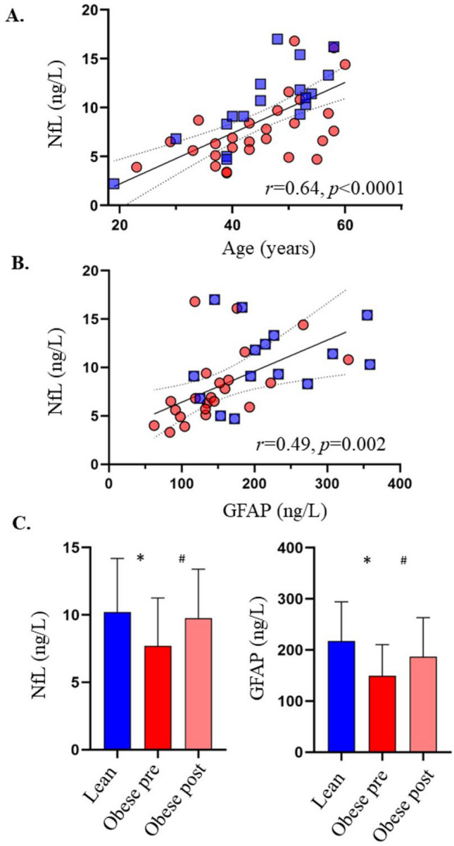
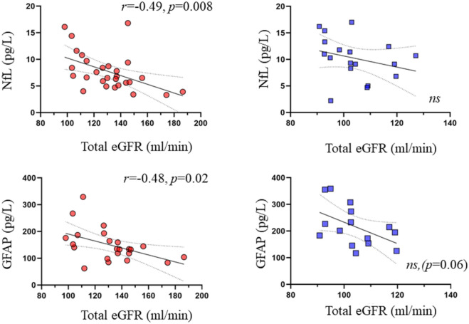
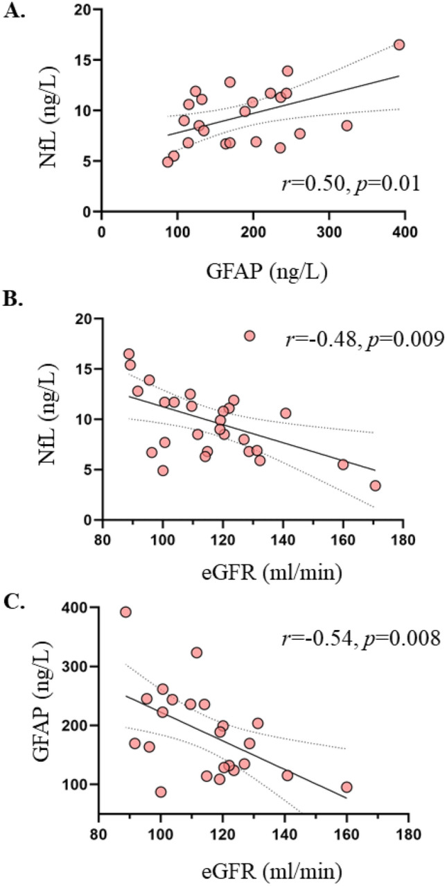
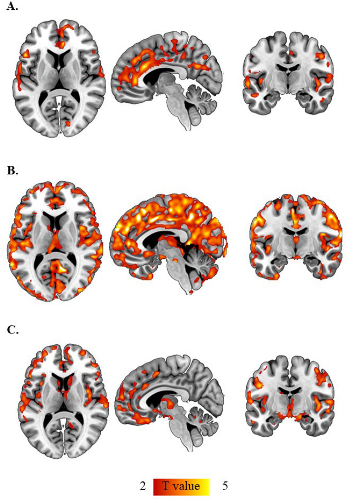
Neurofilament light chain (NfL) is a novel biomarker reflecting neuroaxonal damage and associates with brain atrophy, and glial fibrillary acidic protein (GFAP) is a marker of astrocytic activation, associated with several neurodegenerative diseases. Since obesity is associated with increased risk for several neurodegenerative disorders, we hypothesized that circulating NfL and GFAP levels could reflect neuronal damage in obese patients. 28 morbidly obese and 18 lean subjects were studied with voxel based morphometry (VBM) MRI to assess gray and white matter densities. Serum NfL and GFAP levels were determined with single-molecule array. Obese subjects were re-studied 6 months after bariatric surgery. Morbidly obese subjects had lower absolute concentrations of circulating NfL and GFAP compared to lean individuals. Following bariatric surgery-induced weight loss, both these levels increased. Both at baseline and after weight loss, circulating NfL and GFAP values correlated inversely with eGFR. Cross-sectionally, circulating NfL levels correlated inversely with gray matter (GM) density, and this association remained significant also when accounting for age and total eGFR. GFAP values did not correlate with GM density. Our data suggest that when determining circulating NfL and GFAP levels, eGFR should also be measured since renal function can affect these measurements. Despite the potential confounding effect of renal function on NfL measurement, NfL correlated inversely with gray matter density in this group of subjects with no identified neurological disorders, suggesting that circulating NfL level may be a feasible biomarker of cerebral function even in apparently neurologically healthy subjects.
© 2022. The Author(s).
Conflict of interest statement
The authors declare no competing interests.
Figures
Similar articles
-
Lower White Matter Volume and Worse Executive Functioning Reflected in Higher Levels of Plasma GFAP among Older Adults with and Without Cognitive Impairment.J Int Neuropsychol Soc. 2022 Jul;28(6):588-599. doi: 10.1017/S1355617721000813. Epub 2021 Jun 22. J Int Neuropsychol Soc. 2022. PMID: 34158138 Free PMC article.
-
The relationship between serum neurofilament light chain and glial fibrillary acidic protein with the REM sleep behavior disorder subtype of Parkinson's disease.J Neurochem. 2023 Apr;165(2):268-276. doi: 10.1111/jnc.15780. Epub 2023 Feb 24. J Neurochem. 2023. PMID: 36776136
-
Association of serum neurofilament light chain and glial fibrillary acidic protein levels with cognitive decline in Parkinson's disease.Brain Res. 2023 Apr 15;1805:148271. doi: 10.1016/j.brainres.2023.148271. Epub 2023 Feb 7. Brain Res. 2023. PMID: 36754139
-
Neurofilament light chain as a biomarker in neuromyelitis optica spectrum disorder: a comprehensive review and integrated analysis with glial fibrillary acidic protein.Neurol Sci. 2024 Mar;45(3):1255-1261. doi: 10.1007/s10072-023-07277-8. Epub 2023 Dec 23. Neurol Sci. 2024. PMID: 38141119 Review.
-
Neurofilament light and glial fibrillary acidic protein in mood and anxiety disorders: A systematic review and meta-analysis.Brain Behav Immun. 2025 Jan;123:1091-1102. doi: 10.1016/j.bbi.2024.11.001. Epub 2024 Nov 5. Brain Behav Immun. 2025. PMID: 39510417
Cited by
-
Association of Plasma Biomarkers of Alzheimer Disease With Cognition and Medical Comorbidities in a Biracial Cohort.Neurology. 2023 Oct 3;101(14):e1402-e1411. doi: 10.1212/WNL.0000000000207675. Epub 2023 Aug 14. Neurology. 2023. PMID: 37580163 Free PMC article.
-
Sociodemographic patterns in biomarkers of aging in the Add Health cohort.Biodemography Soc Biol. 2024 Apr-Jun;69(2):57-74. doi: 10.1080/19485565.2024.2334687. Epub 2024 Mar 29. Biodemography Soc Biol. 2024. PMID: 38551453 Free PMC article.
-
Associations between blood selenium and serum neurofilament light chain: results of a nationwide survey.Front Neurol. 2025 Apr 8;16:1490760. doi: 10.3389/fneur.2025.1490760. eCollection 2025. Front Neurol. 2025. PMID: 40264648 Free PMC article.
-
Exploring the Link Between Renal Function Fluctuations Within the Physiological Range and Serum/CSF Levels of NfL, GFAP, tTAU, and UCHL1.Int J Mol Sci. 2025 Jan 17;26(2):748. doi: 10.3390/ijms26020748. Int J Mol Sci. 2025. PMID: 39859463 Free PMC article.
-
Plasma NfL and GFAP for predicting VCI and related brain changes in community and clinical cohorts.Alzheimers Dement. 2025 Jun;21(6):e70381. doi: 10.1002/alz.70381. Alzheimers Dement. 2025. PMID: 40566826 Free PMC article.
References
Publication types
MeSH terms
Substances
LinkOut - more resources
Full Text Sources
Medical
Research Materials
Miscellaneous
