. 2022 Oct 21;145(10):3472-3487.
doi: 10.1093/brain/awac176.

Lipid pathway dysfunction is prevalent in patients with Parkinson's disease

Affiliations

Lipid pathway dysfunction is prevalent in patients with Parkinson's disease

Jasmin Galper et al. Brain. .

Abstract

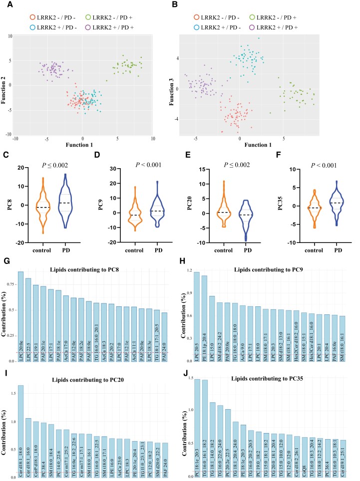
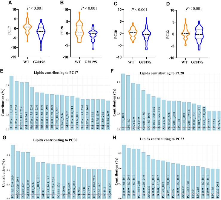
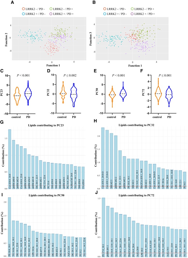
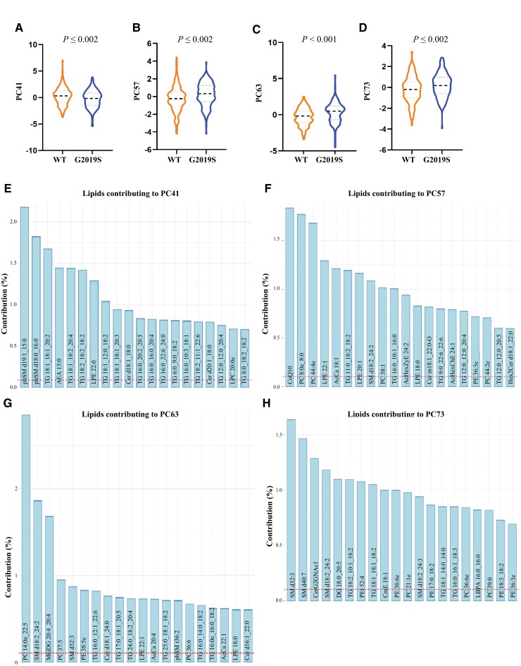
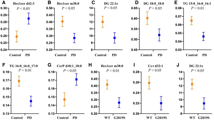
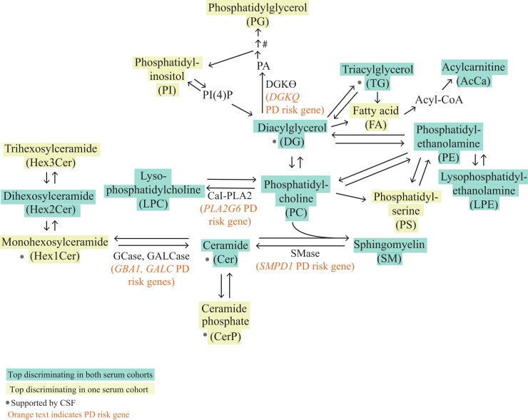
Many genetic risk factors for Parkinson's disease have lipid-related functions and lipid-modulating drugs such as statins may be protective against Parkinson's disease. Moreover, the hallmark Parkinson's disease pathological protein, α-synuclein, has lipid membrane function and pathways dysregulated in Parkinson's disease such as the endosome-lysosome system and synaptic signalling rely heavily on lipid dynamics. Despite the potential role for lipids in Parkinson's disease, most research to date has been protein-centric, with large-scale, untargeted serum and CSF lipidomic comparisons between genetic and idiopathic Parkinson's disease and neurotypical controls limited. In particular, the extent to which lipid dysregulation occurs in mutation carriers of one of the most common Parkinson's disease risk genes, LRRK2, is unclear. Further, the functional lipid pathways potentially dysregulated in idiopathic and LRRK2 mutation Parkinson's disease are underexplored. To better determine the extent of lipid dysregulation in Parkinson's disease, untargeted high-performance liquid chromatography-tandem mass spectrometry was performed on serum (n = 221) and CSF (n = 88) obtained from a multi-ethnic population from the Michael J. Fox Foundation LRRK2 Clinical Cohort Consortium. The cohort consisted of controls, asymptomatic LRRK2 G2019S carriers, LRRK2 G2019S carriers with Parkinson's disease and Parkinson's disease patients without a LRRK2 mutation. Age and sex were adjusted for in analyses where appropriate. Approximately 1000 serum lipid species per participant were analysed. The main serum lipids that distinguished both Parkinson's disease patients and LRRK2 mutation carriers from controls included species of ceramide, triacylglycerol, sphingomyelin, acylcarnitine, phosphatidylcholine and lysophosphatidylethanolamine. Significant alterations in sphingolipids and glycerolipids were also reflected in Parkinson's disease and LRRK2 mutation carrier CSF, although no correlations were observed between lipids identified in both serum and CSF. Pathway analysis of altered lipid species indicated that sphingolipid metabolism, insulin signalling and mitochondrial function were the major metabolic pathways dysregulated in Parkinson's disease. Importantly, these pathways were also found to be dysregulated in serum samples from a second Parkinson's disease cohort (n = 315). Results from this study demonstrate that dysregulated lipids in Parkinson's disease generally, and in LRRK2 mutation carriers, are from functionally and metabolically related pathways. These findings provide new insight into the extent of lipid dysfunction in Parkinson's disease and therapeutics manipulating these pathways may be beneficial for Parkinson's disease patients. Moreover, serum lipid profiles may be novel biomarkers for both genetic and idiopathic Parkinson's disease.

Keywords: LRRK2; Parkinson’s disease; biomarker; lipid; sphingolipid.


Figures

Figure 1
Figure 1
Serum lipid profiles discriminate Parkinson’s disease patients from controls in a multi-ethnic cohort. Using linear discriminant analysis (LDA) on identified principal components, three canonical functions were generated that could significantly discriminate the groups. (A) A scatterplot of the first two functions clearly showed separation between the control and Parkinson’s disease groups and between the LRRK2 mutation and non-mutation Parkinson’s disease groups. (B) A scatterplot of the first and third functions shows separation of all four groups. (C) To determine which lipids may contribute to the discrimination of Parkinson’s disease patients from controls independent of LRRK2 G2019S mutation, LDA using identified principal components was performed and the results indicated control (controls plus LRRK2 carriers without Parkinson’s disease) and Parkinson’s disease (LRRK2 and non-LRRK2 Parkinson’s disease) groups were distinguishable. Multivariate analysis of variance covarying for age and sex identified four principal components, (C) PC8, (D) PC9, (E) PC20 and (F) PC35. (G–J) The top 20 lipid species that contributed to the four principal components discriminating Parkinson’s disease from controls. The dashed red line represents the expected value if the contribution of lipids were uniform. n = 221.
Figure 2
Figure 2
Serum lipid profiles discriminate LRRK2 G2019S carriers from non-LRRK2 G2019S carriers in a multi-ethnic cohort. To determine which lipids may contribute to the discrimination of LRRK2 G2019S mutation carriers from non-LRRK2 G2019S carriers, linear discriminant analysis (LDA) was performed to identify the principal components that significantly differed between these two groups. The LRRK2 mutation group consisted of both asymptomatic carriers and manifesting Parkinson’s disease patients, while the non-LRRK2 mutation group consisted of control and idiopathic Parkinson’s disease patients. LRRK2 mutation carriers could be significantly discriminated from non-LRRK2 mutation carriers. (A–E) Multivariate ANOVA covarying for age and sex identified four principal components, (B) PC17, (C) PC28, (D) PC30 and (E) PC32, were significantly different between LRRK2 and non-LRRK2 mutation groups. (F–H) The top 20 lipid species that contributed to the four principal components discriminating LRRK2 and non-LRRK2 mutation groups. The dashed red line represents the expected value if the contribution of lipids were uniform. n = 221. WT = wild-type.
Figure 3
Figure 3
Serum lipid profiles discriminate Parkinson’s disease patients from controls in the LRRK2 Ashkenazi Jewish cohort. To determine if lipid profiles could distinguish between groups in a second cohort, linear discriminant analysis (LDA) on identified principal components was performed. It was revealed that the three canonical functions generated significantly discriminated the groups. (A) A scatterplot of the first two functions clearly showed separation between the control and Parkinson’s disease groups, and between the LRRK2 mutation and non-mutation Parkinson’s disease groups. (B) A scatterplot of the first and third functions shows separation between Parkinson’s disease groups. To determine lipids that may contribute to the discrimination of Parkinson’s disease patients from controls independent of LRRK2 G2019S mutation, controls (including LRRK2 carriers without Parkinson’s disease) were compared to a Parkinson’s disease group (LRRK2 and non-LRRK2 Parkinson’s disease). (CF) Multivariate ANOVA covarying for age and sex identified four principal components, (C) PC23, (D) PC32, (E) PC50 and (F) PC72, which remained significantly different between the two groups. (GJ) The top 20 lipid species that contributed to the top four principal components that significantly distinguished between controls and Parkinson’s disease. The dashed red line represents the expected value if the contribution of lipids were uniform. n = 315.
Figure 4
Figure 4
Serum lipid profiles discriminate LRRK2 G2019S carriers from non-LRRK2 G2019S carriers in the LRRK2 Ashkenazi Jewish Cohort. Multivariate ANOVA covarying for age and sex identified four principal components, (A) PC41, (B) PC57, (C) PC63 and (D) PC73, that were significantly different between the LRRK2 mutation and non-mutation groups. (EH) The top 20 lipid species that contributed to the top four components which significantly distinguished between LRRK2 mutation and non-mutation groups. The dashed red line represents the expected value if the contribution of lipids were uniform. n = 315. WT = wild-type.
Figure 5
Figure 5
Sphingolipids and glycerolipids and altered in multi-ethnic LRRK2 cohort CSF. Two-factor multivariate analysis covarying for age and sex revealed a significant effect of both Parkinson’s disease and LRRK2 G2019S mutation (both P < 0.05) on CSF lipid profiles. (AG) Post hoc analysis identified sphingolipids and glycerolipids were different between Parkinson’s disease patient and control CSF samples. (HJ) Glycerolipid and sphingolipid species were significantly different between LRRK2 G2019S mutation carriers and non-carriers. n = 88. WT = wild-type.
Figure 6
Figure 6
Implicated lipids are metabolically linked in Parkinson’s disease risk gene pathways. Pathway analysis was performed to generate a metabolic map of the main lipid classes implicated in the discrimination of Parkinson’s disease patients and LRRK2 carriers from controls. The resulting map indicated that most implicated lipids were highly integrated and were in metabolic pathways that included enzymes encoded by Parkinson’s disease risk genes (GBA1, GALC, SMPD1, PLA2G6 and DGKQ). GCase = glucocerebrosidase; GALCase = galactoceramidase; SMase = sphingomyelinase; CaI-PLA2 = 85/88 kDa calcium-independent phospholipase A2; PA = phosphatidic acid; PI(4)P = phosphatidylinositol-4-phosphate. #PG is formed from PA in a three-step reaction pathway.

References

    1. Aharon-Peretz J, Rosenbaum H, Gershoni-Baruch R. Mutations in the glucocerebrosidase gene and Parkinson’s disease in Ashkenazi Jews. N Engl J Med. 2004;351(19):1972–1977. - PubMed
    1. Gan-Or Z, Ozelius LJ, Bar-Shira A, et al. . The p.L302P mutation in the lysosomal enzyme gene SMPD1 is a risk factor for Parkinson disease. Neurology. 2013;80(17):1606–1610. - PMC - PubMed
    1. Chang D, Nalls MA, Hallgrímsdóttir IB, et al. . A meta-analysis of genome-wide association studies identifies 17 new Parkinson’s disease risk loci. Nat Genet. 2017;49(10):1511–1516. - PMC - PubMed
    1. de Carvalho Guimarães B, Valente Pereira AC, da Costa Rodrigues F, et al. . Glucocerebrosidase N370S and L444P mutations as risk factors for Parkinson’s disease in Brazilian patients. Parkinsonism Relat Disord. 2012;18(5):688–689. - PubMed
    1. Simón-Sánchez J, Schulte C, Bras JM, et al. . Genome-wide association study reveals genetic risk underlying Parkinson’s disease. Nat Genet. 2009;41(12):1308–1312. - PMC - PubMed

Publication types