. 2022 Nov 25;43(6):1051-1073.
doi: 10.1210/endrev/bnac012.

Update on Biology and Genomics of Adrenocortical Carcinomas: Rationale for Emerging Therapies

Affiliations

Update on Biology and Genomics of Adrenocortical Carcinomas: Rationale for Emerging Therapies

Antonio Marcondes Lerario et al. Endocr Rev. .

Abstract

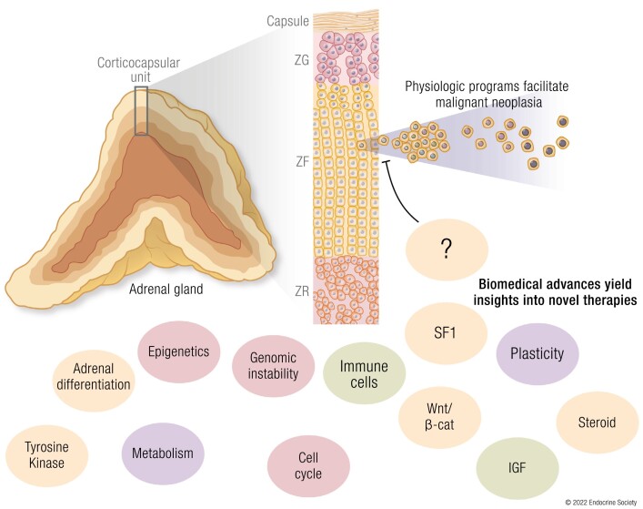
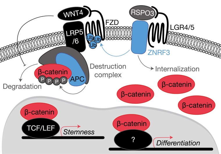
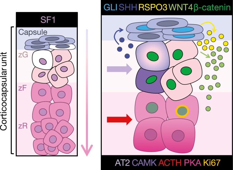
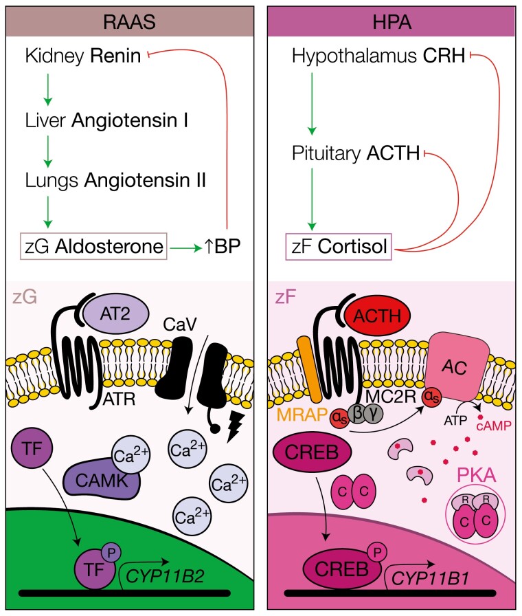
The adrenal glands are paired endocrine organs that produce steroid hormones and catecholamines required for life. Adrenocortical carcinoma (ACC) is a rare and often fatal cancer of the peripheral domain of the gland, the adrenal cortex. Recent research in adrenal development, homeostasis, and disease have refined our understanding of the cellular and molecular programs controlling cortical growth and renewal, uncovering crucial clues into how physiologic programs are hijacked in early and late stages of malignant neoplasia. Alongside these studies, genome-wide approaches to examine adrenocortical tumors have transformed our understanding of ACC biology, and revealed that ACC is composed of distinct molecular subtypes associated with favorable, intermediate, and dismal clinical outcomes. The homogeneous transcriptional and epigenetic programs prevailing in each ACC subtype suggest likely susceptibility to any of a plethora of existing and novel targeted agents, with the caveat that therapeutic response may ultimately be limited by cancer cell plasticity. Despite enormous biomedical research advances in the last decade, the only potentially curative therapy for ACC to date is primary surgical resection, and up to 75% of patients will develop metastatic disease refractory to standard-of-care adjuvant mitotane and cytotoxic chemotherapy. A comprehensive, integrated, and current bench-to-bedside understanding of our field's investigations into adrenocortical physiology and neoplasia is crucial to developing novel clinical tools and approaches to equip the one-in-a-million patient fighting this devastating disease.

Keywords: adrenocortical carcinoma; adrenocortical development and homeostasis; genomics; molecular biomarkers; targeted therapies.


Figures

Graphical Abstract
Graphical Abstract
Figure 1.
Figure 1.
Recurrent somatic alterations activate Wnt/β-catenin signaling in adrenocortical neoplasia. Benign and malignant tumors of the adrenal cortex harbor frequent somatic alterations leading to constitutive activation of the Wnt/β-catenin signaling pathway, classically culminating in high expression of a β-catenin and TCF/LEF-driven stemness program facilitating tumor growth. Activation of this pathway is regulated at several levels, primarily through availability of Wnt receptors (Frizzled receptors, FZD) and stability of β-catenin. Membrane availability of FZDs is regulated by R-spondins (in the adrenal cortex, RSPO3) and E3 ubiquitin ligases such as ZNRF3. In the absence of RSPO3, ZNRF3 ubiquitinates FZDs, targeting these receptors for internalization and degradation. When RSPO3 binds its receptors (in the adrenal cortex, these are LGR4/5), the ZNRF3/LGR/RSPO complex is internalized, permitting the activation of FZDs by Wnt ligands (in the adrenal cortex, this is WNT4). β-catenin stability is regulated by the destruction complex, a large multiprotein complex containing classical tumor suppressor APC. The destruction complex phosphorylates cytoplasmic β-catenin and targets it for degradation. When Wnt ligands such as WNT4 bind FZD receptors, the destruction complex is localized to the cell membrane and can no longer efficiently target β-catenin for degradation. Intracellular β-catenin therefore accumulates and translocates to the nucleus to drive its transcriptional programs. Importantly, in ACC, activating mutations in this pathway are associated with a higher degree of adrenal differentiation, suggesting that β-catenin may engage a herein unknown transcription factor to drive expression of a genome-wide differentiation program. Signaling components encoded by genes targeted for somatic gain of function (GOF) alterations are depicted in red (β-catenin, recurrent mutations prevent phosphorylation), and somatic loss of function (LOF) alterations are depicted in blue (APC, and ZNRF3).
Figure 2.
Figure 2.
Paracrine and endocrine signaling programs support homeostasis and renewal in the adrenal corticocapsular unit. Shown left, stem and progenitor cells (white) residing in the capsule or subcapsular cortex (histological zG) may be deployed for cortical renewal in response to physiologic homeostatic and endocrine demands. Differentiation is centripetal (as indicated by the arrow), and lower zR cells (humans) or lower zF cells (mice, which do not possess a zR) are terminally differentiated and undergo apoptosis at the boundary between the cortex and the medulla. Detailed in panel right, this process is regulated by interplay between capsule- and cortex-derived paracrine factors and systemic endocrine regulators, which together coordinate stem/progenitor cell maintenance, anatomic and functional zonation, lineage conversion, and steroidogenesis. Sonic hedgehog (SHH, dark blue) produced by zG cells centrifugally activates GLI family transcription factors in the capsule (cyan), which drive the expression of RSPO3 (yellow), an essential positive regulator of Wnt signaling in the cortex. Wnt-responsive cells in the cortex (possessing nuclear β-catenin, dark green) produce WNT4 (light green), further perpetuating Wnt signaling throughout the lower zG and upper zF and maintaining SHH expression throughout the zG. Endocrine signaling, through angiotensin II/calmodulin kinase (AT2/CAMK) and adrenocorticotropic hormone/protein kinase A (ACTH/PKA), establish discrete differentiation states required for zone-specific steroidogenesis. Importantly, cells at the zG-zF boundary possess mitotic activity (Ki67, gold) and represent a “transit-amplifying” population that can rapidly expand in response to ACTH. Despite responding to a zF endocrine cue, transit amplification of this population also requires intact Wnt/β-catenin signaling. This current model of adrenocortical homeostasis is supported by numerous studies, as detailed in this review.
Figure 3.
Figure 3.
Endocrine feedback loops and cellular mechanisms supporting aldosterone and cortisol production. The renin-angiotensin-aldosterone system (RAAS) and the hypothalamus-pituitary-adrenal (HPA) axis are the endocrine feedback loops that regulate aldosterone and cortisol production, respectively. These feedback loops are activated by distinct physiologic demands and target diverse cell populations in different zones of the cortex according to their differentiation state (and therefore the expression of hormone receptors). Shown left, the angiotensin II receptor (ATR) is expressed by zG cells. On angiotensin II (ATII or AT2) binding to ATR, membrane depolarization occurs, leading to opening of voltage-gated calcium channel CaV, triggering a calcium-dependent intracellular signaling cascade that activates calmodulin kinase (CAMK), and subsequently transcription factors that drive expression of critical regulators of aldosterone production such as aldosterone synthase, encoded by CYP11B2. Shown right, the adrenocorticotropin (ACTH) receptor MC2R and its accessory protein, MRAP, are expressed by zF cells. On binding of ACTH to MC2R, the Gαs subunit dissociates and activates adenylyl cyclase (AC), which triggers cellular accumulation of cyclic adenosine 5′-monophosphate (cAMP), and dissociation of the protein kinase A (PKA) tetramer with liberation of the catalytic subunits. These subunits phosphorylate and activate cAMP response element binding protein (CREB), enabling transcription of machinery required for glucocorticoid synthesis, for example CYP11B1. Not shown, this signaling program is extinguished by intracellular phosphodiesterases.
Figure 4.
Figure 4.
Adrenocortical carcinoma (ACC) is composed of 3 homogeneous molecular subtypes associated with distinct clinical outcomes. Multiplatform profiling in ACC-TCGA (23) revealed that ACC is composed of 3 molecular subtypes: COC1, COC2, and COC3. COC1 ACC is associated with favorable clinical outcomes (few recurrences and deaths in this group, longest event-free and overall survival), COC2 is associated with intermediate outcomes, and COC3 is associated with dismal clinical outcomes (accounting for up to 40% of all ACC but nearly 70% of recurrences and more than half of deaths) (4, 23). COC2-COC3 ACC are associated with clinically significant cortisol production. On a molecular level, virtually all ACC is characterized by loss of imprinting (LOI) leading to constitutive expression of IGF2; however, COC1-COC3 possess distinct somatic alteration profiles and differential immune infiltration, expression of adrenal differentiation score (ADS) and methylation of CpG islands (CpGi). COC1 ACC possess a higher degree of immune infiltration, lower ADS, minimal CpGi methylation, no recurrent driver somatic alterations, and a chromosomal somatic copy number alteration (SCNA) profile. COC2 ACC also possess a chromosomal SCNA profile. COC2-COC3 ACC are characterized by frequent driver somatic alterations leading to constitutive activation of the Wnt pathway. COC2-COC3 ACC also possess higher ADS (with COC3 ACC at the higher end of this spectrum), suggesting that Wnt pathway activation in these tumors facilitates steroidogenesis. COC3 ACC possess the highest degree of cell cycle activation, enriched for driver alterations promoting constitutive cell cycle activation, and possess a noisy SCNA profile. COC2 ACC possess intermediate levels of CpGi methylation (CIMP-int) while COC3 ACC possess high levels of CpGi methylation (CIMP-high), suggesting that these classes of ACC are characterized by profound disruption in epigenetic patterning. Example event-free survival curves adapted from ACC-TCGA are depicted left. Molecular features are depicted right in the curves (top panel), theoretical heat map (middle panel, columns represent patients; light gray squares indicate “null values,” eg, no recurrence, death, mutation, cortisol production or a quiet SCNA profile, while colored squares indicate the presence of the abnormality), and pseudomicroscope images depict tumor genetic, epigenetic, and cell type heterogeneity (bottom panel).
Figure 5.
Figure 5.
Putative hyperplasia to carcinoma sequence originating from zG-zF boundary cells in COC2-COC3 ACC. COC2-COC3 adrenocortical carcinoma (ACC) are characterized by active Wnt/β-catenin signaling, high levels of adrenal differentiation (measured by ADS), and profound epigenetic rewiring (possessing intermediate and high levels of CpG island methylation genome-wide; ie, CIMP-int and CIMP-high signatures) (23). Intriguingly, despite the well-established role of β-catenin in supporting zG differentiation in physiology, COC2-COC3 tumors also frequently produce glucocorticoids (cortisol) (23). Recent mouse models of adrenocorticotropin (ACTH)-driven zF renewal (32), expanded Wnt/β-catenin signaling driven by ZNRF3 deficiency (65), sustained proliferation triggered by adrenocortical expression of the SV40 Large T antigen (106), or combined simultaneous Wnt/β-catenin and cell cycle activation (107) also demonstrate a unique interplay between Wnt/β-catenin and ACTH/PKA signaling in enabling proliferation of cells residing in the zG-zF boundary. Taken together, these studies support the existence of a small population of cells in the zG-zF boundary that are capable of rapid proliferation in response to sustained Wnt/β-catenin and/or ACTH signaling. We postulate that COC2-COC3 ACC arise from this vulnerable population through recurrent genetic events that drive hyperplasia and malignant transformation (eg, activating alterations in the Wnt pathway and/or driver alterations leading to constitutive cell cycle activation). Given the relatively homogeneous abnormal epigenetic patterns in these 2 groups of tumors, and recent studies suggesting that metastatic ACC do not acquire novel recurrent genetic events (245, 246), we speculate that tumor growth during and after transformation as well as metastatic dissemination are facilitated by epigenetic reprogramming and/or private genetic events.
Figure 6.
Figure 6.
Pleiotropic, oncogenic actions of steroidogenesis factor 1 (SF1) in ACC. High expression of NR5A1 (or its gene product, SF1) is retained in adrenocortical neoplasia, through recurrent genetic and/or alternative noncoding mechanisms (23, 108, 109). Studies using in vitro models bearing endogenous or enforced high SF1 expression have demonstrated that SF1 plays a critical role in cytoskeletal remodeling and cell migration/invasion, glycolytic metabolism, and proliferation (47, 110, 111). Importantly, adult adrenocortical carcinoma (ACC) with constitutive Wnt/β-catenin pathway activation possess higher expression of NR5A1 and the SF1-driven differentiation program (23). Given SF1 and β-catenin are known to cooperate to drive expression of specific gene loci (61-64), it is possible (though not yet proven) that these factors also cooperate to drive adrenal differentiation, offering a possible mechanism for glucocorticoid production in Wnt-pathway–mutated adrenocortical tumors. The inverse relationship between immune infiltration and adrenal differentiation in ACC (23) suggests that this program (alone or synergistically with the Wnt/β-catenin-driven programs) may promote immune cell exclusion. These consequences of SF1’s pleiotropic actions, facilitating tumor development through cell-autonomous and non–cell-autonomous mechanisms, likely underlie the events that facilitate selection for the SF1-driven transcriptional program throughout adrenocortical carcinogenesis.
Figure 7.
Figure 7.
Schematic of cellular pathways that represent promising therapeutic targets for adrenocortical carcinoma (ACC). In COC2-COC3 ACC, Wnt signaling (which is likely at least partially autocrine via WNT4) can be targeted through agents that inhibit Wnt secretion via Porcupine (PORCN), through as yet undiscovered agents that may target tissue-specific Wnt ligands or Frizzled receptors (FZDs), or through agents that restrict the actions of β-catenin in the nucleus (inhibitors of β-catenin’s interactions with TCF/LEF, tissue-specific partners, or histone acetyltransferases such as CBP that facilitate transcriptional programming). High levels of cell cycle activation and genomic instability in COC3 tumors can targeted by traditional cytotoxic agents, targeted radiation, novel CDK inhibitors, and also through novel small molecules that take advantage of vulnerabilities that occur in cells with specific patterns of genomic instability (eg, inhibitors of PARP). We suspect that sole therapy with immune checkpoint blockade is likely to be most effective in patients with tumors that possess preexisting immune checkpoint activation (eg, COC1) and mismatch repair deficiency; however, immune checkpoint blockade may also be effective in patients with COC2-COC3 tumors treated with inhibitors of glucocorticoid secretion or action. Similarly, while inhibiting growth factor signaling has not demonstrated global efficacy as monotherapy (and is likely highly susceptible to acquired resistance), we believe that agents targeting these programs can be combined with other therapies to facilitate tumor regression. Patients with COC2-COC3 ACC or steroidogenic ACC (regardless of androgen or glucocorticoid pattern of secretion) may be susceptible to agents that lead to the cellular accumulation of toxic steroidogenesis byproducts and other reactive oxygen species. Given the physiologic importance of detoxifying steroidogenesis and other cellular process in adrenal cells, these classes of tumors may be vulnerable to agents that restrict detoxification (eg, inhibitors of glutathione peroxidase 4; GPX4) and promote cell death via iron-dependent nonapoptotic mechanisms (ferroptosis). Putative tissue-specific transcriptional programs that coordinate differentiation states favorable for cancer development and evolution (eg, driven by steroidogenesis factor 1; SF1) represent unique vulnerabilities for all classes of ACC with a potentially high therapeutic index, and are active areas of investigation. Importantly, COC2-COC3 ACC exhibit profound epigenetic rewiring that may facilitate cancer cell plasticity and can be targeted through inhibitors of epigenetic programming (eg, inhibitors of EZH2 or DNA methyltransferases [DNMT]). Given the emergent understanding of the interface between cellular metabolism and epigenetic programming, it is also promising to consider therapeutic targeting of cancer cell–specific metabolic states in combination with other strategies.

Similar articles

Cited by

References

    1. Else T, Kim A, Sabolch A, et al. . Adrenocortical carcinoma. Endocr Rev. 2014;35(2):2372-2380. - PMC - PubMed
    1. Terzolo M, Angeli A, Fassnacht M, et al. . Adjuvant mitotane treatment for adrenocortical carcinoma. N Engl J Med. 2007;356(23):2372-2380. - PubMed
    1. Fassnacht M, Dekkers OM, Else T, et al. . European Society of Endocrinology Clinical Practice Guidelines on the management of adrenocortical carcinoma in adults, in collaboration with the European Network for the Study of Adrenal Tumors. Eur J Endocrinol. 2018;179(4):G1-G46. - PubMed
    1. Mohan DR, Lerario AM, Else T, et al. . Targeted assessment of G0S2 methylation identifies a rapidly recurrent, routinely fatal molecular subtype of adrenocortical carcinoma. Clin Cancer Res. 2019;25(11):3276-3288. - PMC - PubMed
    1. Glenn JA, Else T, Hughes DT, et al. . Longitudinal patterns of recurrence in patients with adrenocortical carcinoma. Surgery. 2019;165(1):186-195. - PubMed

Publication types

MeSH terms