Review
. 2023 Mar 9;46(3):zsac113.
doi: 10.1093/sleep/zsac113.

Central sleep apnea: pathophysiologic classification

Affiliations
Review

Central sleep apnea: pathophysiologic classification

Shahrokh Javaheri et al. Sleep. .

Abstract

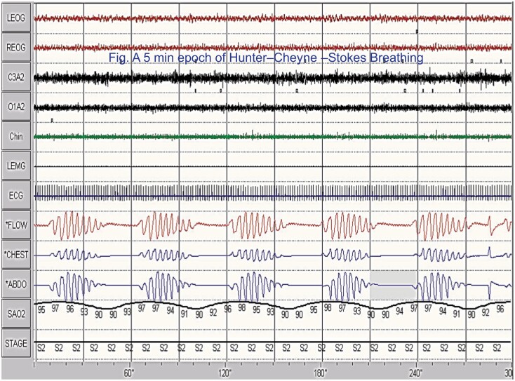
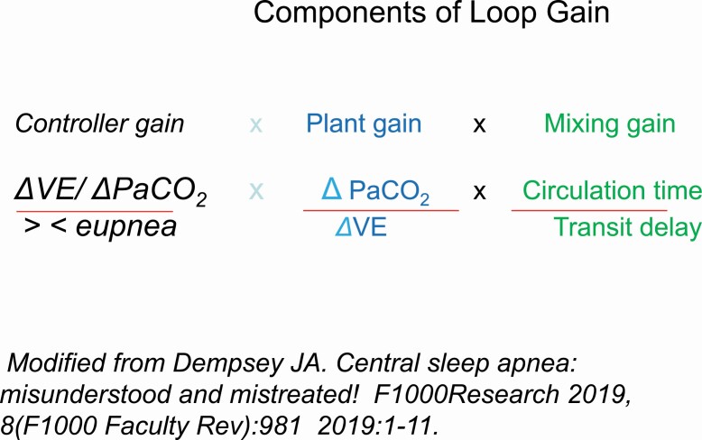
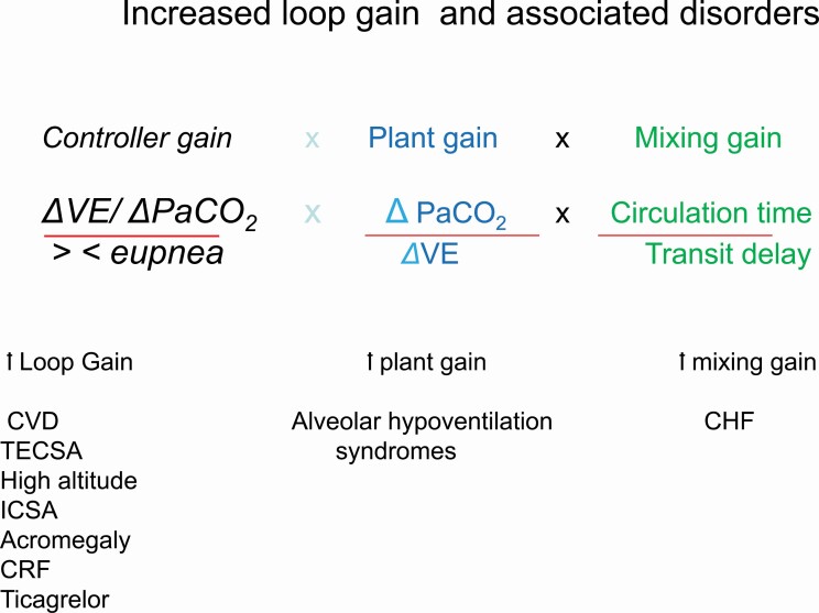
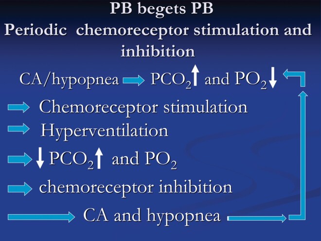
Central sleep apnea is not a single disorder; it can present as an isolated disorder or as a part of other clinical syndromes. In some conditions, such as heart failure, central apneic events are due to transient inhibition of ventilatory motor output during sleep, owing to the overlapping influences of sleep and hypocapnia. Specifically, the sleep state is associated with removal of wakefulness drive to breathe; thus, rendering ventilatory motor output dependent on the metabolic ventilatory control system, principally PaCO2. Accordingly, central apnea occurs when PaCO2 is reduced below the "apneic threshold". Our understanding of the pathophysiology of central sleep apnea has evolved appreciably over the past decade; accordingly, in disorders such as heart failure, central apnea is viewed as a form of breathing instability, manifesting as recurrent cycles of apnea/hypopnea, alternating with hyperpnea. In other words, ventilatory control operates as a negative-feedback closed-loop system to maintain homeostasis of blood gas tensions within a relatively narrow physiologic range, principally PaCO2. Therefore, many authors have adopted the engineering concept of "loop gain" (LG) as a measure of ventilatory instability and susceptibility to central apnea. Increased LG promotes breathing instabilities in a number of medical disorders. In some other conditions, such as with use of opioids, central apnea occurs due to inhibition of rhythm generation within the brainstem. This review will address the pathogenesis, pathophysiologic classification, and the multitude of clinical conditions that are associated with central apnea, and highlight areas of uncertainty.

Keywords: Adaptive-Servo Ventilation (ASV); apneic threshold; bi-level positive pressure therapy (BPAP); central apnea; continuous positive pressure therapy (CPAP); controller gain; hypocapnia; loop gain; plant gain.


Figures

Figure 1.
Figure 1.
A 5 min polysomnographic example of Hunter–Cheyne–Stokes breathing. Tracings are: electro-oculogram (EOG, 1and 2), electroencephalogram (EEG, 3 and 4), chin electromyogram (EMG, 5), Leg EMG (6, idle), ECG (7) airflow measured by thermocouple (8) rib cage (RC, 9), abdominal (ABD, 10), and combined (11th) excursions, and oxyhemoglobin saturation(12). Please note, airflow is absent and channels are flat consistent with central sleep apnea. Also, note the smooth and gradual changes in the thoracoabdominal excursions and in the crescendo and decrescendo arms of the cycle.
Figure 2.
Figure 2.
Depicts the concept of loop gain (LG). When LG is less than 1, the ventilatory response to a disturbance is limited and breathing soon stabilizes (A). In contrast, when the magnitude of the ventilatory response is greater than the magnitude of the initial disturbance, the system overshoots and breathing instability occurs (B). From Javaheri and Dempsey [4].
Figure 3.
Figure 3.
Depicts the 3 components of the loop gain, consisting of the controller gain, plant gain, and the mixing gain. Loop gain represents the overall ventilatory response to a disturbance. The classic 3 components of loop gain include 1, the controller gain, the plant gain, and the mixing gain. Collectively, under normal circumstances, loop gain as a negative feedback system functions to keep arterial blood gases and pH within normal range.The controller gain is the chemoreflex control mediated by carotid bodies and central chemoreceptors. It represents the sensitivity of these receptors to changes in blood gases, above and below eupnea. The plant gain represents the efficiency of the respiratory system to clear CO2. The mixing gain represents the transfer of information, i.e. changes in pulmonary capillary. Modified from Dempsey JA. Central sleep apnea: misunderstood and mistreated! F1000Research 2019, 8(F1000 Faculty Rev):981 2019:1–11.
Figure 4.
Figure 4.
The increased controller gain represents augmented chemoreflex control because of hypersensitivity of the carotid bodies and central chemoreceptors to changes in PaCO2 and PO2. The disorders associated with increased controller gain are noted (Table 1).The increased plant gain is due to increased CO2 clearance for a given change in ventilation. When plant gain is increased a small increase in ventilation, decreases PaCO2 excessively. As a restate PaCO2 is lowered below apneic threshold facilitating central apnea. the disorders associated with increased plant gain are noted (Table 1). The mixing gain is related to increased circulation time unique to heart failure, prolonging the cycle time of periodic breathing.
Figure 5.
Figure 5.
The figure depicts the important role of augmented chemical control in promoting periodic breathing in heart failure. Chemostimulation increases ventilation lowering arterial PaCO2 towards or below apneic threshold causing chemoinhibition with consequent hypopnea and central apnea. With diminished ventilation, PaCO2 increases and PO2 decreases causing chemostimulation and the cycle keeps repeating.
Figure 6.
Figure 6.
An example of sleep-disordered breathing in association with opioid use. The breathing is quite ataxic, in contrast to that in heart failure(Figure 1). CSA = central sleep apnea; OSA = obstructive sleep apnea; Hyp = hypopnea.
Figure 7.
Figure 7.
An example of baclofen induced central sleep apnea. Please note the pattern is similar to that of opioids in Figure 6.

Similar articles

Cited by

References

    1. Skatrud JB, et al. . Interaction of sleep state and chemical stimuli in sustaining rhythmic ventilation. J Appl Physiol Respir Environ Exerc Physiol. 1983;55(3):813–822. doi:10.1152/jappl.1983.55.3.813 - DOI - PubMed
    1. Badr MS, et al. . Central sleep apnea: a brief review. Curr Pulmonol Rep. 2019;8(1):14–21. doi:10.1007/s13665-019-0221-z - DOI - PMC - PubMed
    1. Dempsey JA. . Crossing the apnoeic threshold: causes and consequences. Exp Physiol. 2005;90(1):13–24. doi:10.1113/expphysiol.2004.028985 - DOI - PubMed
    1. Javaheri S, et al. . Central sleep apnea. Compr Physiol. 2013;3(1):141–163. doi:10.1002/cphy.c110057 - DOI - PubMed
    1. Dempsey JA. Central sleep apnea: misunderstood and mistreated!. F1000Res. 2019;8:981. doi:10.12688/f1000research.18358.1 - DOI - PMC - PubMed

Publication types