. 2022 May 12;13(1):2617.
doi: 10.1038/s41467-022-30250-6.

Molecular insights into biogenesis of glycosylphosphatidylinositol anchor proteins

Affiliations

Molecular insights into biogenesis of glycosylphosphatidylinositol anchor proteins

Yidan Xu et al. Nat Commun. .

Abstract

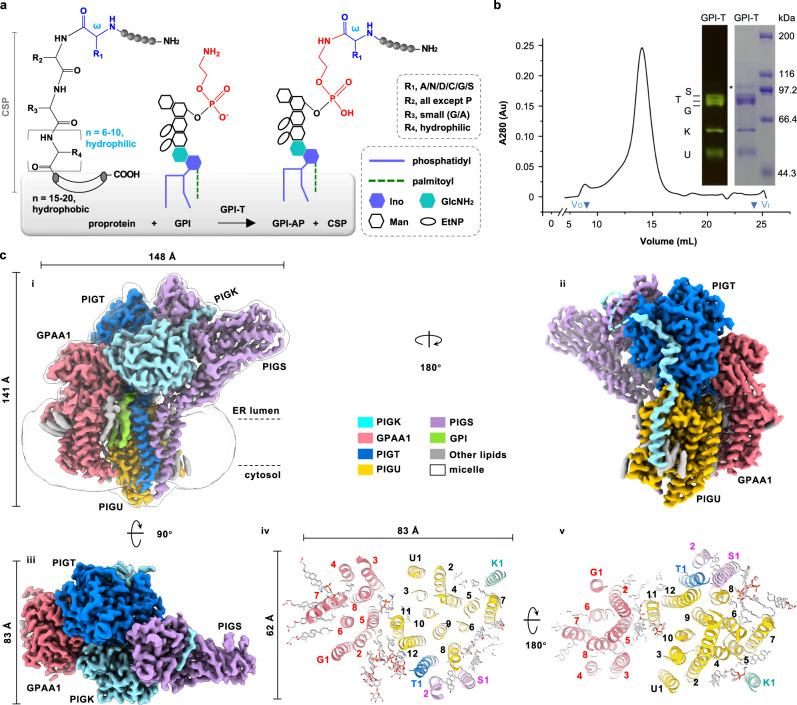
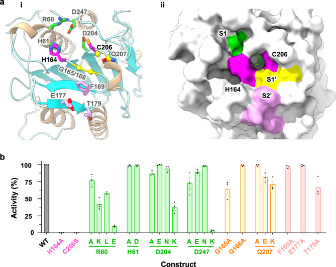
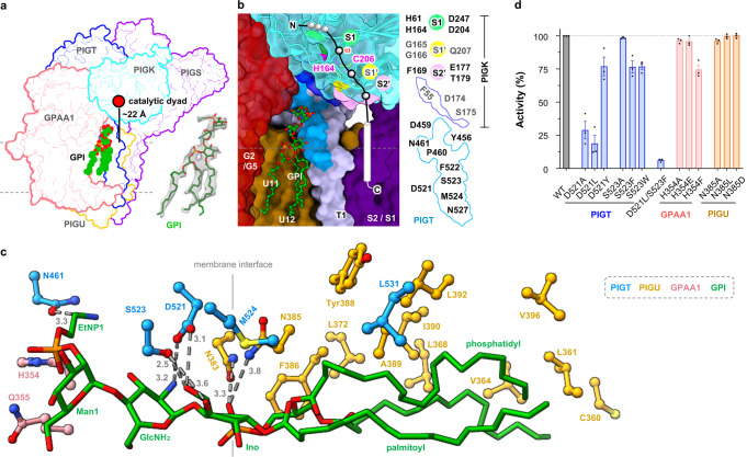
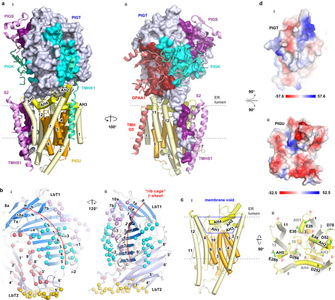
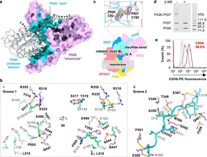
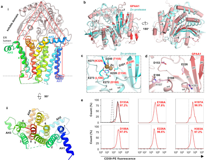
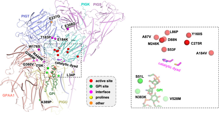
Eukaryotic cells are coated with an abundance of glycosylphosphatidylinositol anchor proteins (GPI-APs) that play crucial roles in fertilization, neurogenesis, and immunity. The removal of a hydrophobic signal peptide and covalent attachment of GPI at the new carboxyl terminus are catalyzed by an endoplasmic reticulum membrane GPI transamidase complex (GPI-T) conserved among all eukaryotes. Here, we report the cryo-electron microscopy (cryo-EM) structure of the human GPI-T at a global 2.53-Å resolution, revealing an equimolar heteropentameric assembly. Structure-based mutagenesis suggests a legumain-like mechanism for the recognition and cleavage of proprotein substrates, and an endogenous GPI in the structure defines a composite cavity for the lipid substrate. This elongated active site, stemming from the membrane and spanning an additional ~22-Å space toward the catalytic dyad, is structurally suited for both substrates which feature an amphipathic pattern that matches this geometry. Our work presents an important step towards the mechanistic understanding of GPI-AP biosynthesis.


Conflict of interest statement

The authors declare no competing interests.

Figures

Fig. 1
Fig. 1. Cryo-EM map of the human GPI-T.
a GPI-T replaces the C-terminal signal peptide (CSP) of proproteins with GPI at the ω residue (blue) by a transamination reaction. Various parts are denoted in the dashed box. EtNP, ethanolamine phosphate; Man, mannose; Ino, inositol; GlcNH2, glucosamine. The preferences for the ω, ω+1, ω+2, and ω+3 sites are indicated in the dashed box with amino acid abbreviated with single letters. The gray shading indicates the endoplasmic reticulum (ER) membrane. b GPI-T shows a near-Gaussian peak on gel filtration and all five subunits are present on an SDS-PAGE (inset) visualized by in-gel fluorescence (left) and Coomassie staining (right). Vo and Vt (triangle) indicate void and total volume, respectively. Background absorbance signals before Vo are not shown fully. G/T/S/K/U refers to the subunits GPAA1/PIGT/PIGS/PIGK/PIGU. The position of each subunit was separately determined by comparing the complex with singly expressed subunits. An asterisk indicates a minor contaminant. Molecular weights of the protein markers are indicated on the right. Shown is a representative result of three independent experiments. Uncropped images are provided in Source Data. c Cryo-EM map (i-iii) and normal view of the transmembrane domain from ER lumen (iv) or cytosol (v). Numbers in iv and v indicate TMHs and G/T/U/S/K refer to GPAA1 and PIGT/U/S/K, respectively. Lipids and detergents are shown as sticks (gray). Subunits and associated cryo-EM densities are color-coded as indicated.
Fig. 2
Fig. 2. Structural and functional resemblance of the PIGK active site to that of legumains.
a Cartoon (wheat, α-helix; cyan, β-strand, i) and surface (ii) representation of the PIGK protease domain with the catalytic dyad (magenta), S1 (green), S1′ (yellow), and S2′ (pink) residues shown as sticks (i) or highlighted in colors (ii). S1/S1′/S2′ sites are superposed from legumain structures. b Functional assay of the active site mutants. Apparent activity (% of wild-type, WT) was measured by immune staining of a reporter GPI-AP (CD59) on the surface of PIGK-KO cells transfected with the indicated mutants of the catalytic dyad and S1/S1′/S2′ residues (color-coded to match those in a). Cells expressing PIGK fused with a thermostable green fluorescence protein were gated, and the surface staining of CD59 was further analyzed by flow cytometry. Data represent mean ± s.e.m. from three independent experiments. Source data are provided.
Fig. 3
Fig. 3. Characterization of a composite GPI-binding site.
a Cryo-EM density that fills a nearly complete GPI was observed in the membrane cavity “underneath” the catalytic dyad. b Expanded view of the composite site encompassed by the indicated TMHs from GPAA1 (G) /PIGU (U) /PIGT (T) /PIGS (S). Evolutionarily conserved residues (Supplementary Fig. 6c, d) in the vicinity are colored marine (PIGT) and blue (PIGK). Subunits are shown as surfaces except that PIGK was additionally shown as ribbon representations with the catalytic dyad and S1/S1′/S2′ residues highlighted in indicated colors. c Interaction between the partial GPI (green) and GPI-T (colored-coded as indicated). Distances (Å) are either indicated by numbers for H-bonding interactions or omitted for hydrophobic interactions (within 5 Å of GPI). A vertical line marks the membrane boundary. Although the major form of the phosphatidyl moiety in mammal cells contains 1-alkyl-2-acyl-glycerol, a diacylglycerol was modeled based on the density. d Apparent activity of GPI-T mutants relative to the wild-type (WT). GPI-T KO cells were gated by TGP fluorescence for subunit expression and analyzed for surface staining of the reporter GPI-AP (CD59) by flow cytometry. Bar graph is color-coded to match the coloring for subunits in (b). Data represent mean ± s.e.m. from three independent experiments. Source data are provided.
Fig. 4
Fig. 4. PIGT and PIGU form a platform for complex assembly.
a PIGT (surface representations, blue) and PIGU (cylinder, yellow/orange) form a docking platform for other subunits (GPAA1, red; PIGS, purple; PIGK, cyan, ribbon). Interaction surfaces on PIGT are shaded to match the color of GPAA1/PIGK/PIGS. b PIGT displays skeleton features. Its luminal domain contains two lobes (LbT1/2). LbT1 (marine) consists of a central “rib-cage” β-sheet (red curve, β1-10) sandwiched by connecting loops, α-helices (α1-4), and β-strands (β7a, 7b, 8a, and 10a). LbT2 (light blue) consists of two layers of β-sheets with a total of 9 β-strands (β1′-9′). Spheres indicate residues that interact with GPAA1 (red), PIGK (cyan), PIGS (purple), and PIGU (orange). c Side (i) and normal (ii) view of PIGU. PIGU features a membrane core region (orange) with short transmembrane helices (TMHs) enclosed by a ring of TMHs (pale yellow). This arrangement creates a membrane void (blue trapezoid) to hold the five amphipathic helixes (AH) 1-5 (yellow). The TMHs and AHs are so arranged such that the C-terminal ends of several α-helices (labeled with black text) expose to the surface (ii). The resulting dipole moments (δ-) and acidic residues (stick representation, green) make the surface electrostatically negative. d “Open-book” representation of the electrostatic potential molecular surface (red, negative; blue, positive; white, neutral) generated using the Adaptive Poisson-Boltzmann Solver module in PyMOL (version 2.3.3).
Fig. 5
Fig. 5. PIGS and PIGT place PIGK and its catalytic dyad in a position suitable for catalysis.
a PIGS (surface, pink) holds PIGK (ribbon) by embracing the protease domain using one of the shamrock grooves and hosting the loop region in another groove. The buried surface is colored cyan. b Detailed interactions between PIGK and PIGS at Groove 1 (i) and Groove 2 (ii). PIGK residues (cyan) are indicated with gray texts and PIGS residues (light purple) are labeled with black texts. Dashed lines (yellow) indicate distances within 3.6 Å. c An inter-subunit disulfide bond (PIGT C182 / PIGK C92) (magenta) nails PIGK (cyan) onto the PIGT (blue) (top). This and the other interactions fix PIGK in a position such that the catalytic dyad (red dot) is ~22 Å “above” the membrane interface (gray shading). This geometry would suit interactions with GPI which is expected to insert into the membrane by the phosphatidyl moiety with its hydrophilic glycan chains (measuring ~25 Å in the extended form) stemming from the membrane to meet with the catalytic dyad. The cryo-EM density for the disulfide bond is shown gray. A GPI-T structure (bottom) color-coded as indicated shows the approximate position of the disulfide bond. d Verification of the PIGK-PIGT disulfide bond. SDS-PAGE in-gel fluorescence shows a high-molecular-weight band (PIGK-PIGT) in the absence of, but not in the presence of, the reducing agent β-mercaptoethanol (β-ME) at the cost of the individual PIGT/PIGK bands. Free PIGT/PIGK bands under non-reducing conditions were probably from uncomplexed PIGT/PIGK proteins due to the uneven expression level of all the five subunits. Other subunits were also co-transfected but were invisible owing to their lack of the TGP-tag. Theoretical molecular weights of home-made fluorescent molecular markers are labeled on the right. Shown is a representative result of three independent experiments. The uncropped image is available in the Source Data file. e Disruption of the PIGT-PIGK disulfide bond by PIGK C92A causes loss of GPI-T activity. PIGK knockout cells expressing TGP-tagged wild-type (black), C92A (red), or a control membrane protein (gray) were gated by TGP fluorescence (for the expression of PIGK), and the sub-population was analyzed for surface staining of the reporter GPI-AP (CD59) by flow cytometry using phycoerythrin (PE)-conjugated antibodies. C92A showed an apparent activity of 61.0 ± 6.3% (s.e.m., n = 3) compared to the wild-type PIGK. A vertical line indicates the threshold for CD59 fluorescence. Shown is a representative result from three independent experiments. Source data are provided.
Fig. 6
Fig. 6. Structural and functional characterization of GPAA1 reveals a protease-like domain.
a Side (i) and normal (ii) view of GPAA1. Numbers indicate transmembrane helices (TMHs) and AH1-3 label the three amphipathic helices (AH). The soluble domain is colored pink and the membrane-associated domain is rainbow-colored (blue, N-terminal; red, C-terminal). b The soluble domain of GPAA1 (red/pink, cylinder) is structurally similar to a Zn-protease AM-1 (cyan, cartoon, PDB ID 2EK8 10.2210/pdb2EK8/pdb) with a Z-score of 20.6 and Cα RMSD of 3.2 Å (from a DALI search). c The Zn-binding site of AM-1 (expanded view of the boxed region in b) consists of two each of aspartate, glutamate, and histidine residues that are not fully conserved in GPAA1 (in brackets). d Arrangement of GPAA1 aspartate/glutamate/histidine (D/E/H) residues in the region corresponding to the Zn-binding site in AM-1. Despite having the same composition, these residues are unlikely to form a Zn-binding site because of different spatial arrangements, especially for the two histidine residues (gray text). e Mutation of the D/E/H residues in (d) did not reduce CD59 staining in the flow cytometry assay. The function of wild-type GPAA1 and mutants were assessed by the surface expression of the GPI-AP reporter (CD59) in GPAA1 knockout cells transfected with appropriate plasmids. Cells were gated by TGP fluorescence for GPAA1 expression and analyzed for CD59 staining using phycoerythrin (PE)-conjugated antibodies. The dotted line (gray) indicates the CD59-staining background level from cells expressing an unrelated TGP-tagged membrane protein (negative control). Solid lines indicate staining of cells transfected with the wild-type (black) or mutant genes (red). A vertical dash line marks the threshold (CD59-gating) determined from the negative control. Shown is a representative result from three independent experiments. Data for all the three experiments are included in the Source Data file.
Fig. 7
Fig. 7. Distribution of genetic mutations on GPI-T.
Mapping the disease mutations (Cα spheres, color-coded by the categories in the gray box) onto the human GPI-T structure (main chain, color-coded cylinder representations; catalytic dyad, magenta stick presentations; GPI, green stick representations). The mutations include PIGK S53F/L86P/A87V/D88N/Y160S/A184V/M246K/C275R that cause a neurodevelopmental syndrome with hypotonia, cerebellar atrophy, and epilepsy, PIGT T183P that causes an intellectual disability syndrome, PIGT E184K/G360V/R448W that are related to the Multiple Congenital Anomalies-Hypotonia Seizures Syndrome 3, , , PIGT E237Q/V528M that cause developmental disorders characterizing learning disability, epilepsy, microcephaly, congenital malformations and mild dysmorphic features, PIGT G366W that is found in patients with epileptic apnea and multiple congenital anomalies, severe intellectual disability, and seizures, PIGS L34P/E308G that are related to a neurological syndrome ranging from fetal akinesia to epileptic encephalopathy, PIGU I70K/N383K that are found in patients with severe intellectual disability, epilepsy, and brain anomalies, and GPAA1 S51L/W176S/A389P that are also related to developmental disorders featuring global developmental delay, epilepsy, cerebellar atrophy, and osteopenia. Left, overview with a box showing the area close to the active site. Right, the expanded view of the boxed region on the left.

Similar articles

Cited by

References

    1. Gamage DG, Hendrickson TL. GPI transamidase and GPI anchored proteins: oncogenes and biomarkers for cancer. Crit. Rev. Biochem. Mol. Biol. 2013;48:446–464. doi: 10.3109/10409238.2013.831024. - DOI - PubMed
    1. Pittet M, Conzelmann A. Biosynthesis and function of GPI proteins in the yeast Saccharomyces cerevisiae. Biochim. Biophys. Acta. 2007;1771:405–420. doi: 10.1016/j.bbalip.2006.05.015. - DOI - PubMed
    1. Kinoshita T. Biosynthesis and biology of mammalian GPI-anchored proteins. Open Biol. 2020;10:190290. doi: 10.1098/rsob.190290. - DOI - PMC - PubMed
    1. Peter O, Menon AK. Thematic review series: Lipid Posttranslational Modifications. GPI anchoring of protein in yeast and mammalian cells, or: how we learned to stop worrying and love glycophospholipids. J. Lipid Res. 2007;48:993–1011. doi: 10.1194/jlr.R700002-JLR200. - DOI - PubMed
    1. Paulick MG, Bertozzi CR. The glycosylphosphatidylinositol anchor: a complex membrane-anchoring structure for proteins. Biochemistry. 2008;47:6991–7000. doi: 10.1021/bi8006324. - DOI - PMC - PubMed

Publication types