. 2022 Aug;158(2):169-180.
doi: 10.1007/s00418-022-02099-9. Epub 2022 May 12.

Aniridia-related keratopathy relevant cell signaling pathways in human fetal corneas

Affiliations

Aniridia-related keratopathy relevant cell signaling pathways in human fetal corneas

André Vicente et al. Histochem Cell Biol. 2022 Aug.

Abstract

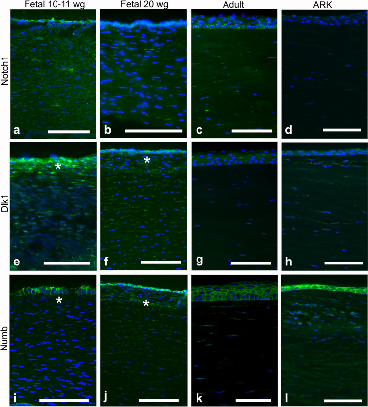
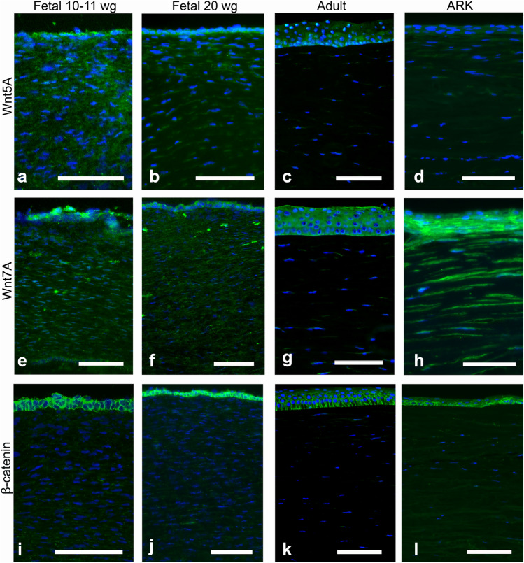
We aimed to study aniridia-related keratopathy (ARK) relevant cell signaling pathways [Notch1, Wnt/β-catenin, Sonic hedgehog (SHH) and mTOR] in normal human fetal corneas compared with normal human adult corneas and ARK corneas. We found that fetal corneas at 20 weeks of gestation (wg) and normal adult corneas showed similar staining patterns for Notch1; however 10-11 wg fetal corneas showed increased presence of Notch1. Numb and Dlk1 had an enhanced presence in the fetal corneas compared with the adult corneas. Fetal corneas showed stronger immunolabeling with antibodies against β-catenin, Wnt5a, Wnt7a, Gli1, Hes1, p-rpS6, and mTOR when compared with the adult corneas. Gene expression of Notch1, Wnt5A, Wnt7A, β-catenin, Hes1, mTOR, and rps6 was higher in the 9-12 wg fetal corneas compared with adult corneas. The cell signaling pathway differences found between human fetal and adult corneas were similar to those previously found in ARK corneas with the exception of Notch1. Analogous profiles of cell signaling pathway activation between human fetal corneas and ARK corneas suggests that there is a less differentiated host milieu in ARK.

Keywords: Adult cornea; Aniridia; Fetal cornea; Notch; Sonic hedgehog; Wnt; mTOR.


Conflict of interest statement

The authors declare no competing or financial interests.

Figures

Fig. 1
Fig. 1
Cross-sections of fetal corneas 10–11 wg (a, e, i) and 20 wg (b, f, j) as well as normal adult corneas (c, g, k) and ARK (d, h, l) labeled with antibodies (green) against Notch1 (ad), Dlk1 (eh), and Numb (il). The corneal epithelium is shown at the top and the stroma below, in all photographs of all figures. Cell nuclei are labeled blue with DAPI (al). Immunolabeling against Notch1 (ad) was detected as streaks in the stroma and strongly around the basal layers of the epithelium in all fetal and adult corneas (ac), whereas in the ARK corneas, Notch1 was absent in the epithelium and only very scarce in the stroma (d). In the 20 wg fetal corneas, the labeling of the epithelium against Notch1 was slightly surpassed by the strong DAPI labeling of the epithelial cell nuclei (b). Dlk1 (eh) labeled the epithelial cells and streaks in the stroma of all fetal corneas, more abundantly in the anterior region (e, f, asterisk), whereas in the adult corneas labeling was only present in the epithelium (g). In the ARK corneas, Dlk1 immunolabeling was present both in the epithelium and in streaks, more pronounced in the anterior pannus (h). In all fetal corneas, Abs against Numb (il), another inhibitor of Notch1, labeled the epithelial cells and streaks in the stroma (i, j, asterisk), but stromal labeling was more abundant in the 20 wg (j) than in the 10–11 wg fetal corneas (i). Numb labeling in adult corneas was present in the epithelial cells and in sporadic streaks in the stroma (k). In ARK, Numb immunolabeling was detected in the epithelium and anterior pannus (l) in a pattern similar to that of Dlk1. Bars, 100 μm
Fig. 2
Fig. 2
Cross-sections of fetal corneas 10–11 wg (a, e, i), 20 wg (b, f, j), normal adult corneas (c, g, k), and ARK (d, h, l) labeled with antibodies (green) against Wnt5a (ad), Wnt7a (eh), and β-catenin (il). Cell nuclei are labeled blue with DAPI (al). The Abs against Wnt5a abundantly labeled both the epithelial cells and streaks in the stroma of the fetal corneas (a, b). The stromal labeling was more abundant in the 10–11 wg (a) than in the 20 wg fetal corneas (b), in which the streaks were more profuse in the anterior region (b). In the adult corneas, only the epithelium was labeled (c), whereas in ARK corneas both the epithelium and the anterior pannus were labeled (d). Immunolabeling against Wnt7a was found in the epithelial cells and as stromal streaks in all fetal samples (e, f). In contrast, in the adult corneas, labeling was present in the epithelium but only in extremely sparse steaks in the stroma (g). In ARK, the epithelium and anterior pannus as well as the rest of the stroma were labeled by Wnt7a (h). β-Catenin immunolabeling was present abundantly in the contours of epithelial cells but only discretely in stromal streaks, in a similar pattern in the 10–11 wg (i) and 20 wg fetal corneas (j). In the adult corneas, labeling was present in the epithelial cells more intensively in the basal region but absent in the stroma (k). The staining pattern in the ARK corneas was similar to that of the fetal corneas, with β-catenin immunolabeling delineating the contours of epithelial cells and present in streaks in the anterior pannus (l). Bars, 100 μm
Fig. 3
Fig. 3
Cross-sections of fetal corneas 10–11 wg (a, e, i, m) and 20 wg (b, f, j, n), adult normal corneas (c, g, k, o), and ARK (d, h, l, p) labeled with antibodies (green) against Hes1 (ad), Gli1 (eh), mTOR (il), and p-rpS6 (mp). Tissue preservation of the fetal tissue was variable. Cell nuclei are labeled blue with DAPI (ap). Abs against Hes1 strongly immunolabeled the epithelial cells of the fetal corneas (a, b). These Abs labeled streaks in the stroma more profusely in the 10–11 wg (a) than in the 20 wg fetal corneas (b) and more intensively in the anterior stroma (b). In contrast, in the adult corneas, immunolabeling was not detected (c), whereas Hes1 was present in the epithelium and in the anterior stroma of the ARK corneas (d). Immunolabeling with Abs against Gli1 was present in the epithelial cells in all fetal corneas (e, f). Labeling in the stroma was present in streaks in all fetal corneas (e, f). Immunolabeling for these Abs was not observed in adult corneas (g). In the ARK corneas, the Abs against Gli1 labeled the epithelium and the anterior pannus (h). The inserts in ah show the epithelium at higher magnification. The Ab against mTOR abundantly labeled the epithelial cells of all fetal corneas (i, j) but marked the epithelium in the 20 wg fetal corneas more intensively (j). The stroma in all fetal corneas was labeled in streaks (i, j) but was more abundantly marked in the 10–11 wg fetal corneas (i). This Ab did not immunolabel the adult corneas (k), but the epithelium and anterior pannus were labeled by this Ab in the ARK corneas (l). The Ab against p-rpS6 labeled the epithelial cells and abundant streaks in the stroma of all fetal corneas in a likewise pattern in both 10–11 wg (m) and 20 wg fetal corneas (n). The stroma in the adult corneas was not labeled and the surface of the epithelium was only scarcely labeled, suggesting sticky adherence to the epithelial surface (o). In the ARK corneas, immunostaining against p-rpS6 was present in the epithelium and anterior pannus (p). Bars, 100 μm
Fig. 4
Fig. 4
Gene expression of Notch1 (NOTCH1), Dlk1 (DLK1), Numb (NUMB) (a), Wnt5A (WNT5A), Wnt7A (WNT7A), β-catenin (CTNNB1) (b), and Hes1 (HES1), mTOR (mTOR), and rps6 (RPS6) (c) in 9–12 wg fetal corneas as compared with adult cornea. There was a 3.89-fold increase in Notch1 gene expression, 1540-fold increase in Dlk1 gene expression, and 0.64-fold decrease in Numb gene expression in 9–12 wg fetal corneas. (a). Expression of Wnt5A, Wnt7A, and β-catenin genes was increased by 1.85-, 3.57-, and 2.05-fold, respectively. (b). Gene expression of Hes1, mTOR, and rps6 was increased by 2.73-, 1.89-, and 1.78-fold, respectively (c). Values are mean ± SD. *p < 0.05; **p < 0.01; ***p < 0.001; ****p < 0.0001

Similar articles

Cited by

References

    1. Abdallah BM, Jensen CH, Gutierrez G, Leslie RG, Jensen TG, Kassem M. Regulation of human skeletal stem cells differentiation by Dlk1/Pref-1. J Bone Miner Res. 2004;19(5):841–852. doi: 10.1359/JBMR.040118. - DOI - PubMed
    1. Ang SJ, Stump RJ, Lovicu FJ, McAvoy JW. Spatial and temporal expression of Wnt and Dickkopf genes during murine lens development. Gene Expr Patterns. 2004;4(3):289–295. doi: 10.1016/j.modgep.2003.11.002. - DOI - PubMed
    1. Bausili MM, Alvarez de Toledo J, Barraquer RI, Michael R, Tresserra F, de la Paz MF. Histopathology findings of corneal buttons in congenital aniridia patients. Ophthalmic Res. 2016;56(4):202–206. doi: 10.1159/000444930. - DOI - PubMed
    1. Belloni E, Muenke M, Roessler E, Traverso G, Siegel-Bartelt J, Frumkin A, Mitchell HF, Donis-Keller H, Helms C, Hing AV, Heng HH, Koop B, Martindale D, Rommens JM, Tsui LC, Scherer SW. Identification of sonic hedgehog as a candidate gene responsible for holoprosencephaly. Nat Genet. 1996;14(3):353–356. doi: 10.1038/ng1196-353. - DOI - PubMed
    1. Borello U, Buffa V, Sonnino C, Melchionna R, Vivarelli E, Cossu G. Differential expression of the Wnt putative receptors Frizzled during mouse somitogenesis. Mech Dev. 1999;89(1–2):173–177. doi: 10.1016/S0925-4773(99)00205-1. - DOI - PubMed