Review
. 2022 May 13;479(9):973-993.
doi: 10.1042/BCJ20210556.

No-nonsense: insights into the functional interplay of nonsense-mediated mRNA decay factors

Affiliations
Review

No-nonsense: insights into the functional interplay of nonsense-mediated mRNA decay factors

Justine Mailliot et al. Biochem J. .

Abstract

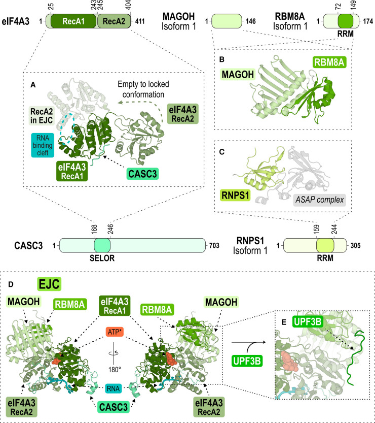
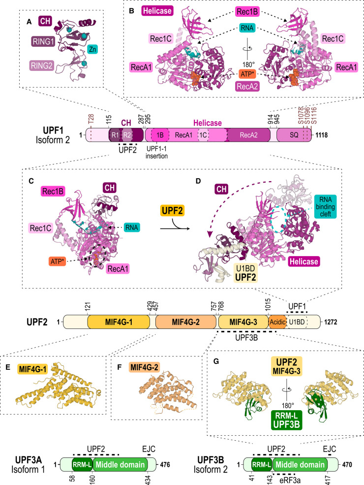
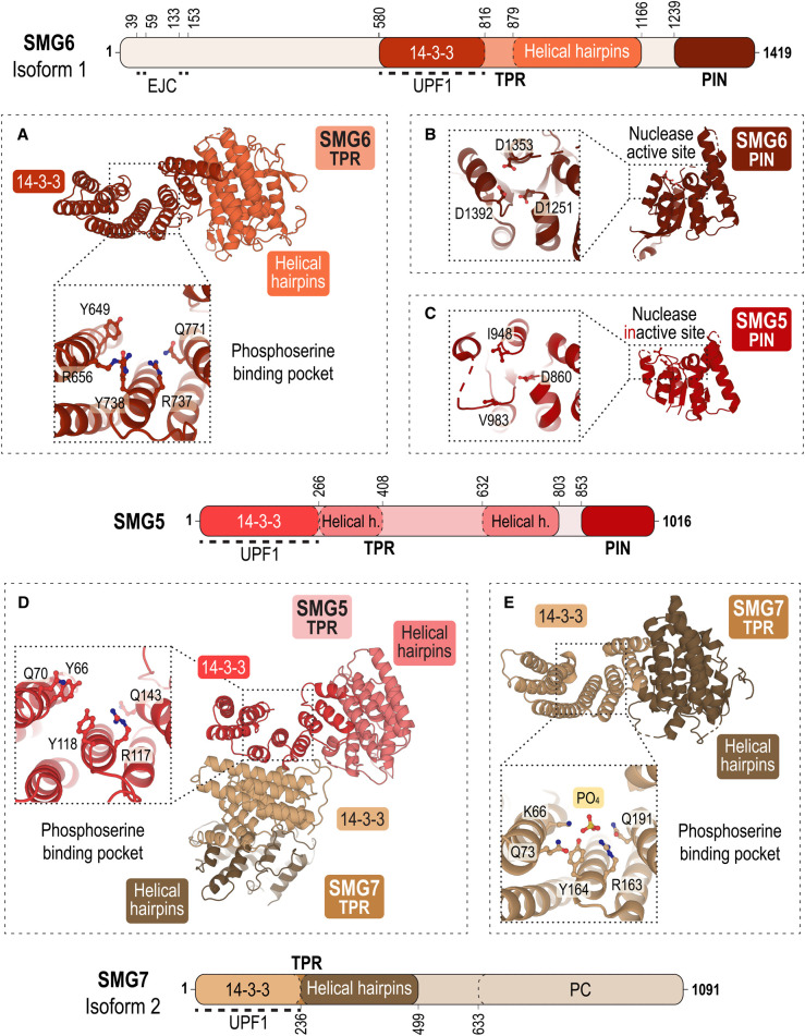
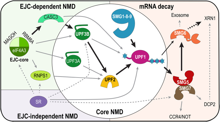
Nonsense-mediated messenger RNA decay (NMD) represents one of the main surveillance pathways used by eukaryotic cells to control the quality and abundance of mRNAs and to degrade viral RNA. NMD recognises mRNAs with a premature termination codon (PTC) and targets them to decay. Markers for a mRNA with a PTC, and thus NMD, are a long a 3'-untranslated region and the presence of an exon-junction complex (EJC) downstream of the stop codon. Here, we review our structural understanding of mammalian NMD factors and their functional interplay leading to a branched network of different interconnected but specialised mRNA decay pathways. We discuss recent insights into the potential impact of EJC composition on NMD pathway choice. We highlight the coexistence and function of different isoforms of up-frameshift protein 1 (UPF1) with an emphasis of their role at the endoplasmic reticulum and during stress, and the role of the paralogs UPF3B and UPF3A, underscoring that gene regulation by mammalian NMD is tightly controlled and context-dependent being conditional on developmental stage, tissue and cell types.

Keywords: exon-junction complexes; nonsense-mediated mRNA decay; regulation of gene expression; translational control; up-frameshift proteins.


Conflict of interest statement

The authors declare that there are no competing interests associated with the manuscript.

Figures

Figure 1.
Figure 1.. The architecture of the exon-junction complex.
Schemes showing the domain architecture of the EJC core proteins eIF4A3, MAGOH and RBM8A, and the EJC-associated proteins CASC3 and RNPS1 which bind in a mutually exclusive manner. In the structures, the protein domains are coloured according to their primary structure scheme. (A) Structure of eIF4A3's RecA1 (forest) and RecA2 (pale green) in the presence of CASC3 (aquamarine) only (PDB ID: 2J0U) [43] or in the context of the EJC, in the presence of RNA (cyan circle) and an ATP-analogue (not shown) (PDB ID: 3EX7) [44]. The green arrow indicates the change of eIF4A3's RecA2 domain between open (opaque) [43] and locked (semi-transparent) [44] conformations upon EJC complex formation. (B) Structure of the heterodimer of MAGOH (lime) and the RRM domain of RBM8A (green) (PDB ID: 1P27) [166]. (C) Structure of the RRM domain of RNPS1 (lemon) bound to the ASAP complex (PDB ID: 4A8X) [167]. (D) Front and back view of the EJC bound with an ATP analogue (ATP*, orange), RNA (cyan) and a CASC3 fragment (PDB ID: 3EX7) [44]. (E) Close-up view showing UPF3B's EBM bound to the EJC (PDB ID: 2XB2) [49].
Figure 2.
Figure 2.. The core NMD factors UPF1, UPF2 and UPF3.
Schemes showing the domain architecture of UPF1, UPF2, UPF3B and its paralog UPF3A. In the structures, the protein domains are coloured according to their primary structure scheme. (A) Structure of UPF1's CH domain (deep purple), harbouring two RING modules (dark and light violet) (PDB ID: 2IYK) [65] coordinating three Zn2+ ions (cyan). (B) Front and back view of UPF1's helicase domain with two tandem RecA1 (pink) and RecA2 (dark pink) domains and the subdomains Rec1B (magenta) and Rec1C (light pink) bound with an ATP-analogue (ATP*, orange) and RNA (cyan) (PDB ID: 2XZO) [31]. (C) Structure of the yeast CH-helicase domains bound with an ATP-analogue (ATP*, orange) and RNA (cyan) (PDB ID: 2XZL) [31]. (D) Structure of the human CH-helicase domains of UPF1 bound with the U1BD of UPF2 (light yellow) (PDB ID: 2WJV) [67]. The arrow indicates the conformational change of the CH domain upon UPF2-binding. (E) Structure of UPF2 MIF4G-1 domain (PDB ID: 4CEM, yellow) [87]. (F) Structure of MIF4G-2 domain (PDB ID: 4CEK, pale orange) [87]. (G) Front and back view of the complex formed by the MIF4G-3 domain of UPF2 (pale yellow) and the RRM-L of UPF3B (green) (PDB ID: 1UW4) [86].
Figure 3.
Figure 3.. Cryo-EM structures of SMG1–8–9 kinase complexes.
Schemes showing the domain architecture of SMG1, SMG8 and SMG9. In the structures, the protein domains are coloured according to their primary structure scheme. (A) Top and side view of the structural model of SMG1 kinase with the N-terminal α-helical solenoid arch-containing HEAT repeats (tv-blue) and the globular C-terminus comprised of the FAT domain (light blue), the kinase domain (blue) and the FAT C-terminal (FATC) domain (dark blue) (PDB ID: 6L53) [106]. (B) Structural model of SMG1-9 (blue grey) bound with ATP and an ATP analogue (ATP*) (both in orange) (PDB ID: 7PW9) [110]. (C) Structural model of SMG1–8–9 bound with ATP (PDB ID: 6SYT) [107]. SMG8 (emerald green) interacts with SMG9. The kinase active site is highlighted with a red box. (D) Close-up view of SMG1's active site with a UPF1 leucine-serine-glutamine (LSQ) peptide (in pink) (PDB ID: 6Z3R) [108]. The serine targeted for phosphorylation (S1078) is highlighted in brown. (E) Close-up view of SMG1's active site with a pyrimidine derivative inhibitor (SMG1i, yellow) (PDB ID: 7PW4) [110]. Boxes highlight binding pocket residues P2213, D2339 and N2356 which are specific for the SMG1 kinase active site [110].
Figure 4.
Figure 4.. The architecture of mRNA decay factors SMG6, SMG5 and SMG7.
Schemes showing the domain architecture of SMG6, SMG5 and SMG7. In the structures, the protein domains are coloured according to their primary structure scheme. (A) Structure of the tetratricopeptide (TPR) domain of SMG6 with a 14-3-3-like module (dark red) involved in binding phosphorylated serine/threonine residues of UPF1 and α-helical hairpins (orange) (PDB ID: 4UM2) [71]. The close-up view shows the phosphoserine-binding pocket of SMG6 with key residues highlighted as sticks. (B) Structure of SMG6's PIN domain (chocolate) (PDB ID: 2HWW) [115]. The close-up view of the nuclease active site highlights the canonical catalytic triad of acidic residues (D1251, D1353, D1392). (C) Structure of SMG5's PIN domain (red) (PDB ID: 2HWY) [115]. The close-up view shows the inactive site in SMG5 comprising only one aspartate (D860, I948, V983). (D) Structure of the heterodimer of the TPR domain of SMG5 with a 14-3-3-like module (red) and α-helical hairpins (light red) and the TPR domain of SMG7 (14-3-3-like module, beige; α-helical hairpins, brown) (PDB ID: 3ZHE) [113]. SMG5 and SMG7 interact via their 14-3-3-like modules. The close-up view shows the phosphoserine-binding pocket of SMG5 with key residues highlighted as sticks. (E) Structure of the TPR domain of SMG7 with its 14-3-3-like module (beige) and α-helical hairpins (brown) (PDB ID: 1YA0) [116]. The close-up view shows the phosphoserine-binding pocket of SMG7 with key residues and a phosphate ion highlighted as sticks.
Figure 5.
Figure 5.. The branched NMD pathways.
Scheme summarising the NMD pathway and its branches converging at UPF1 as the key factor. Black thick arrows indicate the canonical NMD pathway in mammalian cells. The EJC-dependent part of the NMD pathway is shown in a green box, the EJC-independent part in a purple box, the core NMD pathway in a white circle and the different mRNA decay routes in an orange box. The branch requiring UPF1, UPF2 and UPF3B presents the canonical NMD branch conserved from yeast to human. UPF3A is an NMD activator in most UPF3B-deficient contexts. In the UPF3-independent NMD branch, RNPS1 stimulates NMD by recruiting UPF2. UPF2-independent NMD is enhanced by CASC3, which binds UPF3B and UPF1. UPF2 activates the SMG1–8–9 kinase complex to phosphorylate UPF1. mRNA decay relies on the recruitment of SMG6 and SMG5–7 by phospho-UPF1. SMG5–7 interact with factors responsible for mRNA deadenylation (CCR4/NOT complex), mRNA 5′-decapping (DCP2), and activates SMG6 endonuclease. Resulting unprotected mRNA fragments are degraded by the exonuclease XRN1 and the exosome complex. SR proteins (in particular SRSF1) enhance NMD by interacting with the EJC-core and its associated proteins as well as NMD factors. SR proteins can also promote mRNA decay in an EJC-independent manner.

Similar articles

Cited by

References

    1. Nasif, S., Contu, L. and Mühlemann, O. (2018) Beyond quality control: the role of nonsense-mediated mRNA decay (NMD) in regulating gene expression. Semin. Cell Dev. Biol. 75, 78–87 10.1016/j.semcdb.2017.08.053 - DOI - PubMed
    1. Kim, Y.K. and Maquat, L.E. (2019) UPFront and center in RNA decay: UPF1 in nonsense-mediated mRNA decay and beyond. RNA 25, 407–422 10.1261/rna.070136.118 - DOI - PMC - PubMed
    1. Lykke-Andersen, S. and Jensen, T.H. (2015) Nonsense-mediated mRNA decay: an intricate machinery that shapes transcriptomes. Nat. Rev. Mol. Cell Biol. 16, 665–677 10.1038/nrm4063 - DOI - PubMed
    1. Kishor, A., Fritz, S.E. and Hogg, J.R. (2019) Nonsense-mediated mRNA decay: the challenge of telling right from wrong in a complex transcriptome. Wiley Interdiscip. Rev. RNA 10, e1548 10.1002/wrna.1548 - DOI - PMC - PubMed
    1. Kurosaki, T., Popp, M.W. and Maquat, L.E. (2019) Quality and quantity control of gene expression by nonsense-mediated mRNA decay. Nat. Rev. Mol. Cell Biol. 20, 406–420 10.1038/s41580-019-0126-2 - DOI - PMC - PubMed

Publication types