

Extracellular Vesicles and Their Emerging Roles as Cellular Messengers in Endocrinology: An Endocrine Society Scientific Statement
- PMID: 35552682
- PMCID: PMC10686249
- DOI: 10.1210/endrev/bnac009
Extracellular Vesicles and Their Emerging Roles as Cellular Messengers in Endocrinology: An Endocrine Society Scientific Statement
Abstract
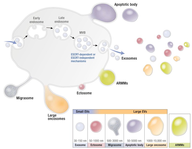
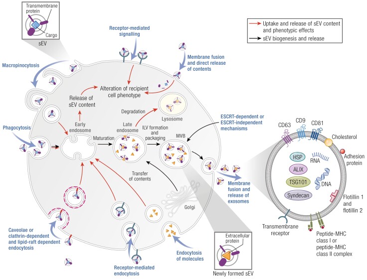
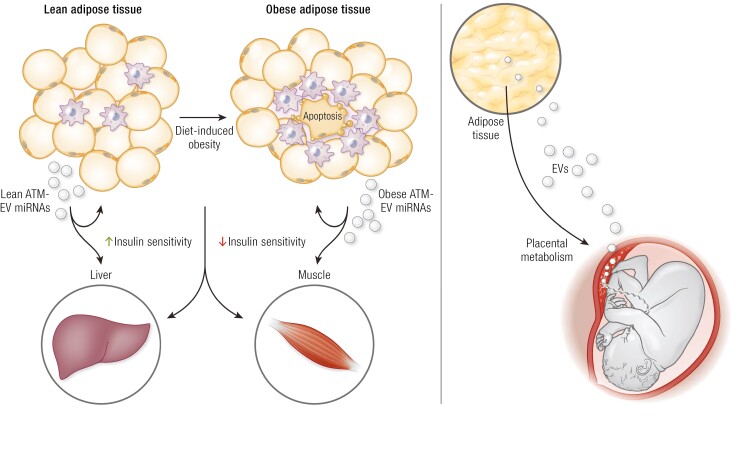
During the last decade, there has been great interest in elucidating the biological role of extracellular vesicles (EVs), particularly, their hormone-like role in cell-to-cell communication. The field of endocrinology is uniquely placed to provide insight into the functions of EVs, which are secreted from all cells into biological fluids and carry endocrine signals to engage in paracellular and distal interactions. EVs are a heterogeneous population of membrane-bound vesicles of varying size, content, and bioactivity. EVs are specifically packaged with signaling molecules, including lipids, proteins, and nucleic acids, and are released via exocytosis into biofluid compartments. EVs regulate the activity of both proximal and distal target cells, including translational activity, metabolism, growth, and development. As such, EVs signaling represents an integral pathway mediating intercellular communication. Moreover, as the content of EVs is cell-type specific, it is a "fingerprint" of the releasing cell and its metabolic status. Recently, changes in the profile of EV and bioactivity have been described in several endocrine-related conditions including diabetes, obesity, cardiovascular diseases, and cancer. The goal of this statement is to highlight relevant aspects of EV research and their potential role in the field of endocrinology.
Keywords: apoptotic body; biogenesis; cellular messenger; ectosome; exosome; extracellular vesicle; microvesicle; migrasome; oncosome; signaling.
© The Author(s) 2022. Published by Oxford University Press on behalf of the Endocrine Society. All rights reserved. For permissions, please e-mail: journals.permissions@oup.com.
Figures


References
-
- Starling EH. The Croonian Lecture; the chemical correlation of the functions of the body. Lancet. 1905;166:339-341.
-
- Salomon C, Rice GE. Role of exosomes in placental homeostasis and pregnancy disorders. Prog Mol Biol Transl Sci. 2017;145:163-179. - PubMed
-
- Mincheva-Nilsson L. Placental exosome-mediated immune protection of the fetus: feeling groovy in a cloud of exosomes. Expert Rev Obstet Gynaecol. 2010;5:619-634.
-
- Théry C, Boussac M, Véron P, et al. Proteomic analysis of dendritic cell-derived exosomes: a secreted subcellular compartment distinct from apoptotic vesicles. J Immunol. 2001;166(12):7309-7318. - PubMed
MeSH terms
Grants and funding
LinkOut - more resources
Full Text Sources
Research Materials
