. 2022 Jul;39(7):1503-1513.
doi: 10.1007/s10815-022-02485-1. Epub 2022 May 12.

Fabrication on the microscale: a two-photon polymerized device for oocyte microinjection

Affiliations

Fabrication on the microscale: a two-photon polymerized device for oocyte microinjection

Suliman H Yagoub et al. J Assist Reprod Genet. 2022 Jul.

Abstract

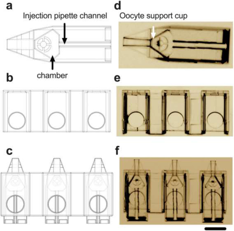
Purpose: Intracytoplasmic sperm injection (ICSI) addresses male sub-fertility by injecting a spermatozoon into the oocyte. This challenging procedure requires the use of dual micromanipulators, with success influenced by inter-operator expertise. We hypothesized that minimizing oocyte handling during ICSI will simplify the procedure. To address this, we designed and fabricated a micrometer scale device that houses the oocyte and requires only one micromanipulator for microinjection.

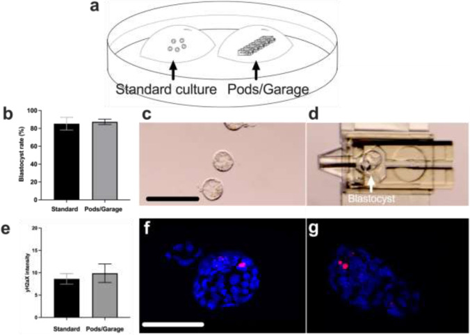
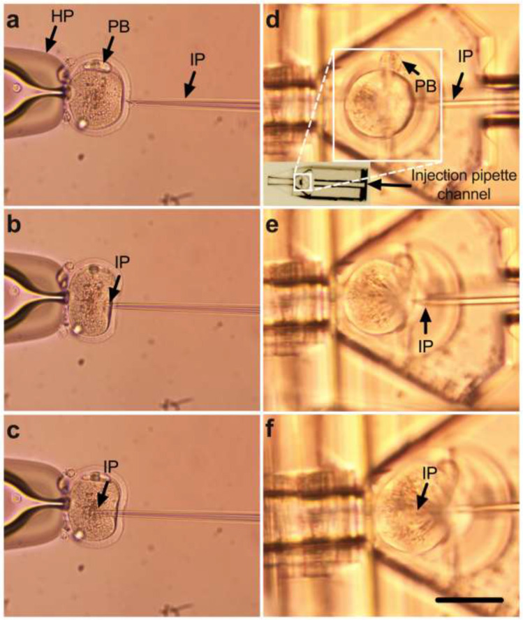
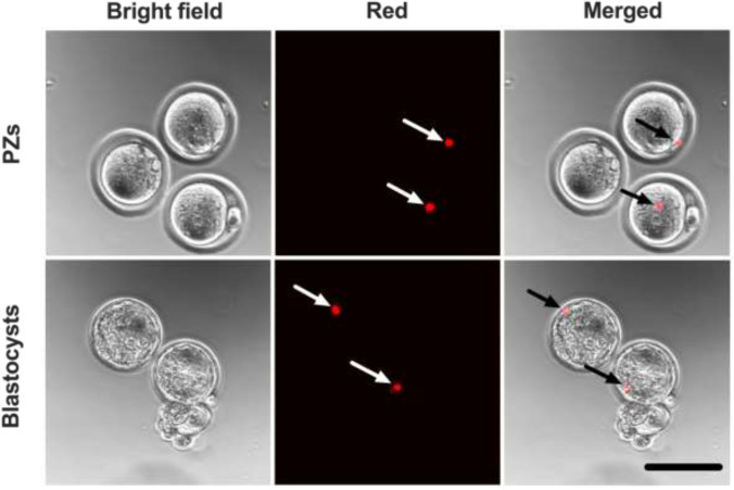
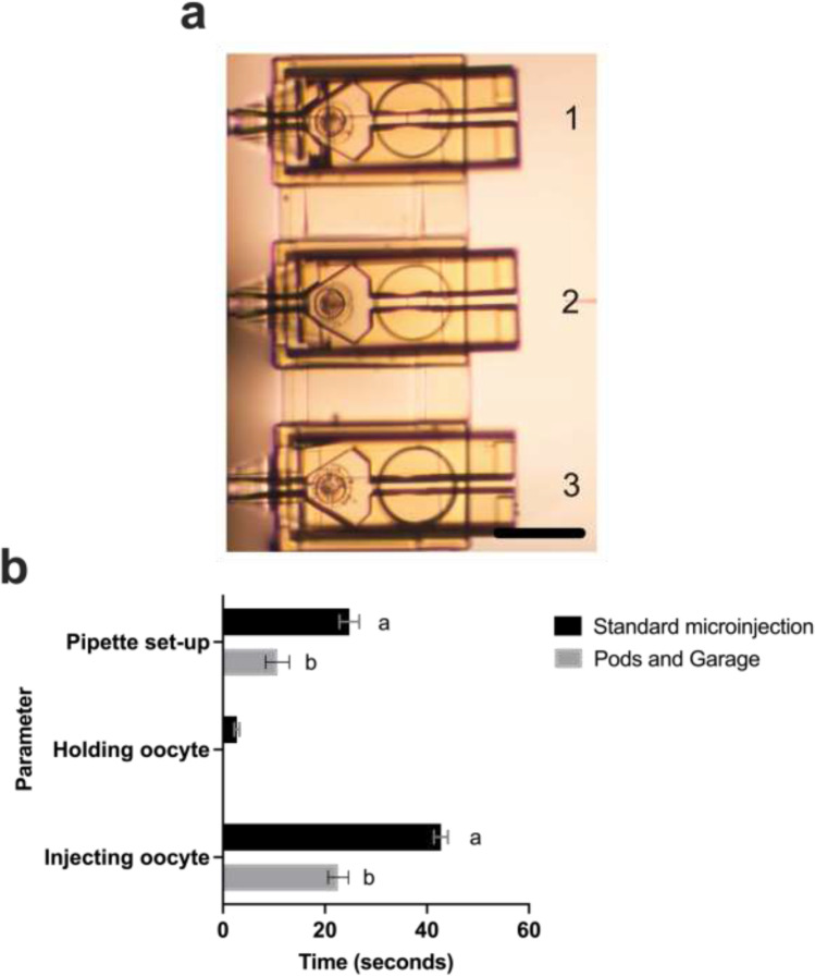
Methods: The device consisted of 2 components, each of sub-cubic millimeter volume: a Pod and a Garage. These were fabricated using 2-photon polymerization. Toxicity was evaluated by culturing single-mouse presumptive zygotes (PZs) to the blastocyst stage within a Pod, with several Pods (and embryos) docked in a Garage. The development was compared to standard culture. The level of DNA damage/repair in resultant blastocysts was quantified (γH2A.X immunohistochemistry). To demonstrate the capability to carry out ICSI within the device, PZs were microinjected with 4-μm fluorescent microspheres and cultured to the blastocyst stage. Finally, the device was assessed for oocyte traceability and high-throughput microinjection capabilities and compared to standard microinjection practice using key parameters (pipette setup, holding then injecting oocytes).

Results: Compared to standard culture, embryo culture within Pods and a Garage showed no differences in development to the blastocyst stage or levels of DNA damage in resultant blastocysts. Furthermore, microinjection within our device removes the need for a holding pipette, improves traceability, and facilitates high-throughput microinjection.

Conclusion: This novel device could improve embryo production following ICSI by simplifying the procedure and thus decreasing inter-operator variability.

Keywords: 3D fabrication; ART; High-throughput microinjection; ICSI; IVF; Infertility.


Conflict of interest statement

J. G. Thompson is a Director and Chief Scientific Officer of Fertilis Pty Ltd. All the other authors declare no competing interests. A PCT patent (PCT/AU2020/051318) has been granted.

Figures

Fig. 1
Fig. 1
Design and fabrication of a device on the micron scale for oocyte microinjection. Three-dimensional schematic of the Pod (a) and Garage (b) and an illustration of three Pods docked within a Garage (c). The Pod (d) and Garage (e) were fabricated using two-photon polymerization (Nanoscribe GmBH, Eggenstein-Leopoldshafen, Germany). In (c) and (f), 3 Pods are docked within a Garage (1500 × 450 × 310 μm; l × w × h). The Pod (725 × 250 × 250 μm) includes access for an injection pipette (injection pipette channel (a and d)) and a chamber with a raised bed that houses and holds the oocyte during microinjection (oocyte support cup (a and d)). Images (d, e, and f) were taken using a 20 × objective with a final magnification of 20 × (Nikon SMZ1500 microscope, Nikon Instruments, Inc., NY, USA). Scale bar = 250 μm
Fig. 2
Fig. 2
Embryo culture within Pods and a Garage has no impact on preimplantation development or DNA integrity within resultant blastocysts. The experimental design used to assess potential embryo toxicity of the Pod and Garage is shown in (a). Mouse presumptive zygotes (PZs) were cultured within a micro-volume of medium overlaid with paraffin oil, with embryo development occurring either on the base of the dish (standard culture) or within the Pods and a Garage (Pods/Garage) (a). Blastocyst rate was calculated from starting number of PZs (b). Representative images of blastocysts developed in standard culture or within a Pod and Garage are shown in (c) and (d), respectively. The integrity of DNA within resultant blastocysts was assessed using γH2A.X immunohistochemistry (e). Representative images of DNA integrity within blastocysts cultured in standard culture or within a Pod and Garage are shown in (f) and (g), respectively [Phospho-histone H2A.X; γH2A.X (red)/4′,6-diamidino-2-phenylindole: DAPI (blue)]. Images were captured using a 10 × objective with a final magnification of 10 × (Nikon SMZ1500 microscope (c and d)) or using a 60 × objective with a final magnification of 60 × (Olympus Fluoview 3000 confocal microscope, DAPI: 358/461 nm and γH2A.X: 591/614 nm (f and g)). All data are presented as mean ± SEM (n = 4 experimental replicates, representative of a total of 200–315 embryos for blastocyst development, and a total of 19–33 embryos for DNA integrity). Data for embryo development was arcsine transformed prior to statistical analysis (b). All data were analyzed using an unpaired Student’s t-test, P > 0.05. Scale bar = 200 μm (c) and 120 μm (f)
Fig. 3
Fig. 3
Compared to standard practice, the microinjection of oocytes within a Pod and Garage avoids the need for a holding pipette, simplifying the procedure. Panels show the microinjection of a mouse metaphase II oocyte using either standard microinjection (ac) or microinjection within a Pod and a Garage (df). In both instances, the oocyte is oriented such that the polar body (PB) is located at the 12 o’clock position to avoid the metaphase plate during microinjection. During standard microinjection, a holding pipette (HP) is used to hold the oocyte (ac). The injection pipette (IP) is pushed against the zona pellucida (a), the ooplasm is then penetrated (b), and microinjection occurs by the application of negative pressure within the IP (c). Oocyte microinjection within a Pod and a Garage is performed using an IP but does not require a HP. The oocyte is loaded into the oocyte support cup (inset) which aligns the oocyte with the injection pipette channel (d). The IP is pushed against the zona pellucida (d), the ooplasm is then penetrated (e), and microinjection occurs by application of negative pressure within the IP (f). Images were captured using a 20 × objective at a final magnification of 20 × (ad) or using a 40 × objective with a final magnification of 40 × (e, f) (Nikon Eclipse TE2000-E microscope). Scale bar = 120 μm
Fig. 4
Fig. 4
Successful completion of preimplantation embryo development following microinjection of presumptive zygotes (PZs) within Pods and a Garage. To demonstrate the utility of the Pod and Garage system for intracytoplasmic sperm injection (ICSI), mouse PZs were injected with fluorescent microspheres (4 μm) while housed within Pods and a Garage. Following microinjection, PZs were cultured to the blastocyst stage. Images were captured using a 10 × objective with a final magnification of 38 × (Olympus Fluoview 10i confocal microscope). The first column on the left shows PZs under bright field, while the second column shows PZs under the red fluorescence channel (660/680 nm) and the third column shows merged images of the bright field and red channels. Arrows point at the location of fluorescent microspheres within the microinjected PZs and resultant blastocysts. Scale bar = 120 μm
Fig. 5
Fig. 5
Multiple Pods docked within a Garage improves traceability and enables high-throughput microinjection of oocytes. Three mouse oocytes were preloaded into individual Pods that were then docked into a Garage (a). Quantification of time required for individual parameters of the microinjection procedure (pipette setup; holding an oocyte; injecting individual oocytes) and comparison between standard microinjection and microinjection within the Pods and a Garage is shown in (b). Images were captured using a 20 × objective with a final magnification of 20 × (Nikon Eclipse TE2000-E microscope). All data are presented as mean ± SEM (pipette setup: n = 3–5 experimental replicates; holding an oocyte: n = 3 experimental replicates with a total of 45 oocytes for the standard group [N/A for Pod/Garage group]; and injection of individual oocytes: n = 3 experimental replicates representative of a total of 18–45 oocytes). Data were analyzed using an unpaired Student’s t-test, P < 0.01. Different superscripts denote statistical difference between procedures (standard microinjection vs within Pods and a Garage) within a parameter. Scale bar = 200 μm

Similar articles

Cited by

References

    1. Kumar N, Singh AK. Trends of male factor infertility, an important cause of infertility: a review of literature. J Hum Reprod Sci. 2015;8(4):191–196. doi: 10.4103/0974-1208.170370. - DOI - PMC - PubMed
    1. Jodar M, Soler-Ventura A, Oliva R. Semen proteomics and male infertility. J Proteomics. 2017;162:125–134. doi: 10.1016/j.jprot.2016.08.018. - DOI - PubMed
    1. Silber S, Van Steirteghem A, Liu J, Nagy Z, Tournaye H, Devroey P. High fertilization and pregnancy rate after intracytoplasmic sperm injection with spermatozoa obtained from testicle biopsy. Hum Reprod. 1995;10(1):148–152. doi: 10.1093/humrep/10.1.148. - DOI - PubMed
    1. Esteves SC, Roque M, Bedoschi G, Haahr T, Humaidan P. Intracytoplasmic sperm injection for male infertility and consequences for offspring. Nat Rev Urol. 2018;15(9):535–562. doi: 10.1038/s41585-018-0051-8. - DOI - PubMed
    1. Lu Z, Zhang X, Leung C, Esfandiari N, Casper RF, Sun Y. Robotic ICSI (intracytoplasmic sperm injection) IEEE Trans Biomed Eng. 2011;58(7):2102–2108. doi: 10.1109/TBME.2011.2146781. - DOI - PubMed

LinkOut - more resources