. 2022 Jul 4;221(3):iyac073.
doi: 10.1093/genetics/iyac073.

DREAM interrupted: severing LIN-35-MuvB association in Caenorhabditis elegans impairs DREAM function but not its chromatin localization

Affiliations

DREAM interrupted: severing LIN-35-MuvB association in Caenorhabditis elegans impairs DREAM function but not its chromatin localization

Paul D Goetsch et al. Genetics. .

Abstract

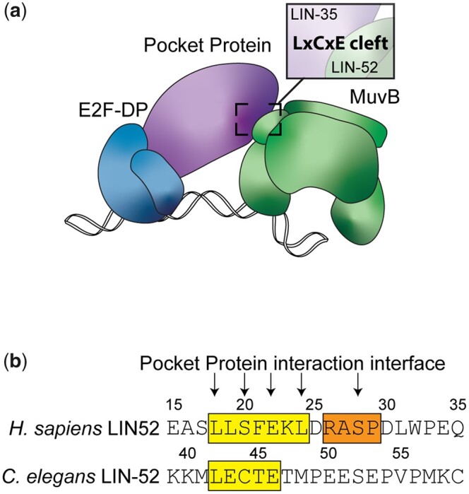
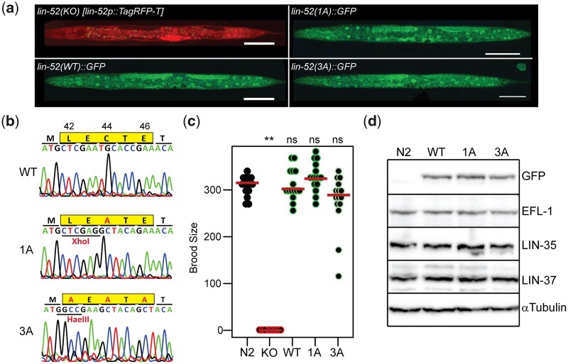
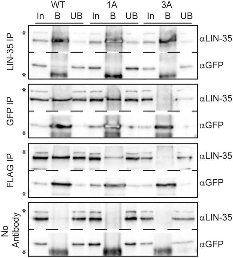
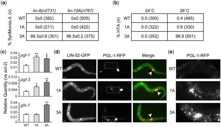
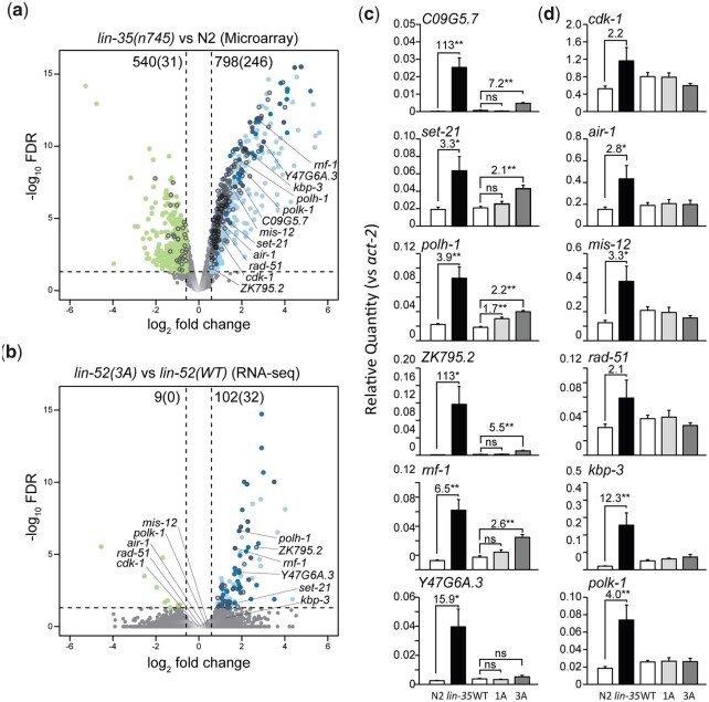
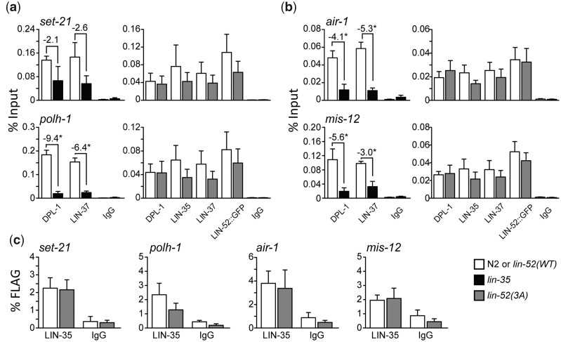
The mammalian pocket protein family, which includes the Retinoblastoma protein (pRb) and Rb-like pocket proteins p107 and p130, regulates entry into and exit from the cell cycle by repressing cell cycle gene expression. Although pRb plays a dominant role in mammalian systems, p107 and p130 are the ancestral pocket proteins. The Rb-like pocket proteins interact with the highly conserved 5-subunit MuvB complex and an E2F-DP transcription factor heterodimer, forming the DREAM (for Dp, Rb-like, E2F, and MuvB) complex. DREAM complex assembly on chromatin culminates in repression of target genes mediated by the MuvB subcomplex. Here, we examined how the Rb-like pocket protein contributes to DREAM formation and function by disrupting the interaction between the sole Caenorhabditis elegans pocket protein LIN-35 and the MuvB subunit LIN-52 using CRISPR/Cas9 targeted mutagenesis. A triple alanine substitution of LIN-52's LxCxE motif severed LIN-35-MuvB association and caused classical DREAM mutant phenotypes, including synthetic multiple vulvae, high-temperature arrest, and ectopic expression of germline genes in the soma. However, RNA-sequencing revealed limited upregulation of DREAM target genes when LIN-35-MuvB association was severed, as compared with gene upregulation following LIN-35 loss. Based on chromatin immunoprecipitation, disrupting LIN-35-MuvB association did not affect the chromatin localization of E2F-DP, LIN-35, or MuvB components. In a previous study, we showed that in worms lacking LIN-35, E2F-DP, and MuvB chromatin occupancy was reduced genome-wide. With LIN-35 present but unable to associate with MuvB, our study suggests that the E2F-DP-LIN-35 interaction promotes E2F-DP's chromatin localization, which we hypothesize supports MuvB chromatin occupancy indirectly through DNA. Altogether, this study highlights how the pocket protein's association with MuvB supports DREAM function but is not required for DREAM's chromatin occupancy.

Keywords: CRISPR/Cas9 genome editing; DREAM; MuvB; pocket protein; transcriptional repression.


Figures

Fig. 1.
Fig. 1.
LIN-35 and MuvB associate via the LxCxE motif of LIN-52. a) Model of the C. elegans DREAM complex bound to DNA: E2F-DP, the pocket protein LIN-35, and the 5-subunit MuvB subcomplex. The highlighted region shows the target region for this study: an LxCxE binding motif in the MuvB subunit LIN-52 that interacts directly with the LIN-35 pocket protein. b) Alignment of Homo sapiens LIN52 and C. elegans LIN-52 sequences. The human LxSxExL and worm LxCxE sequences are highlighted in yellow, and the human DYRK1A consensus phosphorylation sequence is highlighted in orange. Arrows indicate residues involved in the interaction with the pocket protein and amino acid residues converted to alanine in this study.
Fig. 2.
Fig. 2.
Targeted mutagenesis to disrupt DREAM complex formation. a) Live worm fluorescence images of lin-52(KO), lin-52(WT), lin-52(1A), and lin-52(3A) L4 larvae. Composites were artificially straightened. Scale bars, 100 µM. b) Sanger sequencing of the lin-52 LxCxE coding region (highlighted in yellow) in lin-52(WT), lin-52(1A), and lin-52(3A). c) Swarm plots of the brood sizes of WT (N2) worms and lin-52(KO), lin-52(WT), lin-52(1A), and lin-52(3A) transgenic worms. Significance (**P-value < 0.01) was determined by a Wilcoxon-Mann-Whitney test comparing the indicated strains to WT (N2). d) Western blot analysis of DREAM subunits LIN-52 (via GFP tag), EFL-1, LIN-35, and LIN-37 using lysates from WT (N2) worms and lin-52(WT), lin-52(1A), and lin-52(3A) transgenic worms separated by SDS PAGE. Antibodies used are indicated on the right. Alpha-tubulin was used as a loading control. Full blots are shown in Supplementary Fig. 1.
Fig. 3.
Fig. 3.
lin-52 LxCxE binding motif mutants block DREAM formation. Late embryo extracts from lin-52(WT), lin-52(1A), and lin-52(3A) (each tagged with GFP and FLAG) were immunoprecipitated with anti-LIN-35, anti-GFP, and anti-FLAG antibodies, with no antibody serving as a negative control. Proteins bound (B) and unbound (UB) were separated by SDS PAGE, and western blot analysis was performed using the antibodies indicated on the right. 5% of Input (In) is shown on the left. Asterisks indicate nonspecific bands. Full blots are shown in Supplementary Fig. 2.
Fig. 4.
Fig. 4.
Mutagenesis of the lin-52 LxCxE binding motif causes SynMuv B phenotypes. a) Table indicating the percentage Synthetic Multivulval (SynMuv) worms when lin-52(WT), lin-52(1A), and lin-52(3A) were combined with SynMuv A mutant alleles lin-8(n2731) or lin-15A(n767) with standard deviation indicated. The population size (n) is indicated in parentheses. b) Table indicating the percentage of HTA observed in lin-52(WT), lin-52(1A), and lin-52(3A) incubated at 24°C and 26°C. The population size (n) is indicated in parentheses. c) RT-qPCR analysis comparing transcript levels of 3 germline genes (pgl-1, pgl-3, and glh-1) in lin-52(WT) (white), lin-52(1A) (light grey), and lin-52(3A) (dark gray) late embryos. Expression values from 6 biological replicates were averaged and are presented as the relative quantity compared with act-2. Error bars indicate SEM, and significance was determined by a Student’s t-test between transcript levels in mutant (3A or 1A) vs WT (*P-value < 0.05, **P-value < 0.01). d, e) Live worm fluorescence images of lin-52(WT), lin-52(1A), and lin-52(3A) L1 larvae containing a PGL-1::RFP reporter gene. White arrowheads indicate the primordial germ cells Z2 and Z3. Scale bars, 10 µm. The white boxes in (d) indicate regions of ectopic expression of PGL-1::RFP in the intestine of lin-52(1A) and lin-52(3A) shown in (e).
Fig. 5.
Fig. 5.
Disruption of DREAM formation leads to upregulation of a small subset of target genes. a, b) Volcano plots of log2-fold change in transcript levels vs log10 false discovery rate (FDR) of 13,721 genes shared between (a) lin-35(n745) vs N2 L1 microarray expression analysis reported in (Kirienko and Fay 2007) and (b) lin-52(3A) vs lin-52(WT) late embryo RNA-seq. Genes downregulated or upregulated are highlighted, with dark circles indicating upregulated genes observed in both lin-35 and lin-52(3A) data sets. Black circle outlines indicate DREAM target genes, as reported in (Goetsch et al. 2017). The number of genes differentially expressed in each analysis is indicated at the top of each plot, with the number of DREAM target genes differentially expressed indicated in parentheses. Dashed lines indicate the significance cutoff of q = 0.05 (horizontal lines) and a 1.5-fold change in transcript level (vertical lines). Genes selected for RT-qPCR analysis are labeled. c, d) RT-qPCR analysis comparing transcript levels of 6 DREAM target genes that were upregulated in RNA-seq (c) and 6 DREAM target genes that were not upregulated in RNA-seq (d). Transcript levels in late embryos are from WT N2 (white), lin-35 (black), lin-52(WT) (white), lin-52(1A) (light gray), and lin-52(3A) (dark grey). Expression values from 6 biological replicates were averaged and are presented as the relative quantity compared with act-2. Error bars indicate SEM, fold change values > 1.5-fold are provided, and significance was determined by a Student’s t-test between transcript levels in mutant (3A or 1A) vs WT or lin-35 vs N2 (*P-value < 0.05, **P-value < 0.01).
Fig. 6.
Fig. 6.
Analysis of chromatin localization of DREAM subunits at target genes. a, b) ChIP-qPCR of DREAM subunits DPL-1 and LIN-37 in N2 (white) vs lin-35 (black) and DPL-1, LIN-35, LIN-37, and LIN-52 (via GFP tag) in lin-52(WT) (white) vs lin-52(3A) (dark gray) late embryo extracts at 2 DREAM target genes that were upregulated in RNA-seq (a) and 2 DREAM target genes that were not upregulated in RNA-seq (b). IgG was used as a negative control. Signals are presented as percentage of Input DNA, with negative fold-change values >2-fold noted. Error bars indicate SEM. Significance was determined by a Student’s t-test between subunit ChIP values in mutant (lin-35 or lin-52(3A)) vs WT control (N2 or lin-52(WT)) (*P-value < 0.05). c) Sequential ChIP-qPCR of LIN-52 (via FLAG tag) followed by LIN-35 or IgG from lin-52(WT) (white) and lin-52(3A) (dark gray) late embryo extracts at 4 DREAM target genes. Signals are presented as percentage of FLAG IP DNA. Error bars indicate SEM. Additional data are shown in Supplementary Fig. 3.

Similar articles

Cited by

References

    1. Anders S, Pyl PT, Huber W.. HTSeq—a Python framework to work with high-throughput sequencing data. Bioinformatics. 2015;31(2):166–169. - PMC - PubMed
    1. Arribere JA, Bell RT, Fu BXH, Artiles KL, Hartman PS, Fire AZ.. Efficient marker-free recovery of custom genetic modifications with CRISPR/Cas9 in Caenorhabditis elegans. Genetics. 2014;198(3):837–846. - PMC - PubMed
    1. Asthana A, Ramanan P, Hirschi A, Guiley KZ, Wijeratne TU, Shelansky R, Doody MJ, Narasimhan H, Boeger H, Tripathi S, et al.The MuvB complex binds and stabilizes nucleosomes downstream of the transcription start site of cell-cycle dependent genes. Nat Commun. 2022;13(1):526. - PMC - PubMed
    1. Beitel GJ, Lambie EJ, Horvitz HR.. The C. elegans gene lin-9, which acts in an Rb-related pathway, is required for gonadal sheath cell development and encodes a novel protein. Gene. 2000;254(1–2):253–263. - PubMed
    1. Boichuk S, Parry JA, Makielski KR, Litovchick L, Baron JL, Zewe JP, Wozniak A, Mehalek KR, Korzeniewski N, Seneviratne DS, et al.The DREAM complex mediates GIST cell quiescence and is a novel therapeutic target to enhance imatinib-induced apoptosis. Cancer Res. 2013;73(16):5120–5129. - PubMed

Publication types

MeSH terms