. 2022 May 13;13(1):90.
doi: 10.1186/s13244-022-01211-w.

Artifact reduction in contrast-enhanced mammography

Affiliations

Artifact reduction in contrast-enhanced mammography

Gisella Gennaro et al. Insights Imaging. .

Abstract

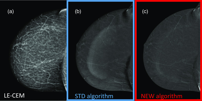
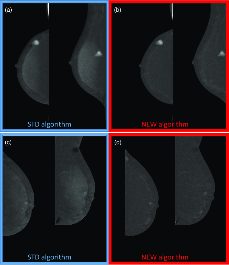
Objective: To evaluate the effectiveness of a new algorithm developed to reduce artifacts in dual-energy subtraction (DES) contrast-enhanced mammography (CEM) images while preserving contrast enhancement of possible lesions.

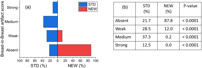
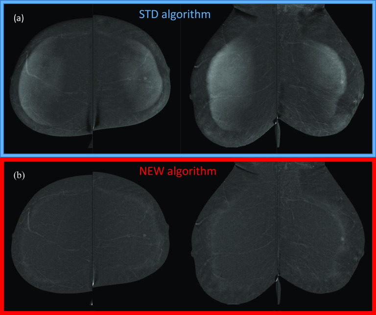
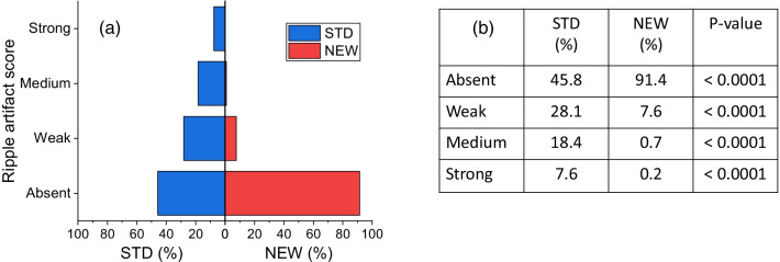
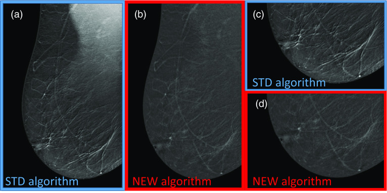
Methods: A retrospective multi-reader paired study was performed by using 134 CEM studies obtained from the first 134 women enrolled in a prospective clinical study aiming to compare the clinical performance of CEM to those of breast MRI in screening of women at increased risk of breast cancer. Four experienced readers compared independently the standard (STD) DES images with those obtained by reprocessing the raw images by a new algorithm (NEW), expected to reduce the DES artifact intensity. The intensity of three types of artifacts (breast-in-breast, ripple, and skinfold enhancement) and the intensity of possible contrast uptake were assessed visually and rated using a categorical ordinal scale. Proportions of images rated by the majority of readers as "Absent", "Weak", "Medium", "Strong" in each artifact intensity category were compared between the two algorithms. P-values lower than 0.05 were considered statistically significant.

Results: The NEW algorithm succeeded in eliminating 84.5% of breast-in-breast artifacts, 84.2% of ripple artifacts, and 56.9% of skinfold enhancement artifacts versus STD DES images, and reduced the artifact intensity in 12.1%, 13.0%, and 28.8% of the images, respectively. The visibility of lesion contrast uptake was the same with the STD and the NEW algorithms.

Conclusion: The new dual-energy subtraction algorithm demonstrated to be effective in reducing/eliminating CEM-related artifacts while preserving lesion contrast enhancement.

Keywords: Artifact; Contrast agent; Mammography.


Conflict of interest statement

The authors declare that they have no competing interests.

Figures

Fig. 1
Fig. 1
a Mirror bar graphs showing the percentages of DES CEM images for which the intensity of breast-in-breast artifact was scored “Absent”, “Weak”, “Medium” or “Strong” by the majority of readers, when the images were obtained with the STD (blue bars) and the NEW (red bars) algorithm, respectively; b results of comparison between proportions associated with STD and NEW algorithms for each category of breast-in-breast artifact and related p-values
Fig. 2
Fig. 2
Postmenopausal woman 56 yo at high risk for breast cancer with large, fatty breasts. a DES CEM images obtained by the STD algorithm showing the breast-in-breast artifact in all the four views; b DES CEM images obtained by the NEW algorithm, where the artifact was completely removed
Fig. 3
Fig. 3
a Mirror bar graphs showing the percentages of DES CEM images for which the intensity of ripple artifact was scored “Absent”, “Weak”, “Medium” or “Strong” by the majority of readers, when the images were obtained with the STD (blue bars) and the NEW (red bars) algorithm, respectively; b results of comparison between proportions associated with STD and NEW algorithms for each category of ripple artifact and related p-values
Fig. 4
Fig. 4
Premenopausal woman 46 yo at high risk for breast cancer with large, fatty breasts. a RMLO DES CEM view obtained by the STD algorithm showing the ripple artifact in the lower part of the image; b RMLO CEM view obtained by the NEW algorithm, where the artifact was completely removed; c Detail of the ripple artifact in the lower part of the RMLO CEM view with the STD algorithm; d Same detail of the lower part of the RMLO CEM view after removal by the NEW algorithm
Fig. 5
Fig. 5
a Mirror bar graphs showing the percentages of DES CEM images for which the intensity of skinfold enhancement was scored “Absent”, “Weak”, “Medium” or “Strong” by the majority of readers, when the images were obtained with the STD (blue bars) and the NEW (red bars) algorithm, respectively; b results of comparison between proportions associated with STD and NEW algorithms for each category of skinfold enhancement artifact and related P values
Fig. 6
Fig. 6
Postmenopausal woman 60 yo at intermediate risk for breast cancer with large, fatty breasts. a RCC LE-CEM view showing a large skinfold close to the chest wall; b RCC DES CEM view obtained by the STD algorithm showing the skinfold enhancement artifact (in addition to the breast-in-breast artifact); c RCC DES CEM view obtained by the NEW algorithm, where the skinfold enhancement is markedly reduced
Fig. 7
Fig. 7
a Mirror bar graphs showing the percentages of DES CEM images for which the intensity of contrast uptake visibility was scored “Absent”, “Weak”, “Medium” or “Strong” by the majority of readers, when the images were obtained with the STD (blue bars) and the NEW (red bars) algorithm, respectively; b results of comparison between proportions associated with STD and NEW algorithms for each category of contrast uptake visibility and related p-values
Fig. 8
Fig. 8
a and b Postmenopausal woman 55 yo at intermediate risk for breast cancer with small, heterogeneously dense breasts and intense contrast uptake corresponding to a 15 mm lesion in the upper-inner quadrant which was proven to be a high grade invasive lobular carcinoma: a RCC and RMLO DES CEM views obtained by the STD algorithm showing a marked breast-in-breast artifact and an intense contrast uptake; b RCC and RMLO DES CEM views obtained by the NEW algorithm without artifacts and with the same contrast uptake. c and d Postmenopausal woman 62 yo woman at intermediate risk for breast cancer with small, heterogeneously dense breasts, showing a tiny contrast uptake corresponding to a 7 mm round lesion behind the nipple: c LCC and LMLO DES CEM views obtained by the STD algorithm, with the breast-in-breast artifact partially overlapped to the lesion; d LCC and LMLO DES CEM views obtained by the NEW algorithm, where the artifact is eliminated while the lesion is more easily detectable
Fig. 9
Fig. 9
Likert scores by the majority of readers used to compare the overall preferences between the two algorithms. The readers rated the image quality of the CEM images processed by the NEW dual-energy subtraction algorithm “better” or “much better” than images processed by the STD algorithm in 97.8% of cases, while remaining 2.8% of cases were judged “equal” with the two algorithms

Similar articles

Cited by

References

    1. Ghaderi KF, Phillips J, Perry H, et al. Contrast-enhanced mammography: current applications and future directions. Radiographics. 2019;39:1907–1920. doi: 10.1148/rg.2019190079. - DOI - PubMed
    1. Sensakovic WF, Carnahan MB, Czaplicki CD, et al. Contrast-enhanced mammography: how does it work? Radiographics. 2021;41:829–839. doi: 10.1148/rg.2021200167. - DOI - PubMed
    1. Patel BK, Lobbes MBI, Lewin J. Contrast enhanced spectral mammography: a review. Semin Ultrasound CT MR. 2018;39:70–79. doi: 10.1053/j.sult.2017.08.005. - DOI - PubMed
    1. Sogani J, Mango VL, Keating D, et al. Contrast-enhanced mammography: past, present, and future. Clin Imaging. 2021;69:269–279. doi: 10.1016/j.clinimag.2020.09.003. - DOI - PMC - PubMed
    1. James JJ, Tennant SL. Contrast-enhanced spectral mammography (CESM) Clin Radiol. 2018;73:715–723. doi: 10.1016/j.crad.2018.05.005. - DOI - PubMed