

Animal Models of COVID-19: Transgenic Mouse Model
- PMID: 35554912
- PMCID: PMC9563002
- DOI: 10.1007/978-1-0716-2111-0_16
Animal Models of COVID-19: Transgenic Mouse Model
Abstract
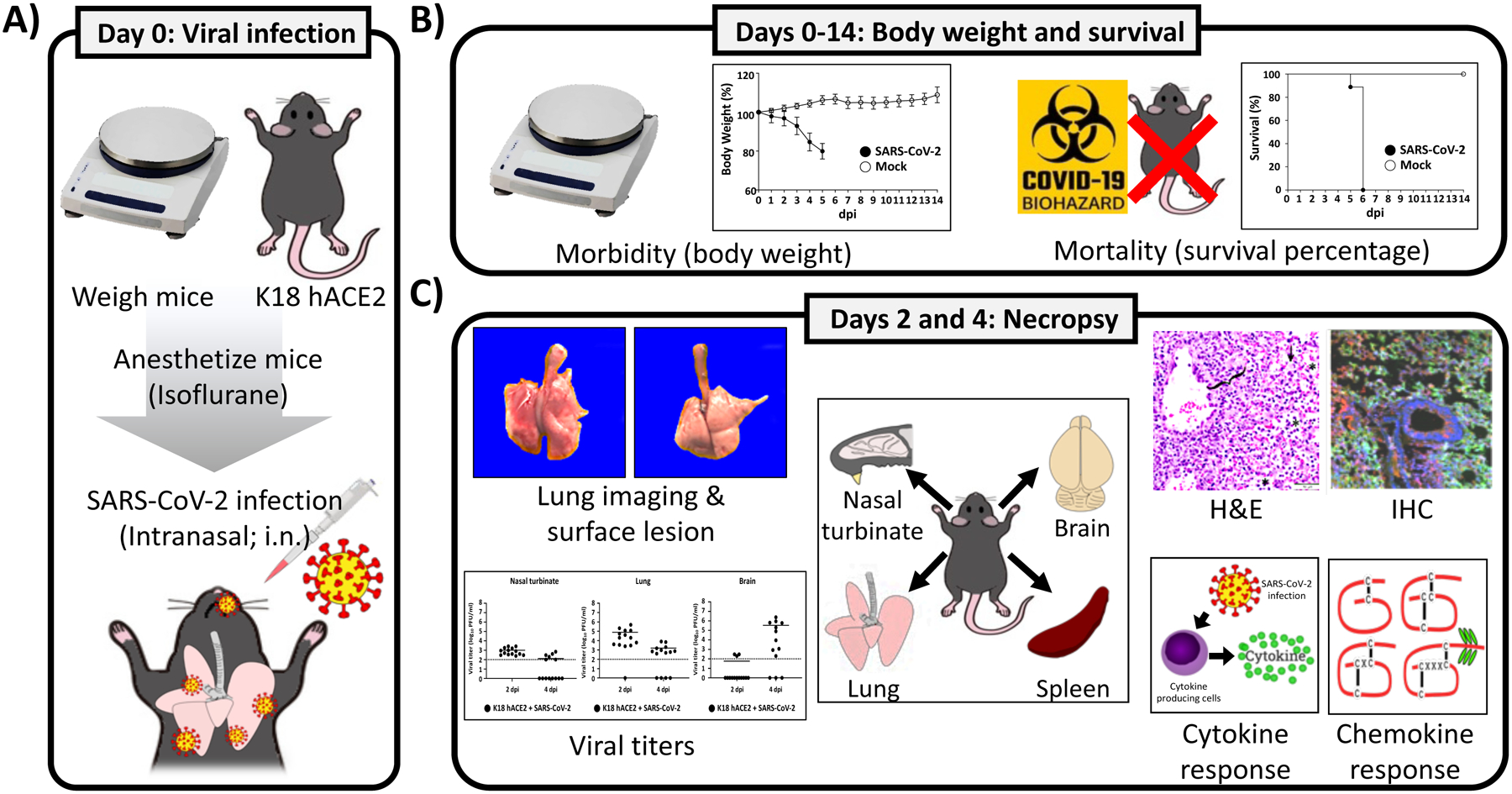
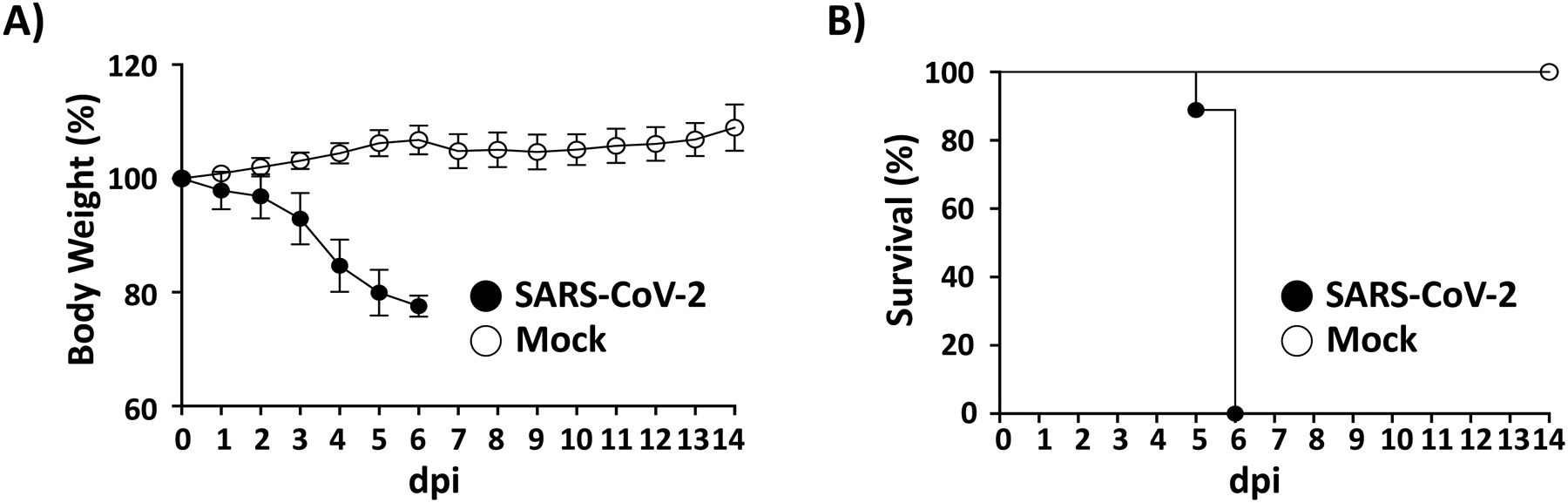
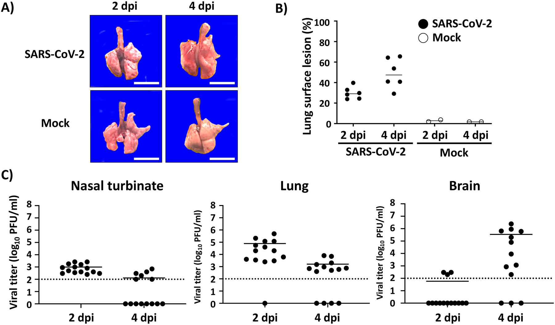
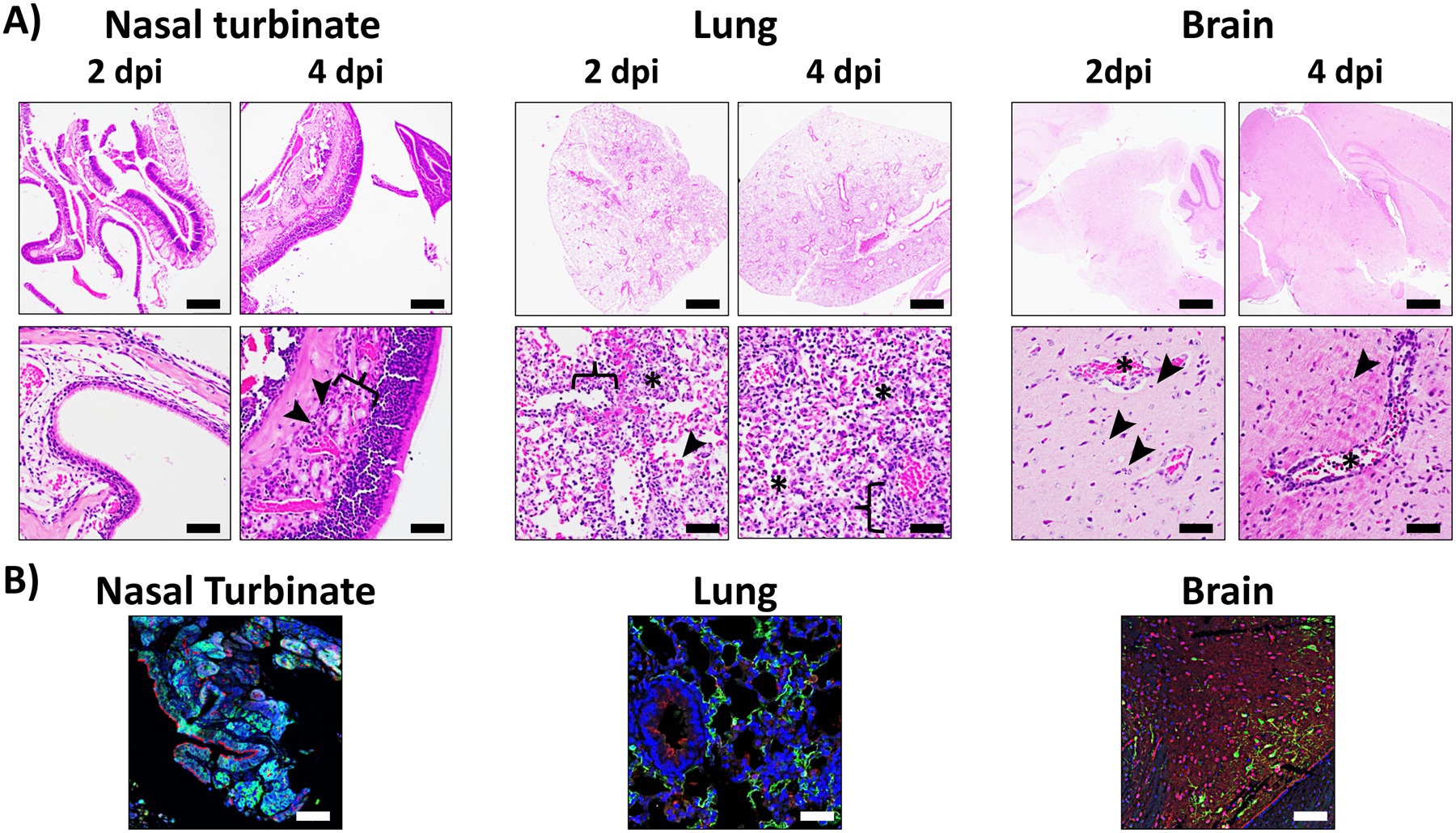
Severe acute respiratory syndrome coronavirus 2 (SARS-CoV-2), the causative agent of coronavirus disease 2019 (COVID-19), emerged in December 2019 in Wuhan, China, and rapidly spread throughout the world, threatening global public health. An animal model is a valuable and a crucial tool that allows understanding of nature in the pathogenesis of SARS-CoV-2 and its associated COVID-19 disease. Here we introduce detailed protocols of SARS-CoV-2 infection and COVID-19 disease using C57BL/6 (B6) transgenic mice expressing the human angiotensin-converting enzyme 2 (hACE2) from the human cytokeratin 18 promoter (K18 hACE2). To mimic natural SARS-CoV-2 infection, K18 hACE2 transgenic mice are infected intranasally under anesthesia. Upon infection, viral pathogenesis is determined by monitoring changes in body weight (morbidity) and monitoring survival (mortality), cytokine/chemokine responses, gross-lung pathology, histopathology, and viral replication in tissues. The presence of the virus and viral replication is evaluated by immunohistochemistry (IHC) and viral titrations, respectively, from the upper (nasal turbinate) and the lower (lungs) respiratory tracts, and nervous system (brain). Also, the immune response to SARS-CoV-2 infection is measured by cytokine/chemokine enzyme-linked immunosorbent assay (ELISA) from lung, spleen and brain homogenates to characterize the cytokine storm that hallmarks as one of the major causes of death caused by SARS-CoV-2 infection. This small rodent animal model based on the use of K18 hACE2 transgenic mice represents an excellent option to understand the pathogenicity of natural SARS-CoV-2 strains and its recently described Variants of Concern (VoC), and will be applicable to the identification and characterization of prophylactic (vaccine) and therapeutic (antiviral and/or neutralizing monoclonal antibodies) strategies for the prevention or treatment of SARS-CoV-2 infection or its associated COVID-19 disease.
Keywords: COVID-19; K18 hACE2 transgenic mice; Mouse model; SARS-CoV-2; hACE2.
© 2022. The Author(s), under exclusive license to Springer Science+Business Media, LLC, part of Springer Nature.
Figures
Similar articles
-
A human-ACE2 knock-in mouse model for SARS-CoV-2 infection recapitulates respiratory disorders but avoids neurological disease associated with the transgenic K18-hACE2 model.mBio. 2025 May 14;16(5):e0072025. doi: 10.1128/mbio.00720-25. Epub 2025 Apr 24. mBio. 2025. PMID: 40272151 Free PMC article.
-
SARS-CoV-2 Causes Lung Infection without Severe Disease in Human ACE2 Knock-In Mice.J Virol. 2022 Jan 12;96(1):e0151121. doi: 10.1128/JVI.01511-21. Epub 2021 Oct 20. J Virol. 2022. PMID: 34668780 Free PMC article.
-
Infectious Clones Produce SARS-CoV-2 That Causes Severe Pulmonary Disease in Infected K18-Human ACE2 Mice.mBio. 2021 Apr 20;12(2):e00819-21. doi: 10.1128/mBio.00819-21. mBio. 2021. PMID: 33879586 Free PMC article.
-
K18- and CAG-hACE2 Transgenic Mouse Models and SARS-CoV-2: Implications for Neurodegeneration Research.Molecules. 2022 Jun 28;27(13):4142. doi: 10.3390/molecules27134142. Molecules. 2022. PMID: 35807384 Free PMC article. Review.
-
Natural and genetically-modified animal models to investigate pulmonary and extrapulmonary manifestations of COVID-19.Int Rev Immunol. 2024;43(1):13-32. doi: 10.1080/08830185.2022.2089666. Epub 2022 Jun 25. Int Rev Immunol. 2024. PMID: 35757923 Review.
Cited by
-
The Fc-effector function of COVID-19 convalescent plasma contributes to SARS-CoV-2 treatment efficacy in mice.Cell Rep Med. 2023 Jan 17;4(1):100893. doi: 10.1016/j.xcrm.2022.100893. Epub 2022 Dec 29. Cell Rep Med. 2023. PMID: 36584683 Free PMC article.
-
Age associated susceptibility to SARS-CoV-2 infection in the K18-hACE2 transgenic mouse model.Geroscience. 2024 Jun;46(3):2901-2913. doi: 10.1007/s11357-024-01102-6. Epub 2024 Feb 22. Geroscience. 2024. PMID: 38388916 Free PMC article.
-
The Role of Viral Infections in the Onset of Autoimmune Diseases.Viruses. 2023 Mar 18;15(3):782. doi: 10.3390/v15030782. Viruses. 2023. PMID: 36992490 Free PMC article. Review.
-
SARS-CoV-2 omicron BA.5 and XBB variants have increased neurotropic potential over BA.1 in K18-hACE2 mice and human brain organoids.Front Microbiol. 2023 Nov 23;14:1320856. doi: 10.3389/fmicb.2023.1320856. eCollection 2023. Front Microbiol. 2023. PMID: 38075874 Free PMC article.
-
A multi-epitope/CXCL11 prime/pull coronavirus mucosal vaccine boosts the frequency and the function of lung-resident memory CD4+ and CD8+ T cells and enhanced protection against COVID-19-like symptoms and death caused by SARS-CoV-2 infection.J Virol. 2023 Dec 21;97(12):e0109623. doi: 10.1128/jvi.01096-23. Epub 2023 Dec 1. J Virol. 2023. PMID: 38038432 Free PMC article.
References
-
- Yang X, Yu Y, Xu J, Shu H, Xia J, Liu H, Wu Y, Zhang L, Yu Z, Fang M, Yu T, Wang Y, Pan S, Zou X, Yuan S, Shang Y (2020) Clinical course and outcomes of critically ill patients with SARS-CoV-2 pneumonia in Wuhan, China: a single-centered, retrospective, observational study. Lancet Respir Med 8(5):475–481. 10.1016/S2213-2600(20)30079-5 - DOI - PMC - PubMed
-
- Subbarao K, McAuliffe J, Vogel L, Fahle G, Fischer S, Tatti K, Packard M, Shieh WJ, Zaki S, Murphy B (2004) Prior infection and passive transfer of neutralizing antibody prevent replication of severe acute respiratory syndrome coronavirus in the respiratory tract of mice. J Virol 78(7):3572–3577. 10.1128/jvi.78.7.3572-3577.2004 - DOI - PMC - PubMed
MeSH terms
Substances
Grants and funding
LinkOut - more resources
Full Text Sources
Medical
Miscellaneous
