Review
. 2022 Jul;474(7):649-663.
doi: 10.1007/s00424-022-02698-4. Epub 2022 May 13.

Paradigm shift: new concepts for HCN4 function in cardiac pacemaking

Affiliations
Review

Paradigm shift: new concepts for HCN4 function in cardiac pacemaking

Konstantin Hennis et al. Pflugers Arch. 2022 Jul.

Abstract

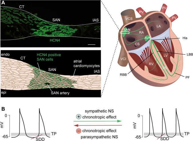
Hyperpolarization-activated cyclic nucleotide-gated (HCN) channels are the molecular correlate of the If current and are critically involved in controlling neuronal excitability and the autonomous rhythm of the heart. The HCN4 isoform is the main HCN channel subtype expressed in the sinoatrial node (SAN), a tissue composed of specialized pacemaker cells responsible for generating the intrinsic heartbeat. More than 40 years ago, the If current was first discovered in rabbit SAN tissue. Along with this discovery, a theory was proposed that cyclic adenosine monophosphate-dependent modulation of If mediates heart rate regulation by the autonomic nervous system-a process called chronotropic effect. However, up to the present day, this classical theory could not be reliably validated. Recently, new concepts emerged confirming that HCN4 channels indeed play an important role in heart rate regulation. However, the cellular mechanism by which HCN4 controls heart rate turned out to be completely different than originally postulated. Here, we review the latest findings regarding the physiological role of HCN4 in the SAN. We describe a newly discovered mechanism underlying heart rate regulation by HCN4 at the tissue and single cell levels, and we discuss these observations in the context of results from previously studied HCN4 mouse models.

Keywords: Autonomic nervous system; Chronotropic effect; HCN4 channel; Heart rate regulation; Hyperpolarization-activated cyclic nucleotide–gated channels; Sinoatrial node (SAN).


Conflict of interest statement

The authors declare no competing interests.

Figures

Fig. 1
Fig. 1
HCN4 is expressed in the sinoatrial node. (A) Right, schematic diagram of the cardiac conduction system (green). The primary pacemaker site is the sinoatrial node (SAN). The atrioventricular node (AVN) is the only electrically conductive connection between the atria and the ventricles. The bundle of His (His) splits into a left and right bundle branch (LBB/RBB, left/right bundle branch), which spread out to the left and right Purkinje fiber (PF) network. Abbreviations: LA, left atrium; RA, right atrium; PV, pulmonary veins; VCS, superior vena cava; VCI, inferior vena cava; CS, coronary sinus; RV, right ventricle; LV, left ventricle; VS, ventricular septum. (A) Left, upper panel: distribution of HCN4 (green) in a transverse section of the murine SAN. HCN4 is expressed across the entire SAN region. Abbreviations: CT, crista terminalis; IAS, interatrial septum. Scale bar: 100 µm. Lower panel: schematic illustration of the original image shown in the upper panel. (B) Action potential recordings of isolated SAN cells demonstrating the chronotropic effect at the single cell level. Input from the sympathetic nervous system accelerates SDD and increases the firing rate of pacemaker cells, whereas input from the parasympathetic nervous system slows down SDD and decelerates the firing rate. Abbreviations: SDD, slow diastolic depolarization; TP, threshold potential; NS, nervous system
Fig. 2
Fig. 2
CDR and hysteresis of HCN4 control the firing mode of SAN cells. (A) Action potential recording of an isolated pacemaker cell showing the typical alternation between firing and nonfiring. The mean membrane potential is more depolarized during firing (~ − 55 mV, green line) and more hyperpolarized during nonfiring (~ − 75 mV, red line). At the same time, slow drifts in membrane potential occur. The firing mode is characterized by a slow, progressive hyperpolarization (Δ =  − 7 mV) until firing stops (1), leading to an abrupt drop to significantly more hyperpolarized potentials. Conversely, during nonfiring, a slow and progressive depolarization occurs until the threshold for firing is reached (2), and the membrane potential abruptly jumps to substantially more depolarized values. Due to hysteresis of HCN4, the changes in membrane potential have important consequences for the voltage-dependent activation of the channel. (B) Original steady-state activation curves recorded from HCN4 channels heterologously expressed in HEK239 cells without cAMP in the intracellular solution. The long-lasting, mean membrane potentials during firing (− 55 mV) and nonfiring (− 75 mV) are mimicked by the holding potential (HP). At a relatively depolarized holding potential of − 55 mV, the activation curve is positioned at extremely hyperpolarized voltages (left curve). Conversely, at a relatively hyperpolarized holding potential of − 75 mV, the activation curve is positioned at extremely depolarized voltages (right curve). Points (1) and (2) on the activation curves reflect the time points (1) and (2) of the action potential measurements shown in panel (A). At the end of firing (1), the activation curve is shifted to the left and the membrane potential is relatively positive, resulting in a small number of open HCN4 channels. The consequent lack of a sufficiently depolarizing If current causes or supports the transition of pacemaker cells to the nonfiring mode. At the end of nonfiring (2), the activation curve is shifted to the right and the membrane potential is relatively negative, leading to a substantial increase in the number of open HCN4 channels, thereby causing or supporting the return of pacemaker cells to the firing mode. (C) Original activation curves of HCN4 channels recorded in the presence of 100 µM cAMP in the intracellular solution. Compared to panel (B), both curves are markedly shifted to the right. Under comparable conditions, SAN cells do not switch into the nonfiring mode, and the mean membrane potential permanently remains at depolarized values (− 55 mV), leading to a sufficient number of open HCN4 channels to maintain continuous firing
Fig. 3
Fig. 3
Tonic entrainment in the SAN. (A) Scheme of a SAN cell visualizing the signal transduction pathway following stimulation by the ANS. Gs protein-coupled beta-1-adrenergic receptors (green) are activated by norepinephrine (NE) released from sympathetic nerve terminals. Subsequent Gαs signaling stimulates adenylyl cyclases (ACs, gray) to synthetize cAMP, which directly activates HCN4 channels. Conversely, Gi protein-coupled M2 muscarinic receptors (red) are activated by acetylcholine (ACh) released from vagal nerve terminals. Gαi signaling inhibits ACs and thereby reduces the intracellular cAMP level, leading to a decrease in HCN4 activity. Similar effects are evoked by TRIP8bnano, a synthetic peptide that binds to the CNBD of HCN4 and inhibits cAMP-dependent activation of the channel. (B) cAMP-dependent activation of HCN4 reduces the number of nonfiring cells in the SAN, which stabilizes the network rhythm during HR acceleration. (C) Reduction in cAMP-dependent activation of HCN4 increases the number of nonfiring cells in the SAN. Overshooting inhibition leads to bradycardia and SAN dysrhythmia due to destabilization of the network rhythm. (D) The tonic entrainment process takes place between firing cells (left) and neighboring nonfiring cells (right). Pacemaker cells in the nonfiring mode are more hyperpolarized and electrotonically draw the flow of cations from more depolarized neighboring cells in the firing mode via gap junctions. This slightly depolarizes the nonfiring cells (green arrow) and hyperpolarizes the firing cells to the same extent (red arrow)
Fig. 4
Fig. 4
Cardiac phenotype of HCN4FEA mice. (A) Telemetric ECG trace of an HCN4FEA mouse showing severe sinus dysrhythmia. (B) Mean (left), minimum (middle), and maximum (right) heart rate of WT (black) and HCN4FEA mice (green) calculated from 72-h telemetric ECG recordings. (C) Heart rate histograms determined from 72-h recordings. In HCN4FEA mice, the average HR and full HR range is shifted towards lower HR values, demonstrating intrinsic bradycardia. (D) Comet-shaped Poincaré plots display high beat-to-beat dispersion in HCN4FEA mice (green). (E) Tachograms of WT (black) and HCN4FEA mice (green) before and after consecutive injections of propranolol and atropine. (F) Optical imaging measurements of biatrial SAN explants reveal prolonged sinoatrial conduction time (SACT) in HCN4FEA mice. (G) Quantification of SACT determined from optical measurements as shown in panel (F). (H) Combined telemetric blood pressure (upper panel) and ECG recordings (RR intervals, lower panel) used to determine baroreflex sensitivity in vivo. (I) Plot of systolic blood pressure (SBP) and corresponding RR intervals (upper panel) demonstrates a steeper slope of the RR/SBP relationship in HCN4FEA mice (green), reflecting inappropriately enhanced HR responses of the SAN to vagal nerve activity in HCN4FEA mice. Lower panel: quantification of the slope of RR/SBP relations in WT (black) and HCN4FEA mice (green). (J) Telemetric ECG trace of an HCN4FEA mouse during episodes with junctional escape rhythm (JER). (K) Telemetric ECG trace of an HCN4FEA mouse during episodes with isorhythmic AV dissociation (IAVD). Figure is modified from Fenske et al. [22]

References

    1. Alig J, Marger L, Mesirca P, Ehmke H, Mangoni ME, Isbrandt D. Control of heart rate by cAMP sensitivity of HCN channels. Proc Natl Acad Sci U S A. 2009;106(29):12189–12194. doi: 10.1073/pnas.0810332106. - DOI - PMC - PubMed
    1. Azene EM, Xue T, Marban E, Tomaselli GF, Li RA. Non-equilibrium behavior of HCN channels: insights into the role of HCN channels in native and engineered pacemakers. Cardiovasc Res. 2005;67(2):263–273. doi: 10.1016/j.cardiores.2005.03.006. - DOI - PubMed
    1. Baruscotti M, Bucchi A, Viscomi C, Mandelli G, Consalez G, Gnecchi-Rusconi T, Montano N, Casali KR, Micheloni S, Barbuti A, DiFrancesco D. Deep bradycardia and heart block caused by inducible cardiac-specific knockout of the pacemaker channel gene Hcn4. Proc Natl Acad Sci U S A. 2011;108(4):1705–1710. doi: 10.1073/pnas.1010122108. - DOI - PMC - PubMed
    1. Baudot M, Torre E, Bidaud I, Louradour J, Torrente AG, Fossier L, Talssi L, Nargeot J, Barrere-Lemaire S, Mesirca P, Mangoni ME. Concomitant genetic ablation of L-type Cav1.3 (alpha1D) and T-type Cav3.1 (alpha1G) Ca(2+) channels disrupts heart automaticity. Sci Rep. 2020;10(1):18906. doi: 10.1038/s41598-020-76049-7. - DOI - PMC - PubMed
    1. Behar J, Ganesan A, Zhang J, Yaniv Y. The autonomic nervous system regulates the heart rate through cAMP-PKA dependent and independent coupled-clock pacemaker cell mechanisms. Front Physiol. 2016;7:419. doi: 10.3389/fphys.2016.00419. - DOI - PMC - PubMed

Publication types

Substances

LinkOut - more resources