. 2022 Jun;12(6):e2594.
doi: 10.1002/brb3.2594. Epub 2022 May 12.

Overexpression of BDNF in the ventrolateral periaqueductal gray regulates the behavior of epilepsy-migraine comorbid rats

Affiliations

Overexpression of BDNF in the ventrolateral periaqueductal gray regulates the behavior of epilepsy-migraine comorbid rats

Long Wang et al. Brain Behav. 2022 Jun.

Abstract

Objective: To investigate the effects of brain-derived neurotrophic factor (BDNF) overexpression in the ventrolateral periaqueductal gray (vlPAG) on behavioral changes in epilepsy-migraine comorbid rats.

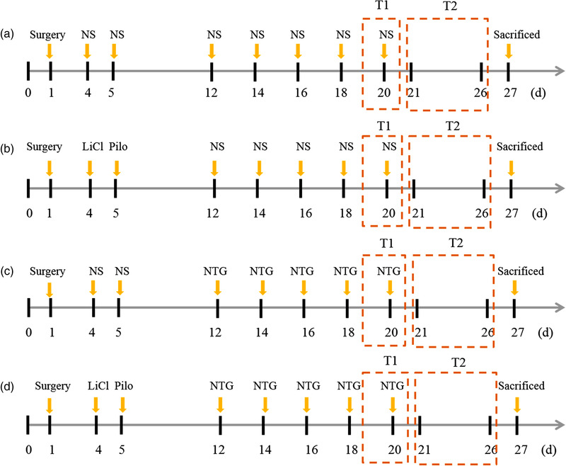
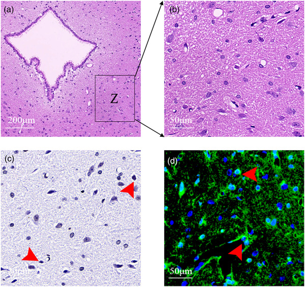
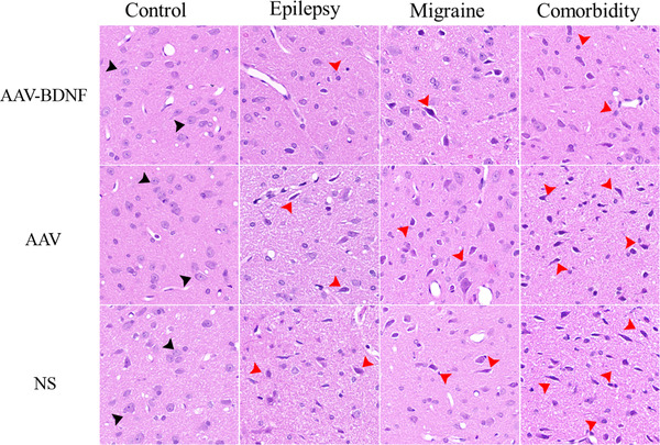
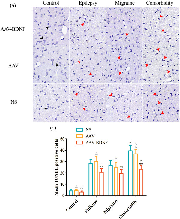
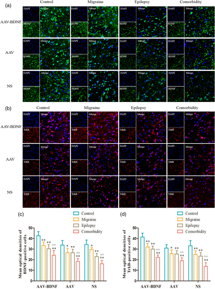
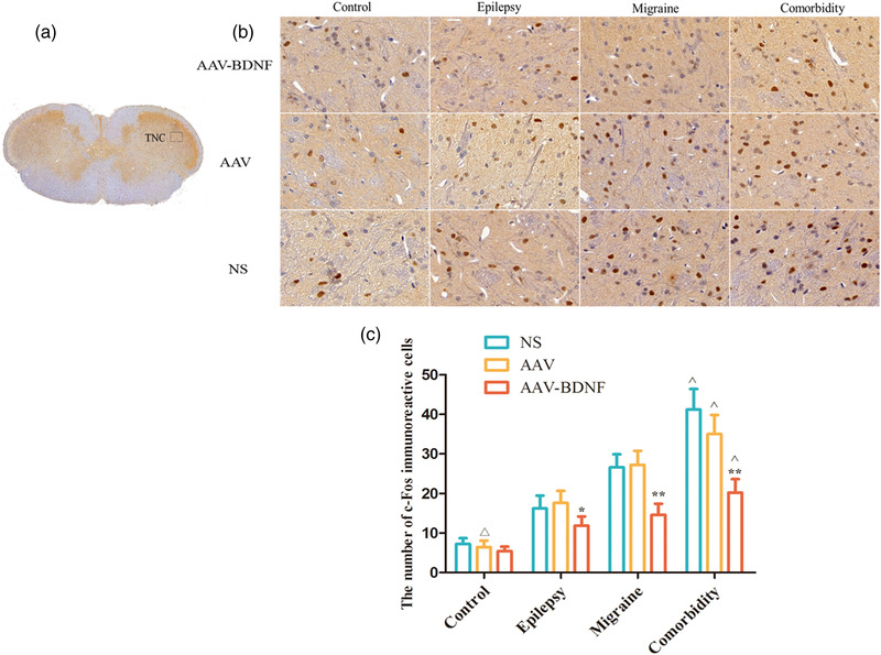
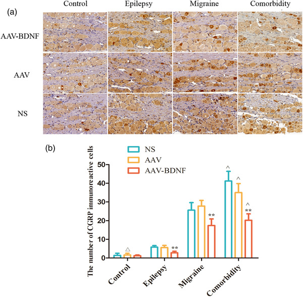
Method: We used an adeno-associated virus (AAV)-mediated vector to supplement BDNF in the vlPAG area prior to the establishment of a pilocarpine-nitroglycerin (Pilo-NTG) combination-induced comorbid model of epilepsy and migraine. Seizure- and migraine-related behaviors were analyzed. Cell loss and apoptosis in vlPAG were detected through hematoxylin-eosin (HE) and TUNEL staining. Immunofluorescence staining analyses were employed to detect expressions of BDNF and its receptor, tyrosine kinase B (TrkB), in vlPAG. Immunohistochemical staining was conducted to detect expressions of c-Fos and calcitonin gene-related peptide (CGRP) in the trigeminal nucleus caudalis (TNC) and trigeminal ganglion (TG).

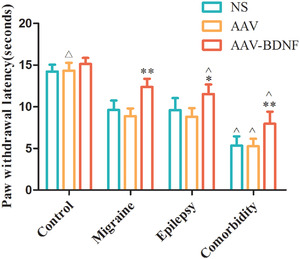
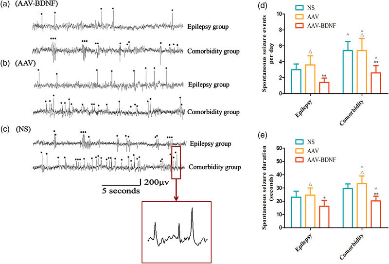
Results: Comparing to control group, AAV-BDNF injected comorbid group showed lower pain sensitivity, scratching head, and spontaneous seizures accompanied by the downregulation of c-Fos labeling neurons and CGRP immunoreactivity in the TNC and TG. However, these changes were still significantly higher in the comorbid group than those in both epilepsy and migraine groups under the same intervention.

Conclusion: These data demonstrated that supplying BDNF to vlPAG may protect structural and functional abnormalities in vlPAG and provide an antiepileptic and analgesic therapy.

Keywords: BDNF; comorbidity; epilepsy; migraine; ventrolateral periaqueductal gray.


Figures

FIGURE 1
FIGURE 1
Experimental procedure. Pretreatment with AAV vector or NS: Microinjection of AAV vector and NS by surgery prior to modeling in each group. Status epilepticus (SE) was induced by i.p. injection of lithium chloride and pilocarpine at 4 and 5 days, respectively, in both epilepsy group (B) and comorbid group (D), while injecting the equal NS in the control group (A) and migraine group (C). Recurrent migraine‐like attacks were induced by i.p. injection of nitroglycerin (NTG) q.o.d. within 12–20 days in both C and D groups, while the equal NS in A and B group. T1: test 1, head scratching count and pain sensitivity test; T2: test 2, Electroencephalogram (EEG) implantation and detection. The rats were sacrificed and midbrain, high cervical spinal cord, trigeminal ganglion were removed for HE, TUNEL, immunohistochemical staining
FIGURE 2
FIGURE 2
Graphic of the vlPAG area for cell counting and immunofluorescence intensity determination. Image B is the enlarged region of frame Z in image A with ×200 magnification, which was the area for cell counting and immunofluorescence intensity assay in HE, TUNEL and immunofluorescence staining. Image C shows the results of TUNEL staining in the same specimen as B. Image C shows the results of TUNEL staining in the same specimen as B. TUNEL‐positive cells are irregular with dense dark purple nuclei (red arrows). Image D represents the results of BDNF immunofluorescence staining in the same specimen as B. Red arrows indicate cells expressing BDNF
FIGURE 3
FIGURE 3
PWL changes in each treatment group. PWL was not significantly different among the control subgroups ( > 0.05). AAV–BDNF injected rats in both the monomorphic and comorbidity groups exhibited significantly lengthened PWL compared to NS or AAV vector‐injected animals. (*< 0.05, ˆ< 0.05). In the comorbidity groups, three subgroups exhibited significantly lengthened PWL than that of the corresponding monomorphic and control subgroups (ˆ< 0.05). Data are expressed as Mean ± SEM. A One‐way ANOVA was performed. n = 7 per group
FIGURE 4
FIGURE 4
The change of scratching frequency in each treatment group. The subgroup rats in the control group showed less head scratching frequency with no significant difference ( > 0.05). AAV–BDNF injected rats in both the monomorphic and comorbidity groups exhibited significantly increased number of head scratching compared to NS or AAV vector‐injected animals (*< 0.05, **< 0.01). In the comorbidity group, the number of head scratching of the three subgroups were higher than that of the corresponding subgroups in both monomorphic and control groups (ˆ< 0.01). Data are expressed as Mean ± SEM. A One‐way ANOVA was performed. n = 7 per group
FIGURE 5
FIGURE 5
Overexpression of BDNF in vlPAG alleviates the Interepileptic peak discharges and spontaneous seizure events in epilepsy–migraine comorbid rats. Typical EEG recordings from epilepsy group and epilepsy–migraine comorbid group with AAV–BDNF (A), AAV (B), and NS (C) intervention. Epileptic peak in EEG is characterized by sharp peak and high amplitude (asterisks on A–C), and the typical peak is demonstrated in big square box enlarged from that in small square frame (C). (D, E) Statistical analysis showed significantly reduced the spontaneous seizures and seizure duration in the AAV–BDNF injection subgroup than those in the AAV or NS injection subgroups (*< 0.05, **< 0.01). There was no significant difference between AAV and NS subgroups ( > 0.05). In addition, under the same intervention, the spontaneous seizures and seizure duration in the comorbidity group were higher than those in the epilepsy group (ˆ< 0.05). Data was Mean ± SEM. A One‐way ANOVA was conducted. n = 7 per group
FIGURE 6
FIGURE 6
HE staining of the vlPAG area. More normal‐shaped neurons (black arrows) were observed in the AAV–BDNF subgroups as compared to those in AAV or NS subgroups in all model rats. The damaged neurons showed deeply staining and the injured neurons were deeply stained and shrunken spindle cells, especially in comorbidity group (red arrows). n = 5 per group. Scale bar = 50 μm
FIGURE 7
FIGURE 7
BDNF overexpression reduces neuronal cell loss. (A) The number of TUNEL‐positive cells in model groups was significantly increased than those in the control group, and they presented irregular shape with condensed purple brown nucleus (red arrows). (B) Few TUNEL‐positive cells were observed between AAV and NS subgroups with no significant difference ( > 0.05). The average number of TUNEL‐positive cells in AAV and NS injection subgroups was significantly higher than that in AAV–BDNF injection subgroups (*< 0.05, **< 0.01). In addition, in the comorbidity subgroup, the average number of TUNEL‐positive cells was significantly higher than that of the corresponding monomorphic subgroup (ˆ< 0.05). Data was Mean ± SEM. A one‐way ANOVA was performed. n = 7 per group. Scale bar = 50 μm
FIGURE 8
FIGURE 8
Optical densities of BDNF‐ and TrkB‐positive neurons in vlPAG. (A,B) Immunofluorescence staining showed that BDNF and TrkB puncta appeared uniformly along the cytomembrane in all subgroups. (C,D) The average optical densities of BDNF‐ and TrkB‐positive cells in all model subgroups were significantly lower than those in the corresponding control subgroups (*< 0.05, **< 0.01). In addition, the mean optical densities of BDNF‐ and TrkB‐positive cells in the comorbidity subgroup were significantly lower than that of the corresponding monomorphic subgroup (ˆ< 0.05, ˆˆ< 0.01). Scale bar (upper and lower panel) = 100 μm, Scale bar (middle panel) = 50 μm. Data was Mean ± SEM. A one‐way ANOVA was performed. n = 7 per group
FIGURE 9
FIGURE 9
BDNF treatment reduces c‐Fos immunoreactive cells in TNC. (A) c‐Fos immunoreactive cells were investigated in TNC (black rectangle). (B) Immunohistochemical staining showed that c‐Fos puncta were presented in the brownish yellow or nigger‐brown nucleus. (C) The expression of c‐Fos immunoreactive neurons was less among control subgroups with no significant difference ( > 0.05). In all model groups, the average c‐Fos immunoreactive cells in AAV–BDNF injection subgroups were significantly lower than those in the corresponding AAV or NS injection subgroups (*< 0.05, **< 0.01). The average c‐Fos immunoreactive cells in theomorbidity subgroups were significantly higher than that of the corresponding monomorphic subgroups (ˆ< 0.05). Data was Mean ± SEM. A one‐way ANOVA was performed. n = 7 per group. Scale bar = 50 μm
FIGURE 10
FIGURE 10
BDNF treatment reduces CGRP immunoreactive cells in TG. (A) Immunohistochemical staining showed that CGRP puncta were presented in the brownish yellow or nigger‐brown nucleus. (B) The expression of CGRP immunoreactive neurons was less among control subgroups with no significant difference ( > 0.05). In all model groups, the average CGRP immunoreactive cells in AAV–BDNF injection subgroups were significantly lower than those in the corresponding AAV or NS injection subgroups (*< 0.05, **< 0.01). The average CGRP immunoreactive cells in theomorbidity subgroups were significantly higher than that of the corresponding monomorphic subgroups (ˆ< 0.05). Data was Mean ± SEM. A One‐Way ANOVA was performed. n = 7 per group. Scale bar = 50 μm

Similar articles

Cited by

References

    1. Akerman, S. , Holland, P. R. , & Goadsby, P. J. (2007). Cannabinoid (CB1) receptor activation inhibits trigeminovascular neurons. The Journal of Pharmacology and Experimental Therapeutics, 320(1), 64–71. - PubMed
    1. Bartsch, T. , Knight, Y. E. , & Goadsby, P. J. (2004). Activation of 5‐HT(1B/1D) receptor in the periaqueductal gray inhibits nociception. Annals of Neurology, 56(3), 371–381. - PubMed
    1. Bovolenta, R. , Zucchini, S. , Paradiso, B. , Rodi, D. , Merigo, F. , Navarro Mora, G. , Osculati, F. , Berto, E. , Marconi, P. , Marzola, A. , Febene, P. A. , & Simonato, M. (2010). Hippocampal FGF‐2 and BDNF overexpression attenuates epileptogenesis‐associated neuroinflammation and reduces spontaneous recurrent seizures. Journal of Neuroinflammation, 7, 81. - PMC - PubMed
    1. Casarotto, P. C. , de Bortoli, V. C. , Correa, F. M. , Resstel, L. B. , & Zangrossi, H. Jr. (2010). Panicolytic‐like effect of BDNF in the rat dorsal periaqueductal grey matter: The role of 5‐HT and GABA. The International Journal of Neuropsychopharmacology, 13(5), 573–582. - PubMed
    1. Chanda, M. L. , Tuttle, A. H. , Baran, I. , Atlin, C. , Guindi, D. , Hathaway, G. , Israelian, N. , Levenstadt, J. , Low, D. , Macrae, L. , O'Shea, L. , Silver, A. , Zendegui, E. , Lenselink, A. M. , Spijker, S. , Ferrari, M. D. , van den Maagdenberg, A. M. J. M. , & Mogil, J. S. (2013). Behavioral evidence for photophobia and stress‐related ipsilateral head pain in transgenic Cacna1a mutant mice. Pain, 154(8), 1254–1262. - PubMed

Substances