. 2022 Apr 26:13:857455.
doi: 10.3389/fimmu.2022.857455. eCollection 2022.

Rapid Inflammasome Activation Is Attenuated in Post-Myocardial Infarction Monocytes

Affiliations

Rapid Inflammasome Activation Is Attenuated in Post-Myocardial Infarction Monocytes

Hector Giral et al. Front Immunol. .

Abstract

Inflammasomes are crucial gatekeepers of the immune response, but their maladaptive activation associates with inflammatory pathologies. Besides canonical activation, monocytes can trigger non-transcriptional or rapid inflammasome activation that has not been well defined in the context of acute myocardial infarction (AMI). Rapid transcription-independent inflammasome activation induced by simultaneous TLR priming and triggering stimulus was measured by caspase-1 (CASP1) activity and interleukin release. Both classical and intermediate monocytes from healthy donors exhibited robust CASP1 activation, but only classical monocytes produced high mature interleukin-18 (IL18) release. We also recruited a limited number of coronary artery disease (CAD, n=31) and AMI (n=29) patients to evaluate their inflammasome function and expression profiles. Surprisingly, monocyte subpopulations isolated from blood collected during percutaneous coronary intervention (PCI) from AMI patients presented diminished CASP1 activity and abrogated IL18 release despite increased NLRP3 gene expression. This unexpected attenuated rapid inflammasome activation was accompanied by a significant increase of TNFAIP3 and IRAKM expression. Moreover, TNFAIP3 protein levels of circulating monocytes showed positive correlation with high sensitive troponin T (hsTnT), implying an association between TNFAIP3 upregulation and the severity of tissue injury. We suggest this monocyte attenuation to be a protective phenotype aftermath following a very early inflammatory wave in the ischemic area. Damage-associated molecular patterns (DAMPs) or other signals trigger a transitory negative feedback loop within newly recruited circulating monocytes as a mechanism to reduce post-injury tissue damage.

Keywords: IRAKM; TNFAIP3 (A20); acute myocardial infarction; inflammasome; interleukin 18; monocytes; refractory behavior.


Conflict of interest statement

The authors declare that the research was conducted in the absence of any commercial or financial relationships that could be construed as a potential conflict of interest.

Figures

Figure 1
Figure 1
Rapid inflammasome activation is attenuated in monocyte subpopulations during AMI patients. Monocyte subpopulations simultaneously treated 45 minutes with 500ng/ml LPS and 2mM ATP, or ATP only, or in the presence of 10μM MCC950 were evaluated using CASP1 activation assay with FAM-YVAD-FMK peptide and flow cytometry measurement of median fluorescence intensity (MFI). (A) All three monocyte subpopulations from healthy donors induced CASP1 activation upon rapid inflammasome activation but only classical and intermediate monocytes showed MCC950-mediated inhibition. Intermediate monocytes showed the strongest CASP1 activation (n = 14). Measurements of CASP1 activity from (B) classical, (C) intermediate and (D) non-classical monocytes of healthy donors or CAD and AMI patients. Rapid inflammasome activation was significantly attenuated in classical and intermediate monocytes from CAD and AMI patients compared to healthy monocytes (n = 10-20). # vs vehicle and § vs LPS+ATP within same subpopulation and subject group; * comparison between disease groups; *, # or § p < 0.05; **, ## or §§ p ≤ 0.01; ***, ### or §§§ p ≤ 0.001.
Figure 2
Figure 2
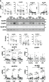
Basal expression of inflammasome-associated genes differs between human monocyte subpopulations. Basal expression of various inflammasome-associated genes in untreated monocyte subpopulations from healthy donors. (A) mRNA expression measured by qPCR (n = 8-15) and (B) protein levels by immunoblotting (n = 4-11) revealed striking differences for NLRP3 and 12, as well as CASP1 between subpopulations. (C) Comparing healthy to CAD or AMI monocytes revealed increased gene expression of NLRP3 in classical monocytes and of NLRP6 in non-classical monocytes of AMI patients; * compared to classical; # compared to healthy; * or # p < 0.05; ** or ## p ≤ 0.01; *** or ###p ≤ 0.001.
Figure 3
Figure 3
IL18 release is predominant in classical monocytes upon rapid inflammasome activation and is greatly attenuated in AMI patients. ELISA measurement of inflammatory cytokines IL18 and IL-1β released from monocyte subpopulations treated as described in Figure 1 . (A) Classical monocytes from healthy donors showed clear predominance for IL18 release compared to the two other subpopulations (n=5-7). IL18 release was completely inhibited by MCC950 treatment. IL-1β was released at a much lower extent than IL18, but still significantly different between classical and non-classical monocytes. (B) Classical monocytes from AMI patients showed a significant impairment of both IL18 and IL-1β release. Decreased IL18 release was not observed in monocytes from CAD patients. (C) LDH release as a measure of pyroptotic cell death was significantly induced upon rapid inflammasome activation in classical monocytes of healthy subjects and CAD patients but substantially attenuated in monocytes from AMI patients. # vs vehicle; § vs LPS+ATP; *, # or § p < 0.05; **, ## or §§ p ≤ 0.01; ***, ### or §§§ p ≤ 0.001.
Figure 4
Figure 4
Differential expression profile of IL18 gene as a potential explanation for variation of inflammatory interleukin release. (A) Basal mRNA and (B) protein expression profiles of IL18 and IL18BP in monocyte subpopulations of healthy donors. IL18 expression was significantly higher in classical monocytes while intracellular glycosylated IL18BP protein levels (~45-50 kDa) were higher in intermediate and non-classical monocytes (n = 9-11). At least two bands were detected that would correspond to different IL18BP isoforms. (C) Unlike IL18, IL18BP release into the supernatant from monocyte subpopulations upon treatments, was unaffected by inflammatory stimulation (n = 6). (D) Therefore, active free IL18 using calculated IL18BP sequestration resulted in practically the same IL18 concentrations measured in Figure 3A ; * compared to classical; *p < 0.05; **p ≤ 0.01; ***p ≤ 0.001.
Figure 5
Figure 5
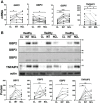
Differential expression levels of genes regulating inflammasome activation between monocyte subpopulations. (A) mRNA expression and (B) protein levels of GBP2, 3 and 5 displayed downregulation in classical monocytes from healthy donors more significantly at protein level. TNFAIP3 had significantly higher mRNA expression in classical monocytes but TNFAIP3 protein levels were higher in both intermediate and non-classical monocytes; * comparison between subpopulations. *p < 0.05; **p ≤ 0.01; ***p ≤ 0.001.
Figure 6
Figure 6
Upregulation of negative feedback loop regulators TNFAIP3 and IRAKM linked to attenuated inflammatory phenotype in AMI CD14+ monocytes. (A) Both TNFAIP3 and IRAKM mRNA was significantly increased in circulating CD14+ monocytes from AMI patients compared to healthy subjects or CAD patients (n = 8-27). (B) In contrast to IRAKM, the mRNA expression of TNFAIP3 was even further upregulated in lesional monocytes compared to peripheral cells within the same AMI patients (n = 7). (C) TNFAIP3 protein levels were also significantly higher in monocytes from AMI patients. (D) Moreover, TNFAIP3 protein showed a direct correlation with high sensitive Troponin T (hsTnT) levels. *p < 0.05; **p ≤ 0.01; ***p ≤ 0.001.
Figure 7
Figure 7
Graphical summary of rapid inflammasome activation events in monocytes during acute myocardial infarction. Rapid inflammasome activation happens after simultaneous dual signal, DAMP-induced TLR priming and extracellular ATP sensing by P2RX7 at the injury site. TLR signaling triggers removal of ubiquitin modification or phosphorylation (red tags) of basal NLRP3 that repress inflammasome complex formation. The same signaling cascades also induces post-translational modifications in other residues that enhances inflammasome activation (green tags). P2RX7 ion-channel activation triggers intracellular K+ efflux, which induces NLRP3 multimerization. Active inflammasome complex recruits pro-CASP1, its catalytic activation and subsequent pro-IL18 cleavage and release. Rapid inflammasome activation and IL18 release happens in classical monocytes (Mo) at the site of the occluded vessel or in the ischemic area at the early phase of AMI, within the first hours after infarction (left panel). This pathway releases very low levels of IL-1β because IL-1β is transcription dependent. NLRP3 specific inhibitor, MCC950, can block this process. Circulating monocytes in the blood away from the site of ischemic lesion are potentially only able to sense DAMPs but not the short-lived ATP. Activation of only TLR signal induces upregulation of the anti-inflammatory mediators TNFAIP3 and IRAKM. TNFAIP3 potentially modulates a concomitant decline of inflammasome priming alongside a significant decrease of acute IL18 release in a protective mechanism against systemic inflammatory hyper-activation (right panel). The given time frames in this graphical summary are just an estimated guess of the authors.

References

    1. Shi C, Pamer EG. Monocyte Recruitment During Infection and Inflammation. Nat Rev Immunol (2011) 11:762–74. doi: 10.1038/nri3070 - DOI - PMC - PubMed
    1. Arfvidsson J, Ahlin F, Vargas KG, Thaler B, Wojta J, Huber K. Monocyte Subsets in Myocardial Infarction: A Review. Int J Cardiol (2017) 231:47–53. doi: 10.1016/j.ijcard.2016.12.182 - DOI - PubMed
    1. Ziegler-Heitbrock L, Hofer TP. Toward a Refined Definition of Monocyte Subsets. Front Immunol (2013) 4:23. doi: 10.3389/fimmu.2013.00023 - DOI - PMC - PubMed
    1. Carlin LM, Stamatiades EG, Auffray C, Hanna RN, Glover L, Vizcay-Barrena G, et al. . Nr4a1-Dependent Ly6C(low) Monocytes Monitor Endothelial Cells and Orchestrate Their Disposal. Cell (2013) 153:362–75. doi: 10.1016/j.cell.2013.03.010 - DOI - PMC - PubMed
    1. Swirski FK, Nahrendorf M. Leukocyte Behavior in Atherosclerosis, Myocardial Infarction, and Heart Failure. Science (2013) 339:161–6. doi: 10.1126/science.1230719 - DOI - PMC - PubMed

Publication types

MeSH terms