. 2022 Apr 26:13:859420.
doi: 10.3389/fmicb.2022.859420. eCollection 2022.

The Interaction of Influenza A NS1 and Cellular TRBP Protein Modulates the Function of RNA Interference Machinery

Affiliations

The Interaction of Influenza A NS1 and Cellular TRBP Protein Modulates the Function of RNA Interference Machinery

Qi Wang et al. Front Microbiol. .

Abstract

Influenza A virus (IAV), one of the most prevalent respiratory diseases, causes pandemics around the world. The multifunctional non-structural protein 1 (NS1) of IAV is a viral antagonist that suppresses host antiviral response. However, the mechanism by which NS1 modulates the RNA interference (RNAi) pathway remains unclear. Here, we identified interactions between NS1 proteins of Influenza A/PR8/34 (H1N1; IAV-PR8) and Influenza A/WSN/1/33 (H1N1; IAV-WSN) and Dicer's cofactor TAR-RNA binding protein (TRBP). We found that the N-terminal RNA binding domain (RBD) of NS1 and the first two domains of TRBP protein mediated this interaction. Furthermore, two amino acid residues (Arg at position 38 and Lys at position 41) in NS1 were essential for the interaction. We generated TRBP knockout cells and found that NS1 instead of NS1 mutants (two-point mutations within NS1, R38A/K41A) inhibited the process of microRNA (miRNA) maturation by binding with TRBP. PR8-infected cells showed masking of short hairpin RNA (shRNA)-mediated RNAi, which was not observed after mutant virus-containing NS1 mutation (R38A/K41A, termed PR8/3841) infection. Moreover, abundant viral small interfering RNAs (vsiRNAs) were detected in vitro and in vivo upon PR8/3841 infection. We identify, for the first time, the interaction between NS1 and TRBP that affects host RNAi machinery.

Keywords: RNA interference machinery; TRBP; antiviral RNAi response; influenza A virus; non-structural protein 1 of IAV.


Conflict of interest statement

The authors declare that the research was conducted in the absence of any commercial or financial relationships that could be construed as a potential conflict of interest.

Figures

Figure 1
Figure 1
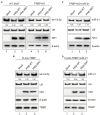
IAV NS1 interacts with TRBP. (A) Plasmids encoding PR8 NS1 and FLAG-TRBP or FLAG-EGFP or empty vector were co-transfected into 293 T cells for 48 h (lanes 1–6). 293 T cells were transfected with FLAG-TRBP or FLAG-EGFP or empty vector for 24 h before infected with PR8-WT (MOI = 1) for 24 h (lanes 7–12). Immunoprecipitations were performed with anti-FLAG antibody. FLAG-tagged proteins, NS1, and β-actin were detected with specific antibodies. (B) Plasmids encoding WSN NS1 and FLAG-TRBP or FLAG-EGFP or empty vector were co-transfected into 293 T cells for 48 h (lanes 1–6). 293 T cells were transfected with FLAG-TRBP or FLAG-EGFP or empty vector for 24 h before infected with WSN-WT (MOI = 1) for 24 h (lanes 7–12). Immunoprecipitations were performed and processed as in (A). (C,D) The NS1-specific interaction with TRBP persists in the presence of RNase. RNase A (10 mg/ml) and RNase III (5 U/ml) were treated with cell lysates. Immunoprecipitations were performed with anti-FLAG antibody. Samples were analyzed by SDS–PAGE with the indicated antibodies.
Figure 2
Figure 2
GST pull-down assays of NS1 and TRBP. (A) Purification of GST and GST-TRBP proteins. (B) GST pull-down assay showing the interaction between NS1 and TRBP. Lysates of 293 T cells transfected with PR8 NS1 or WSN NS1 or infected with PR8-WT (MOI = 1) or WSN-WT (MOI = 1) were incubated with an equal amount of GST or GST-TRBP bound to glutathione-sepharose 4B beads with RNase A (10 mg/ml) and RNase III (5 U/ml) treatment. Samples were analyzed by SDS–PAGE with the indicated antibodies.
Figure 3
Figure 3
R38A and K41A mutations in NS1 abolish the NS1-TRBP interaction. (A) Domain architecture of PR8 NS1 protein (NS1-WT) and the generated mutant constructs (N-NS1, C-NS1, NS1 38/41). (B) Immunoprecipitation of N-NS1 protein with the TRBP protein. Plasmids encoding FLAG-TRBP or FLAG-EGFP and N-NS1 were co-transfected into 293 T cells for 48 h. Immunoprecipitations were performed with anti-FLAG antibody. FLAG-tagged proteins, N-NS1, and β-actin were detected with specific antibodies. (C) Immunoprecipitation of C-NS1 protein with the TRBP protein. Plasmids encoding FLAG-TRBP or FLAG-EGFP and C-NS1 were co-transfected into 293 T cells for 48 h and processed as in (B). (D) Plasmids encoding FLAG-TRBP or FLAG-EGFP and PR8 NS1-WT or PR8 NS1 38/41 were co-transfected into 293 T cells for 48 h and processed as in (B). (E) Plasmids encoding FLAG-TRBP or FLAG-EGFP and WSN NS1-WT or WSN NS1 38/41 were co-transfected into 293 T cells for 48 h and processed as in (B). (F) 293 T cells were transfected with FLAG-TRBP or FLAG-EGFP for 24 h before infected with PR8-WT (MOI = 1) or PR8/3841 (MOI = 1) for 24 h and processed as in (B).
Figure 4
Figure 4
The dsRBD-A and dsRBD-B of TRBP are critical for interacting with NS1. (A) Domain architecture of TRBP protein (TRBP-WT) and the generated mutant constructs (TA, TB, and TC). (B) Immunoprecipitation of PR8 NS1 protein with different mutants of TRBP proteins. PR8 NS1 and the indicated FLAG-tagged constructs were co-transfected into 293 T cells, followed by FLAG immunoprecipitation. FLAG-tagged proteins, NS1, and β-actin were detected with specific antibodies. (C) The weak interaction between Dicer and NS1. 293 T cells were transfected with FLAG-Dicer or FLAG-T7 for 24 h before infected with PR8-WT (MOI = 1) for 24 h and processed as in (B). (D) NS1 participates in the complex with TRBP and Dicer. 293 T cells were co-transfected with FLAG-Dicer and His-EGFP or His-TRBP for 24 h before infected with mock or PR8-WT (MOI = 1) or PR8/3841 (MOI = 1) and then processed as in (B).
Figure 5
Figure 5
TRBP affects miRNAs production. (A) RISC-associated proteins of 293 T and TRBP-KO cells were detected by Western blotting. (B) Northern blotting analysis of miR-126a-3p and let-7a-5p expression levels in the indicated cells. The relative expression levels of miRNAs are normalized to that of U6 small nuclear RNA and to the miRNA level from the WT 293 T cells. (C) TRBP promotes endogenous let-7a-5p expression. FLAG-TRBP was transfected into TRBP-KO cells for 48 h. Northern blotting and Western blotting were performed to detect let-7a-5p, U6, TRBP, and β-actin expressions in the indicated cells. The relative expression level of let-7a-5p is normalized to that of U6 small nuclear RNA and to the miRNA level from the TRBP-KO cells. (D) TRBP promotes exogenous miR-21 expression. TRBP-KO cells were transfected with a miR-21 expressing plasmid and FLAG-TRBP or FLAG-EGFP for 48 h. Northern blotting and Western blotting were performed to detect expression levels of miR-21, U6, FLAG-tagged proteins and β-actin. The relative expression level of miR-21 is normalized to that of U6 small nuclear RNA and to the miRNA level from the miR-21-transfected TRBP-KO cells.
Figure 6
Figure 6
NS1-TRBP interaction reduces miRNAs production. (A) The expression level of let-7a-5p is downregulated in PR8-infected WT cells. WT 293 T cells and TRBP-KO cells were infected with mock or PR8-WT (MOI = 1) or PR8/3841 (MOI = 1) for 48 h. Northern blotting and Western blotting were performed to detect expression levels of let-7a-5p, U6, NS1, and β-actin. The relative expression level of let-7a-5p is normalized to that of U6 small nuclear RNA and to the miRNA level from the uninfected WT 293 T cells or the uninfected TRBP-KO cells. (B) NS1-TRBP interaction reduces the expression of endogenous let-7a-5p. TRBP-KO cells were transfected with FLAG-TRBP for 24 h before infected with PR8-WT or PR8/3841 for 24 h. Northern blotting detection of let-7a-5p and U6 expressions. The relative expression level of let-7a-5p is normalized to that of U6 small nuclear RNA and to the miRNA level from the uninfected cells. Western blotting detection of TRBP, NS1, and β-actin proteins. (C, D) NS1-TRBP interaction reduces the expression of exogenous miR-21. (C) TRBP-KO cells were transfected with a miR-21 expressing plasmid for 24 h before infected with mock or PR8-WT or PR8/3841 for 24 h. (D) TRBP-KO cells were co-transfected with FLAG-TRBP and miR-21 expressing plasmid for 24 h before infected with mock or PR8-WT or PR8/3841 for 24 h. Northern blotting detection expressions of miR-21 and U6. The relative expression level of miR-21 is normalized to that of U6 small nuclear RNA and to the miRNA level from the uninfected cells. Western blotting detection of TRBP, NS1, and β-actin proteins.
Figure 7
Figure 7
R38A-K41A substitutions in NS1 abolish the inhibition of shRNA-mediated RNAi. (A) Relative mRNA levels of EGFP as quantified by qRT-PCR. 293 T cells were co-transfected with a plasmid encoding EGFP and EGFP-specific shRNA (shEGFP) or luciferase-specific shRNA (shLUC) as a negative control and then infected with PR8-WT (MOI = 1) or PR8/3841 (MOI = 1). After 48 h post-transfection, total RNAs were extracted, and the level of EGFP mRNA was examined by qRT-PCR. The experiments were repeated three times independently. The mRNA level of EGFP+shLUC group was set as 1. ***p < 0.001 (Student’s t-test). ns indicates no significance. (B) Western blotting detection of EGFP, NS1, and β-actin proteins.
Figure 8
Figure 8
The abundant vsiRNAs produced in mammalian cells and mice with PR8/3841 infection. (A) Properties of vsiRNAs (per million total mature miRNAs) sequenced from 293 T cells without or with AGO-IP (at 1 dpi). Size distribution, abundance, the 5′-nucleotide preference of vsiRNAs (per million total 18–28 nt reads) with PR8/3841 infection. (B) Properties of vsiRNAs (per million total mature miRNAs) sequenced from adult C57BL/6 or BALB/c mice (at 4 dpi) infected with PR8-WT or PR8/3841. Size distribution, abundance, the 5′-nucleotide preference of vsiRNAs (per million total 18–28 nt reads) with PR8-WT or PR8/3841 infection. (C) Relative abundance of 21–23 nt vsiRNA hotspots mapped to PR8-WT or PR8/3841 genomic RNAs, presented from the 3′ end to the 5′ end.
Figure 9
Figure 9
Schematic model of the suppressor roles of NS1 in RNAi. Left panel: (A) NS1 inhibits miRNAs production by interacting with TRBP. (B) NS1 inhibits shRNA-mediated RNAi by interacting with TRBP. Right panel: (A) NS1 inhibits siRNA production by directly binding to dsRNAs. (B) NS1 suppresses the cleavage activity of Dicer by interacting with TRBP. (C) NS1 suppresses siRNA loading onto AGO2 by directly binding to siRNAs.

Similar articles

Cited by

References

    1. Ampomah P. B., Lim L. H. K. (2020). Influenza A virus-induced apoptosis and virus propagation. Apoptosis 25, 1–11. doi: 10.1007/s10495-019-01575-3, PMID: - DOI - PubMed
    1. Bamunuarachchi G., Pushparaj S., Liu L. (2021). Interplay between host non-coding RNAs and influenza viruses. RNA Biol. 18, 767–784. doi: 10.1080/15476286.2021.1872170, PMID: - DOI - PMC - PubMed
    1. Bernstein E., Caudy A. A., Hammond S. M., Hannon G. J. (2001). Role for a bidentate ribonuclease in the initiation step of RNA interference. Nature 409, 363–366. doi: 10.1038/35053110, PMID: - DOI - PubMed
    1. Bornholdt Z. A., Prasad B. V. (2008). X-ray structure of NS1 from a highly pathogenic H5N1 influenza virus. Nature 456, 985–988. doi: 10.1038/nature07444, PMID: - DOI - PMC - PubMed
    1. Bucher E., Hemmes H., de Haan P., Goldbach R., Prins M. (2004). The influenza A virus NS1 protein binds small interfering RNAs and suppresses RNA silencing in plants. J. Gen. Virol. 85, 983–991. doi: 10.1099/vir.0.19734-0, PMID: - DOI - PubMed