. 2018 Dec 14;8(73):41858-41871.
doi: 10.1039/c8ra07858k. eCollection 2018 Dec 12.

Renoprotective effect of JinQi-JiangTang tablet on high-fat diet and low-dose streptozotocin-induced type 2 diabetic rats

Affiliations

Renoprotective effect of JinQi-JiangTang tablet on high-fat diet and low-dose streptozotocin-induced type 2 diabetic rats

Bingjie Jiang et al. RSC Adv. .

Abstract

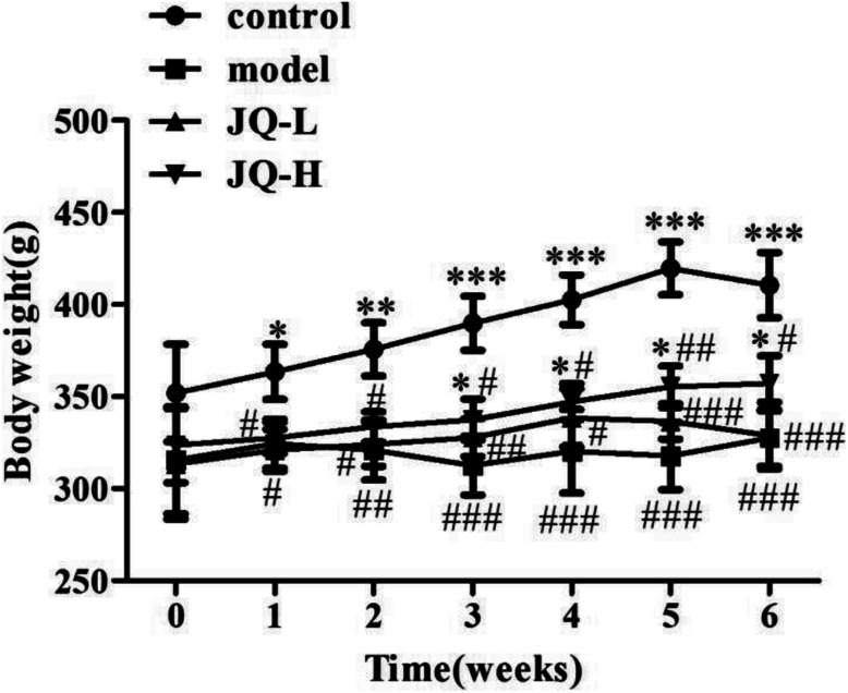
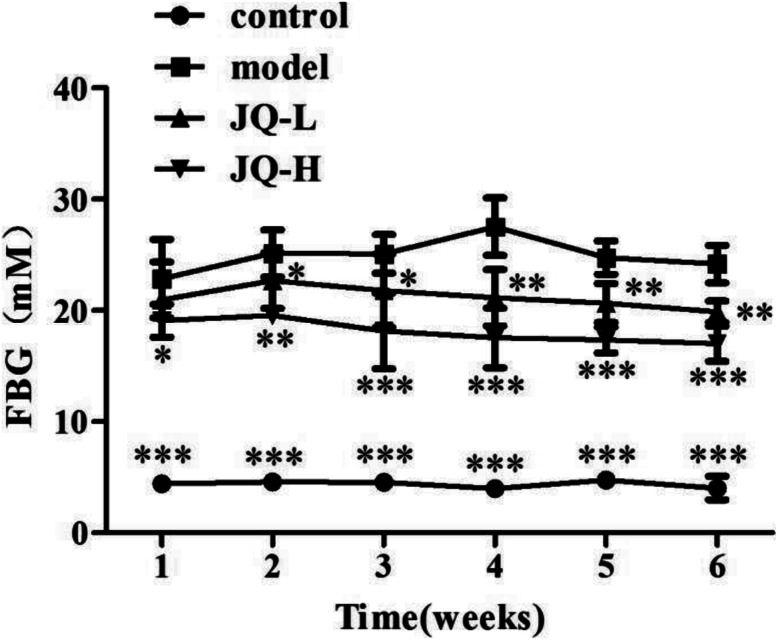
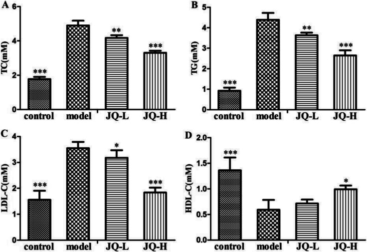
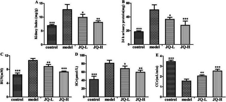
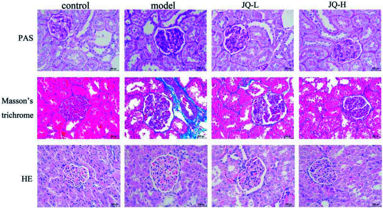
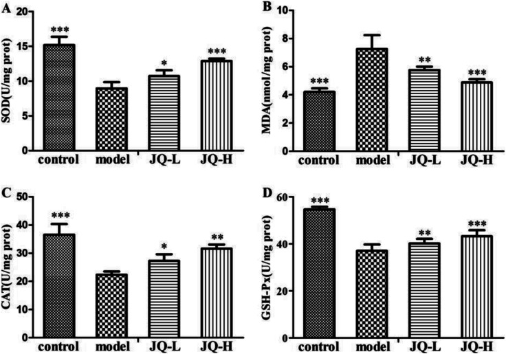
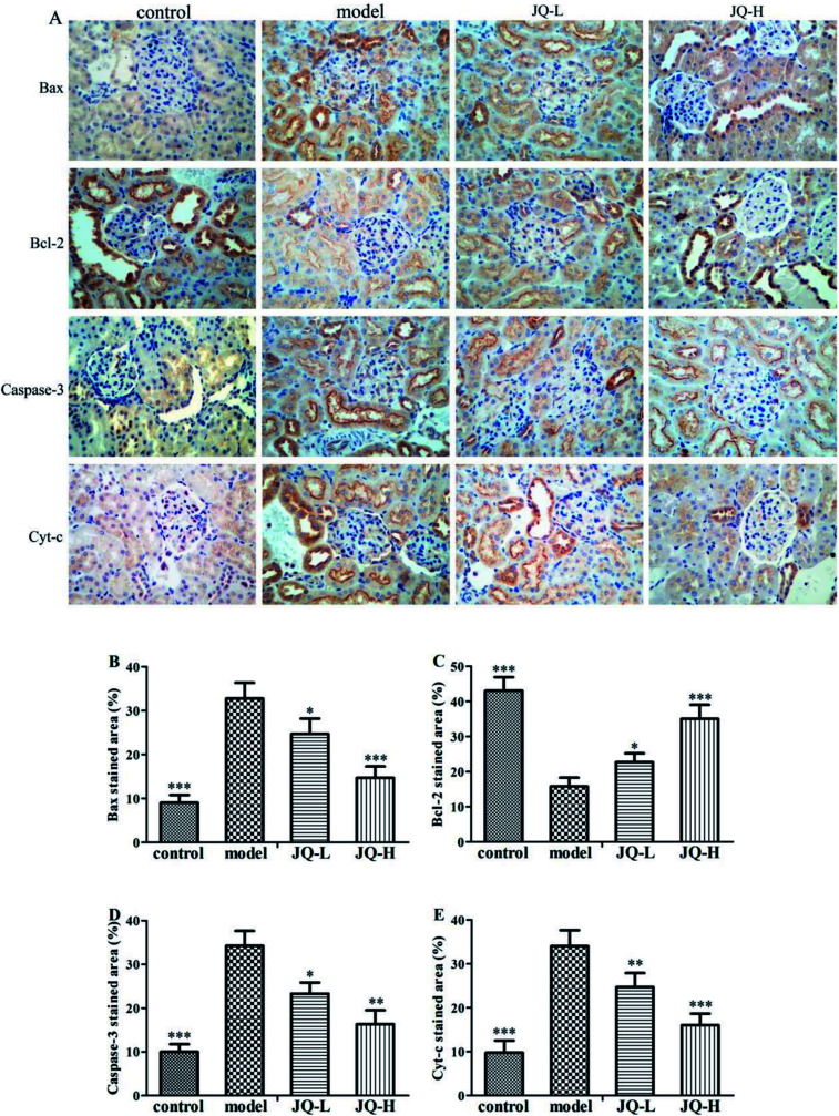
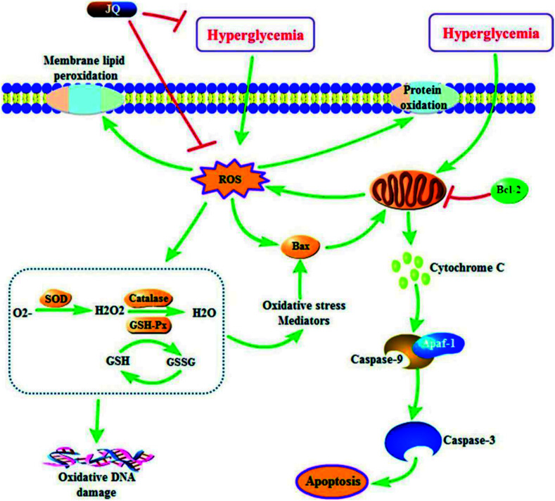
JinQi-JiangTang tablet (JQ), a traditional Chinese patent medicine, have been commonly applied to clinical therapy in type 2 diabetic patients. The present study was undertaken to investigate the renoprotective effect of JQ on type 2 diabetic rats. The type 2 diabetic rat model was successfully induced by a high-fat and high-sugar diet combined with a single low-dose of streptozotocin. Intervention with JQ could significantly diminish the body weight loss, reduce the levels of fasting blood glucose, 24 hour urinary protein, blood urea nitrogen and serum creatinine in STZ-induced diabetic rats. JQ improved the creatinine clearance in diabetic rats. What's more, the levels of total cholesterol, triglyceride and low-density lipoprotein cholesterol were markedly reduced following JQ treatment, while the level of high-density lipoprotein cholesterol was elevated. Moreover, JQ significantly improved the activity of superoxide dismutase, catalase and glutathione peroxidase, whereas decreased the level of lipid peroxidation malondialdehyde in renal tissue of diabetic rats. Furthermore, immunohistochemical analysis showed that JQ significantly downregulated the expression of Bax, Caspase-3 and Cytochrome c and upregulated Bcl-2 protein expression in the renal tissue of diabetic rats, which was considered as the major pathogeneses of apoptosis. These data demonstrated that JQ exhibited a renoprotective effect through blood glucose control, alleviating lipid metabolism, anti-oxidative stress and anti-apoptosis activities.


Conflict of interest statement

The authors declare they have no conflicts of interests.

Figures

Fig. 1
Fig. 1. Schematic diagram of experimental protocol.
Fig. 2
Fig. 2. Chromatograms of reference standards and JQ by UPLC-Q-TOF/MS. (A) Total ion current (TIC) chromatogram of JQ in positive ion mode; (B) total ion current (TIC) chromatogram of reference standards in positive ion mode; (C) total ion current (TIC) of JQ in negative ion mode; (D) total ion current (TIC) of reference standards in negative ion mode.
Fig. 3
Fig. 3. The chemical structure of the identified compounds in JQ.
Fig. 4
Fig. 4. Effect of JQ on body weight during the treatment period. Data were expressed as mean ± SEM (n = 10, each group). *P < 0.05, **P < 0.01, ***P < 0.001, compared with model group; #P < 0.05, ##P < 0.01, ###P < 0.001, compared with normal control group.
Fig. 5
Fig. 5. Effect of JQ on fasting blood glucose in diabetic rats. Data were expressed as mean ± SEM (n = 10, each group). *P < 0.05, **P < 0.01, ***P < 0.001, compared with model group. Control: the rats were treated with vehicle; model: the STZ-induced diabetic rats were treated with vehicle; JQ-L: the STZ-induced diabetic rats were treated at dose of 1.01 g kg−1 JQ; JQ-H: the STZ-induced diabetic rats were treated at dose of 4.04 g kg−1 JQ.
Fig. 6
Fig. 6. Effect of JQ on lipid profiles in STZ-induced diabetic rats. (A) Blood total cholesterol level; (B) blood triglyceride level; (C) blood low-density lipoprotein cholesterol level; (D) blood high-density lipoprotein cholesterol level in different rats groups. Data were expressed as mean ± SEM (n = 10, each group). *P < 0.05, **P < 0.01, ***P < 0.001, compared with model group.
Fig. 7
Fig. 7. Effect of JQ on renal functional parameters in STZ-induced diabetic rats. (A) Kidney weight to body weight ratio (kidney index); (B) 24 hour urinary protein; (C) blood urea nitrogen (BUN); (D) serum creatinine (SCr); (E) creatinine clearance (CCr) in different groups. Data were expressed as mean ± SEM (n = 10, each group). *P < 0.05, **P < 0.01, ***P < 0.001, compared with model group.
Fig. 8
Fig. 8. Representative histological changes in renal sections of periodic acid-Schiff staining, Masson's trichrome staining and Hematoxylin and eosin staining are shown (original magnification, ×400).
Fig. 9
Fig. 9. Effect of JQ on activity of antioxidant enzymes in STZ-induced diabetic rats. (A) Effect of JQ on SOD activity; (B) effect of JQ on MDA activity; (C) effect of JQ on CAT activity; (D) effect of JQ on GSH-Px activity in different rats groups. Data were expressed as mean ± SEM (n = 10, each group). *P < 0.05, **P < 0.01, ***P < 0.001, compared with model group.
Fig. 10
Fig. 10. JQ downregulated the expression of Bax, Caspase-3 and Cyt-c and upregulated the expression of Bcl-2 in the renal tissue of diabetic rats. (A) Photomicrographs of Bax, Bcl-2, Caspase-3 and Cyt-c immune-stained renal sections of different groups (original magnification, ×400). Image analysis of (B) Bax, (C) Bcl-2, (D) Caspase-3 and (E) Cyt-c immune-staining represented as positive areas (%). Data were expressed as mean ± SEM (n = 10, each group). *P < 0.05, **P < 0.01, ***P < 0.001, compared with model group.
Fig. 11
Fig. 11. A proposed schematic diagram for the renoprotective mechanism of JQ on STZ-induced type 2 diabetic rats. ROS, reactive oxygen species; Bcl-2, B cell lymphoma 2; Bax, Bcl-2 associate X protein; SOD, superoxide dismutase; GSH, glutathione; GSH-Px, glutathione peroxidase; GSSG, oxidized glutathione.

Similar articles

Cited by

References

    1. Ma R. C. W. Diabetologia. 2018;61:1249–1260. doi: 10.1007/s00125-018-4557-7. - DOI - PubMed
    1. Tuttle K. R. Bakris G. L. Bilous R. W. Chiang J. L. de Boer I. H. Goldstein-Fuchs J. Hirsch I. B. Kalantar-Zadeh K. Narva A. S. Navaneethan S. D. Neumiller J. J. Patel U. D. Ratner R. E. Whaley-Connell A. T. Molitch M. E. Am. J. Kidney Dis. 2014;64:510–533. doi: 10.1053/j.ajkd.2014.08.001. - DOI - PubMed
    1. Wolf G. J. Am. Soc. Nephrol. 2003;14:1396–1405. doi: 10.1097/01.ASN.0000065639.19190.CF. - DOI - PubMed
    1. Jefferson J. A. Shankland S. J. Pichler R. H. Kidney Int. 2008;74:22–36. doi: 10.1038/ki.2008.128. - DOI - PubMed
    1. Guha M. Xu Z. G. Tung D. Lanting L. Natarajan R. FASEB J. 2007;21:3355–3368. doi: 10.1096/fj.06-6713com. - DOI - PubMed