

The Equine Temporomandibular Joint: Comparisons Between Standard and Needle Arthroscopic Examination of Cadaver Specimens and Standing Horses
- PMID: 35558885
- PMCID: PMC9087581
- DOI: 10.3389/fvets.2022.876041
The Equine Temporomandibular Joint: Comparisons Between Standard and Needle Arthroscopic Examination of Cadaver Specimens and Standing Horses
Abstract
Background: Definitive diagnosis of equine temporomandibular joint osteoarthritis (TMJ-OA) may require advanced diagnostic imaging. Arthroscopy is a modern, minimally invasive, diagnostic, and treatment modality. Standing arthroscopic treatment of joint disease is a relatively recent advance in equine surgery, despite which there are few published comparisons between the available arthroscopic systems.
Objective: To compare and contrast two arthroscopic systems for assessing the equine temporomandibular joint compartments in cadavers and standing horses.
Study design: Experimental study.
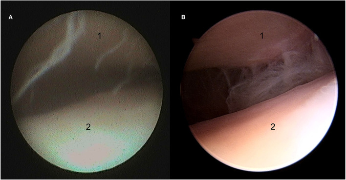
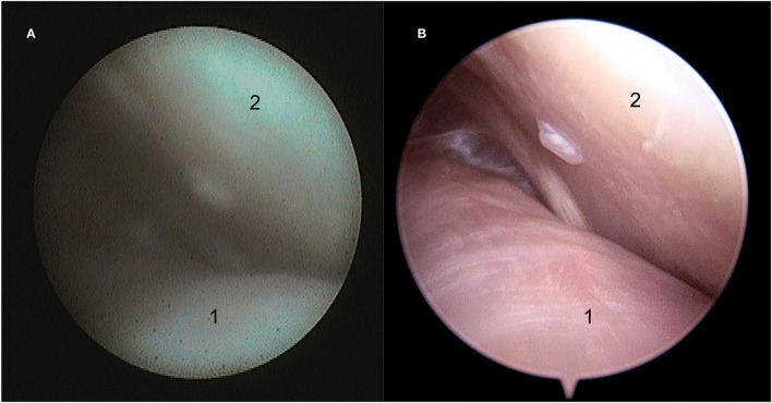
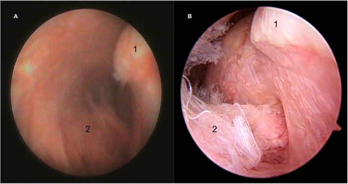
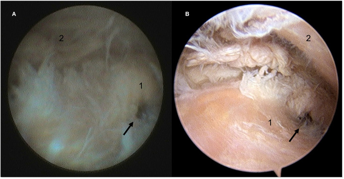
Methods: Phase I involved the assessment of the discotemporal joint (DTJ) and discomandibular (DMJ) joint compartments of both temporomandibular joints (TMJ) of 14 cadaveric equine heads using a caudally placed arthroscopy portal. Joints were initially examined using the needle arthroscope and the results compared to the findings of examination using a 2.5 mm 30° arthroscope system (standard). Three healthy horses were subsequently examined to determine the validity of the procedure in live animals in Phase II.
Results: Needle and standard arthroscopy, in combination with mandibular manipulation, allowed evaluation of the caudal aspects of both joint compartments of the TMJ in Phase I. However, the extreme margins of the joint were more commonly visualized using standard arthroscopy. Live horses in phase II were restrained in stocks and both the rostral and caudal aspects of the DTJ and DMJ compartments of both TMJs were examined successfully understanding sedation and local analgesia. The use of a modified Guenther speculum allowed the mandible to be manipulated and offset, which facilitated a complete examination of the joint compartments. Despite adverse behavior encountered during the procedure in one horse, no surgical complications ensued.
Main limitations: Not blinded-bias; learning curve.
Conclusions: The needle arthroscope system is a relatively inexpensive diagnostic tool, which can be used to evaluate the TMJ in the absence of advanced diagnostic imaging such as computed tomography or magnetic resonance imaging. However, if arthroscopic treatment is required after advanced imaging and pre-operative diagnosis, superior image quality and ease of manipulation may favor the use of the standard equipment.
Keywords: arthroscopy; fiberoptic; needle arthroscopy; temporomandibular joint; video arthroscopy.
Copyright © 2022 Carmalt and Pimentel.
Conflict of interest statement
The authors declare that the research was conducted in the absence of any commercial or financial relationships that could be construed as a potential conflict of interest.
Figures
References
LinkOut - more resources
Full Text Sources