

Altered Expressions of NF1 and NF1-Related microRNAs as Biomarkers in the Diagnosis of Undifferentiated Pleomorphic Sarcoma
- PMID: 35559021
- PMCID: PMC9086456
- DOI: 10.3389/fgene.2022.870191
Altered Expressions of NF1 and NF1-Related microRNAs as Biomarkers in the Diagnosis of Undifferentiated Pleomorphic Sarcoma
Abstract
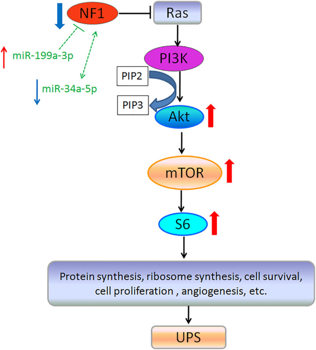
Objective: Undifferentiated pleomorphic sarcoma (UPS) is a highly malignant, aggressive, and pleomorphic subtype of soft tissue sarcoma in adults. However, UPS is difficult to be diagnosed due to the lack of specific morphological and immunophenotypic features. Here, we aimed to identify new biomarkers for the diagnosis of UPS. Methods: The mRNA and protein expression of neurofibromin 1 (NF1) in 68 pairs of UPS and adjacent normal tissues were detected by qRT-PCR and immunohistochemistry, and the correlation between the NF1 protein expression and clinicopathological characteristics was analyzed. Then, differentially expressed microRNAs (DE miRNAs) were identified between the UPS tumor tissue and matched adjacent normal tissue using Hisep sequencing, Gene Ontology (GO), and Kyoto Encyclopedia of Genes and Genomes (KEGG). The DE miRNAs of the regulating NF1 gene were also identified using the TargetScan and miRanda databases and validated by qRT-PCR. Results: Compared with the adjacent normal tissue, both mRNA and protein expressions of NF1 in the UPS tumor tissue were significantly decreased, and the positive rate of NF1 protein was associated with the tumor size, metastasis, and recurrence. A total of 125 known DE miRNAs were identified from the screened miRNAs based on | log2(Fold Change) ≥5 and p-value < 0.05 (A total of 82 upregulated and 43 downregulated DE miRNAs in the UPS tissue). Target genes regulated by the DE miRNAs were enriched in pathways of metabolisms, RNA degradation, PI3K-Akt, and Rap1 pathway. In total, 11 miRNAs which were predicted to regulate the NF1 gene were screened. After verification, the relative expressions of hsa-miR-199a-3p and hsa-miR-34a-5p were increased and decreased in the UPS tumor tissue compared with those in the adjacent normal tissue, respectively. Conclusion: NF1 and NF1-related microRNAs including hsa-miR-199a-3p and hsa-miR-34a-5p may be novel biomarkers in the diagnosis of undifferentiated pleomorphic sarcoma (UPS).
Keywords: NF1; biomarkers; microRNA; soft tissue sarcomas; undifferentiated pleomorphic sarcoma.
Copyright © 2022 Zhang, Huang, Ma and Niu.
Conflict of interest statement
The authors declare that the research was conducted in the absence of any commercial or financial relationships that could be construed as a potential conflict of interest.
Figures


Similar articles
-
miRNA-seq analysis of human vertebrae provides insight into the mechanism underlying GIOP.Bone. 2019 Mar;120:371-386. doi: 10.1016/j.bone.2018.11.013. Epub 2018 Nov 29. Bone. 2019. PMID: 30503955
-
Differentiating soft tissue leiomyosarcoma and undifferentiated pleomorphic sarcoma: A miRNA analysis.Genes Chromosomes Cancer. 2014 Aug;53(8):693-702. doi: 10.1002/gcc.22179. Epub 2014 Apr 26. Genes Chromosomes Cancer. 2014. PMID: 24771630
-
Identification of key microRNAs associated with diffuse large B-cell lymphoma by analyzing serum microRNA expressions.Gene. 2018 Feb 5;642:205-211. doi: 10.1016/j.gene.2017.11.022. Epub 2017 Nov 8. Gene. 2018. PMID: 29128636
-
Dysregulation of miRNAs in Soft Tissue Sarcomas.Cells. 2024 Nov 8;13(22):1853. doi: 10.3390/cells13221853. Cells. 2024. PMID: 39594601 Free PMC article. Review.
-
Current research and management of undifferentiated pleomorphic sarcoma/myofibrosarcoma.Front Genet. 2023 Feb 16;14:1109491. doi: 10.3389/fgene.2023.1109491. eCollection 2023. Front Genet. 2023. PMID: 36873946 Free PMC article. Review.
Cited by
-
Recurrent mucinous carcinoma with sarcomatoid and sarcomatous mural nodules: a case report and literature review.Front Oncol. 2024 Jun 6;14:1387700. doi: 10.3389/fonc.2024.1387700. eCollection 2024. Front Oncol. 2024. PMID: 38903727 Free PMC article.
-
Association between microRNA 671 polymorphisms and the susceptibility to soft tissue sarcomas in a Chinese population.Front Oncol. 2022 Aug 9;12:960269. doi: 10.3389/fonc.2022.960269. eCollection 2022. Front Oncol. 2022. PMID: 36016604 Free PMC article.
-
Undifferentiated high-grade pleomorphic sarcoma of the common bile duct: A case report and review of literature.World J Gastrointest Oncol. 2024 May 15;16(5):2253-2260. doi: 10.4251/wjgo.v16.i5.2253. World J Gastrointest Oncol. 2024. PMID: 38764812 Free PMC article.
References
LinkOut - more resources
Full Text Sources
Research Materials
Miscellaneous