. 2022 May 13;8(19):eabm9881.
doi: 10.1126/sciadv.abm9881. Epub 2022 May 13.

FasL microgels induce immune acceptance of islet allografts in nonhuman primates

Affiliations

FasL microgels induce immune acceptance of islet allografts in nonhuman primates

Ji Lei et al. Sci Adv. .

Abstract

Islet transplantation to treat insulin-dependent diabetes is greatly limited by the need for maintenance immunosuppression. We report a strategy through which cotransplantation of allogeneic islets and streptavidin (SA)-FasL-presenting microgels to the omentum under transient rapamycin monotherapy resulted in robust glycemic control, sustained C-peptide levels, and graft survival in diabetic nonhuman primates for >6 months. Surgical extraction of the graft resulted in prompt hyperglycemia. In contrast, animals receiving microgels without SA-FasL under the same rapamycin regimen rejected islet grafts acutely. Graft survival was associated with increased number of FoxP3+ cells in the graft site with no significant changes in T cell systemic frequencies or responses to donor and third-party antigens, indicating localized tolerance. Recipients of SA-FasL microgels exhibited normal liver and kidney metabolic function, demonstrating safety. This localized immunomodulatory strategy succeeded with unmodified islets and does not require long-term immunosuppression, showing translational potential in β cell replacement for treating type 1 diabetes.


Figures

Fig. 1.
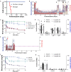
Fig. 1.. Local presentation of SA-FasL via synthetic microgels for islet transplantation.
(A) Synthetic biotin-PEG microgels, generated by microfluidic polymerization, capture, and present SA-FasL. SA-FasL–presenting microgels and islets are immobilized on the surface of the omentum of NHPs using autologous plasma and thrombin, where they induce immune acceptance by potentially eliminating T effector cells and generating T regulatory cells. (B) Schema describing donor and diabetic MHC mismatched recipient NHPs and treatment protocol. SA-FasL–presenting microgels with transient rapamycin (RAPA) monotherapy (SA-FasL Microgels, n = 4); control microgels with transient rapamycin monotherapy (Microgels, n = 3).
Fig. 2.
Fig. 2.. SA-FasL–presenting microgels induce islet allograft acceptance.
(A) Kaplan-Meier survival curves of islet allografts in SA-FasL Microgel (n = 4) and Microgel (n = 3); Mantel-Cox test, P = 0.010. Mean graft survival times: SA-FasL Microgel, >180 days; Microgel, 27.7 days. (B) Nonfasting blood glucose levels (mean, blue line; SEM, gray shadow, left axis) and daily total EIR (mean, red bars; lower SEM, dark red bars, right axis) for SA-FasL Microgel subjects. Animals exhibited high blood glucose levels and external insulin demand after STZ induction but before transplant (defined as post-STZ). After cotransplantation of islets and SA-FasL microgels (Tx), animals rapidly became normoglycemic and had significantly reduced EIR. Animals reverted to hyperglycemic state after graft removal (blood glucose levels, purple lines, left axis; total EIR, tan bars, right axis). (C) Blood glucose levels for SA-FasL Microgel subjects after intravenous infusion of glucose before (pre-STZ) and after diabetes induction (post-STZ), at 3 and 6 months after transplantation, and after graft removal. (D) Insulin (left axis) and C-peptide (right axis) levels in serum for SA-FasL Microgel animals under fasting (F) and post-stimulation (S) before (naïve) and after diabetes induction, at 3 and 6 months after transplantation, and post-graft removal (PGR). (E) Nonfasting blood glucose levels (blue line; SEM gray shadow, left axis) and daily total EIR (mean, red bars; lower SEM, dark red bars, right axis) for Microgel subjects. After restoring normoglycemia following transplantation (Tx), animals became hyperglycemic and required higher external insulin around 1 month after transplantation. (F) Blood glucose after intravenous infusion in Microgel during prediabetic state (Pre-STZ), after diabetes induction (Post-STZ), and at 1 month after transplantation. (G) Insulin (left axis) and C-peptide (right axis) levels in serum for Microgel animals under fasting (F) and post-stimulation (S) before and after diabetes induction and at 1 month after transplantation.
Fig. 3.
Fig. 3.. Histological analyses of grafts showing increased FoxP3+ cells in recipients of SA-FasL–presenting microgels.
(A) Hematoxylin and eosin (H&E) staining of excised omentum biopsies at takedown demonstrates the presence of islet-like clusters (outlined in white dashed line) in SA-FasL Microgel subject with minimal immune cell infiltration (left image: bar, 20 μm; middle image: bar, 50 μm). Representative image from Microgel (right image: bar, 50 μm) shows islet-like cluster with significant cell infiltration. (B) Immunostaining for FoxP3 [green: FoxP3; blue: 4′,6-diamidino-2-phenylindole (DAPI); bar: 50 μm], a marker of Tregs, showing increased numbers of FoxP3+ at the graft site of animals receiving SA-FasL microgels compared to those receiving microgels. FoxP3+ cells were in closed apposition to microgels (dashed white lines). (C and D) Quantification of FoxP3+ cells over total cell counts and mean intensity in sections from Microgel and SA-FasL Microgel animals demonstrating increased frequency of Tregs in SA-FasL Microgel subjects (nested two-tailed t test). Plots show data points for each animal in a different color, with a minimum of three representative images analyzed per animal.
Fig. 4.
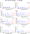
Fig. 4.. Local delivery of SA-FasL microgels does not alter peripheral blood lymphocyte populations.
(A to J) Numbers of circulating (A) CD3+, (B) CD20+, (C) CD4+, and (D) CD8+ cells, as well as naïve (Tn) (E) CD4+ and (F) CD8+, effector memory (EM) (G) CD4+ and (H) CD8+, and central memory (CM) (I) CD4+ and (J) CD8+ lymphocytes in SA-FasL Microgel–treated subjects (dashed blue lines, mean, SEM, n = 3 to 4) and Microgel-receiving recipient (red solid lines, mean, SEM, n = 3) animals.
Fig. 5.
Fig. 5.. SA-FasL microgel recipients display no significant changes in pre- to posttransplant anti-donor MHC antibodies, CD4+ and CD8+ T cell proliferative responses to donor and third-party stimulators, and IFN-γ–secreting cell numbers.
(A and B) IgG responses (mean, individual points) to donor-specific MHC (A) class I and (B) class II epitopes for SA-FasL Microgel (blue, n = 3 to 4) and Microgel (red, n = 3) subjects, demonstrating no donor-specific activation in treated subjects (class I: P = 0.2904, class II: P = 0.0754). No positive donor-specific antibodies to MHC I were detected in Microgel NHPs (P = 0.3292). Nonetheless, these control subjects generated antibodies against donor MHC II (P = 0.0326). (C to F) Mixed lymphocyte reaction (mean, individual points) to CD4+ (C) donor and (D) third-party stimulators and CD8+ (E) donor and (F) third-party antigens for SA-FasL Microgel (blue, n = 3 to 4) and Microgel (red, n = 3) subjects showing no differences in responses for the latter (CD4+ donor: P = 0.4200; CD4+ third-party: P = 0.6949; CD8+ donor: P = 0.4145; CD8+ third-party: P = 0.7858). Microgel animals exhibited no responses against donor (P = 0.6082) or third-party (P = 0.0555) antigens in CD4+ compartment, but CD8+ T cell responses were elevated against both donor (P = 0.0057) and third-party (P = 0.0216) stimulators. (G and H) ELISpot IFN-γ counts (mean, individual points) for (G) donor and (H) third-party stimulation for the SA-FasL Microgel (blue, n = 3 to 4) and Microgel (red, n = 3) subjects. No differences in frequency of IFN-γ–secreting cells in circulation between pre- and posttransplant time points for SA-FasL Microgel (donor: P = 0.2485; third-party: P = 0.1445) or Microgel subjects (donor: P = 0.0824; third-party: P = 0.1413). Repeated-measures ANOVA was used with pairwise comparisons to pretransplant/day 0 values using Dunnett’s test.

References

    1. Devendra D., Liu E., Eisenbarth G. S., Type 1 diabetes: Recent developments. BMJ 328, 750–754 (2004). - PMC - PubMed
    1. Benoit S. R., Hora I., Albright A. L., Gregg E. W., New directions in incidence and prevalence of diagnosed diabetes in the USA. BMJ Open Diabetes Res. Care 7, e000657 (2019). - PMC - PubMed
    1. Thompson D. M., Meloche M., Ao Z., Paty B., Keown P., Shapiro R. J., Ho S., Worsley D., Fung M., Meneilly G., Begg I., Al Mehthel M., Kondi J., Harris C., Fensom B., Kozak S. E., Tong S. O., Trinh M., Warnock G. L., Reduced progression of diabetic microvascular complications with islet cell transplantation compared with intensive medical therapy. Transplantation 91, 373–378 (2011). - PubMed
    1. Shapiro A. M., Geng Hao E., Lakey J. R., Finegood D. T., Rajotte R. V., Kneteman N. M., Defining optimal immunosuppression for islet transplantation based on reduced diabetogenicity in canine islet autografts. Transplantation 74, 1522–1528 (2002). - PubMed
    1. Billingham R. E., Brent L., Medawar P. B., Actively acquired tolerance of foreign cells. Nature 172, 603–606 (1953). - PubMed