Review
. 2022 Jul 1;323(1):H1-H15.
doi: 10.1152/ajpheart.00093.2022. Epub 2022 May 13.

Myocardial injury, troponin release, and cardiomyocyte death in brief ischemia, failure, and ventricular remodeling

Affiliations
Review

Myocardial injury, troponin release, and cardiomyocyte death in brief ischemia, failure, and ventricular remodeling

John M Canty Jr. Am J Physiol Heart Circ Physiol. .

Abstract

Troponin released from irreversibly injured myocytes is the gold standard biomarker for the rapid identification of an acute coronary syndrome. In acute myocardial infarction, necrotic cell death is characterized by sarcolemmal disruption in response to a critical level of energy depletion after more than 15 min of ischemia. Although troponin I and T are highly specific for cardiomyocyte death, high-sensitivity assays have demonstrated that measurable circulating levels of troponin are present in many normal subjects. In addition, transient as well as chronic elevations have been demonstrated in many disease states not clearly associated with myocardial ischemia. The latter observations have given rise to the clinical concept of myocardial injury. This review will summarize evidence supporting the notion that circulating troponin levels parallel the extent of myocyte apoptosis in normal ventricular remodeling and in pathophysiological conditions not associated with infarction or necrosis. It will review the evidence that myocyte apoptosis can be accelerated by diastolic strain from elevated ventricular preload and systolic strain from dyskinesis after brief episodes of ischemia too short to cause a critical level of myocyte energy depletion. We then show how chronic, low rates of myocyte apoptosis from endogenous myocyte turnover, repetitive ischemia, or repetitive elevations in left ventricular diastolic pressure can lead to significant myocyte loss in the absence of neurohormonal stimulation. Finally, we posit that the differential response to strain-induced injury in heart failure may determine whether progressive myocyte loss and heart failure with reduced ejection fraction or interstitial fibrosis and heart failure with preserved ejection fraction become the heart failure phenotype.

Keywords: HFpEF; HFrEF; apoptosis; ischemia; troponin.


Conflict of interest statement

No conflicts of interest, financial or otherwise, are declared by the author.

Figures
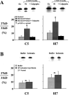
Figure 1.
Figure 1.
The critical role of left ventricular (LV) preload elevation on troponin I proteolysis in the buffer-perfused Langendorff rat heart. A: Western blot analysis of two troponin I antibodies (C5 and 8I7) from normal control excised tissue (Ex) vs. isolated rat hearts subjected to 40 min of preload elevation to an LV end-diastolic pressure (LVEDP) of 25 mmHg. Preload elevation with normal perfusion (Nl) increased the 26-kDa degradation band. The increase in TnI degradation was blocked by inhibiting µ calpain with calpeptin. *P < 0.05 vs. excised. B: troponin I degradation after 20 min of ischemia-reperfusion in isolated rat hearts was prevented by venting the LV to prevent excessive increases in LVEDP during reperfusion of the Langendorff heart preparation. From Feng et al. (34) and reproduced with the permission of the American Heart Association.
Figure 2.
Figure 2.
Troponin I (cTnI) release and myocyte apoptosis after “reversible” regional ischemia in pigs with stunned myocardium. A: serum TnI measurements (log scale) in systemic and coronary venous samples (great cardiac vein) before and after a 10-min LAD occlusion. Regional LAD wall thickening became dyskinetic during the occlusion and gradually returned to normal after reperfusion consistent with stunned myocardium (data not shown). The 99th percentile upper reference limit (URL) for TnI is depicted by the dotted line. There was a delayed increase in TnI which exceeded the normal range within 1 h after reperfusion with levels after 24 h reaching ∼1,000 ng/L although regional function at this time was normal. B: myocyte apoptosis by TUNEL staining was transiently increased in the ischemic LAD region at 1 h but returned to normal 24 h after brief ischemia. Values are means ± SE. †P <0.05 vs. remote. Modified from Weil et al. (43) and reproduced with permission of the American College of Cardiology.
Figure 3.
Figure 3.
Troponin release after brief coronary occlusions of 30–90 s in patients. This slide summarizes serial troponin release up to 4 h following a brief angioplasty balloon occlusion in patients with normal coronary arteries or nonobstructive coronary artery disease. Box and whiskers plots depict troponin normalized to the baseline level before coronary occlusion. Top, left: relative troponin in subjects not receiving a balloon occlusion. The remaining panels summarize measurements up to 4 h after release of a 30-s occlusion (top, right), a 60-s occlusion (bottom, left), or a 90-s occlusion (bottom, right). Like the data following a 10-min occlusion in swine (Fig. 2), high-sensitivity (hs) troponin I assays (red bars and data points Siemens Centaur assay: green bars and data points Abbott ARCHITECT STAT assay) and troponin T (blue bars and data points Roche Elecsys 2010 assay) demonstrated a delayed rise above baseline values that exceeded the 99th URL in a significant number of patients (data not shown). Longer-term measurements beyond 4 h were not performed. These data further support the notion that troponin is released following brief ischemia of a duration compatible with angina. Values are presented as medians and interquartile ranges. From Arnadottir et al. (46), and reproduced with permission of the American Heart Association.
Figure 4.
Figure 4.
Apoptosis, troponin I release and stretch-induced stunning. A: measurements of serum TnI (aorta-blue; coronary sinus red) at baseline and selected time points following a transient 1-h elevation in left ventricular (LV) end-diastolic pressure to 35 mmHg in response to an increase in afterload with phenylephrine (PE). Troponin rose above the 99th URL within 30 min and reached ∼1,400 ng/L after 24 h. Western blot analysis of tissue at 3 h showed TnI proteolysis, light microscopy showed no evidence of necrosis or infarction, and microsphere flow measurements showed no evidence of ischemia (data not shown). B: myocyte apoptosis was increased sixfold in tissue harvested at 3 h and returned to normal levels 24 h after transient preload elevation. Values are means ± SD. *P < 0.05 vs. normal control; †P < 0.05 vs. 24-h post-PE. Adapted from Weil et al. (54) and published with permission of the American College of Cardiology.
Figure 5.
Figure 5.
Myocyte apoptosis and regional myocyte loss in hibernating myocardium. A: histological slides of interstitial connective tissue staining and myocyte cellular size. Chronic repetitive ischemia distal to a severe left anterior descending (LAD) stenosis resulted in hibernating myocardium. This was associated with a small increase in reticular collagen (blue) indicating interstitial fibrosis and a prominent increase in myocyte size indicating regional cellular hypertrophy. This occurred in the absence of anatomic hypertrophy and normally perfused remote myocytes were not enlarged and similar to sham controls. B: myocyte cellular hypertrophy reflected a loss of myocytes as myocyte nuclear density in the hibernating region was reduced as compared with sham controls. There was no evidence of anatomic left ventricular hypertrophy C: chronic repetitive ischemia in collateral-dependent myocardium was associated with an increase in myocyte apoptosis by fluorescence TUNEL staining (green myocyte nucleus). After 3 mo of a chronic stenosis, myocyte apoptosis increased approximately sixfold over sham controls. *P < 0.05, hibernating vs. sham. D: when swine with hibernating myocardium were subjected to β-adrenergic stimulation with isoproterenol to assess contractile reserve, heart rate increased to ∼200 beats/min for 20 min. Two hours after recovery, there was a prominent increase in troponin I. Mean TnI (first generation assay) increased from 350 ± 180 (SE) ng/L to 3,530 ± 560 ng/L (P < 0.001, n = 8). Thus, like brief supply-induced ischemia, transient increases in demand resulting in subendocardial ischemia caused troponin I release. Data in A–C adapted from Lim et al. (16) and D from Valeti et al. (61). Used with permission.
Figure 6.
Figure 6.
Myocyte loss and myocyte cellular hypertrophy in the absence of anatomic hypertrophy after repetitive pressure overload (RPO). A: after only 2 wk of RPO, there was an ∼20% reduction in myocyte nuclear density and a corresponding increase in myocyte diameter. *P < 0.05, RPO vs. control. B: reduction in myocyte nuclear density and cellular hypertrophy occurred in the absence of anatomic left ventricular hypertrophy as assessed by the serial left ventricular (LV) mass-to-body weight ratio from multidetector CT as well as postmortem measurements. These findings are consistent with the notion that repetitive stretch-induced apoptosis led to considerable global myocyte loss and concentric LV remodeling in this model. Adapted from Weil et al. (74) and published with permission of the American College of Cardiology.
Figure 7.
Figure 7.
Interstitial fibrosis and reduced diastolic left ventricular (LV) compliance following 2 wk of repetitive pressure overload (RPO). Swine were subjected to a daily 2-h phenylephrine infusion to increase LV end-diastolic pressure to 30–35 mmHg. A: summarizes the increase in connective tissue found after 2 wk. Interstitial fibrosis averaged 6.5% of LV area in controls (blue) and increased to 12.5% after 2 wk of RPO (red). *P < 0.05, RPO vs. control. B: increase in connective tissue markedly reduced LV diastolic compliance after 2 wk of RPO (red) vs. initial control measurements (blue) assessed using estimates of LV end-diastolic volume (LVEDV) based on echocardiography. Directionally similar changes were obtained using LV diastolic pressure-volume relations using an admittance catheter. C: while left ventricular ejection fraction was markedly depressed after the initial episode of pressure overload (blue), it completely recovered after 24 h (*P < 0.05, PE vs. baseline). After 2 wk of RPO (red), the development of interstitial fibrosis reduced stretch-induced myocyte injury. There was no longer a deterioration in EF during or after pressure overload. In addition, as illustrated in D, the development of interstitial fibrosis with RPO caused a marked attenuation of troponin I release when LVEDP was transiently elevated after 2 wk. These data indicate that the development of reduced LV compliance limits LV filling yet prevents myocyte injury from excessive diastolic myocyte strain. *P < 0.05, PE vs. corresponding baseline. Values are means ± SE. Modified from Weil et al. (74) and republished with permission of the American College of Cardiology.
Figure 8.
Figure 8.
Tenant and Wiggers’ (81) original description of the effects of brief ischemia on cardiac contraction. Left: innovative optical myograph used to assess cardiac contraction (strain) during an acute occlusion of the left anterior descending coronary artery. Middle: original recordings of aortic pressure and dynamic myocardial strain throughout that cardiac cycle on a beat-to-beat basis during a coronary occlusion. Vertical bars indicate 1) end-diastole, 2) onset of ejection, 3) end-systole or aortic valve closure, and 4) onset of diastole and mitral valve opening. Systolic shortening at rest (C) transitions to lengthening or dyskinesis during brief ischemia (D). Right: left ventricular pressure below which are strain measurements demonstrating the rapid and progressive reductions in systolic shortening on a beat-to-beat basis during 125 s of ischemia. Dyskinesis and systolic lengthening consistently developed by 1 min of ischemia. Adapted from Tennant and Wiggers (81) and reproduced with permission of the American Physiological Society.
Figure 9.
Figure 9.
Differential effects of ischemia and preload-induced myocardial injury on the functional phenotype of heart failure. Preload elevation and ischemia both lead to troponin release in association with myocyte apoptosis by TUNEL staining. While single episodes are reversible, repetitive injury from either stress leads to myocyte loss. In the case of preload elevation, the diastolic strain also results in marked interstitial fibrosis. This may arise as a consequence of an increased inflammatory response to myocyte injury or activation of myofibroblasts in response to mechanical stimuli. The development of fibrosis prevents progressive myocyte loss but results in a physiological phenotype of HFpEF with concentric left ventricular (LV) remodeling and reduced LV compliance. Over long periods of time, labile systolic hypertension, reduced aortic compliance, and aging likely contribute to episodic preload elevation in humans. In contrast, the development of apoptosis in response to repetitive nontransmural ischemia appears to be insufficient to elicit a robust fibrotic response. As a result, myocyte loss persists and cellular hypertrophy predominates. When this impacts a large amount of viable myocardium or is associated with irreversible injury from a myocardial infarction, global LV function deteriorates and neurohormonal activation leads to further myocyte injury and loss throughout the heart. Over time, this leads to left ventricular dilatation and ischemic cardiomyopathy with a phenotype of HFrEF. HFpEF, heart failure with preserved ejection fraction; HFrEF, heart failure with reduced ejection fraction.

Similar articles

Cited by

References

    1. Beltrami CA, Finato N, Rocco M, Feruglio GA, Puricelli C, Cigola E, Quaini F, Sonnenblick EH, Olivetti G, Anversa P. Structural basis of end-stage failure in ischemic cardiomyopathy in humans. Circulation 89: 151–163, 1994. doi:10.1161/01.cir.89.1.151. - DOI - PubMed
    1. Olivetti G, Quaini F, Sala R, Lagrasta C, Corradi D, Bonacina E, Gambert SR, Cigola E, Anversa P. Acute myocardial infarction in humans is associated with activation of programmed myocyte cell death in the surviving portion of the heart. J Mol Cell Cardiol 28: 2005–2016, 1996. doi:10.1006/jmcc.1996.0193. - DOI - PubMed
    1. Anversa P, Kajstura J. Myocyte cell death in the diseased heart. Circ Res 82: 1231–1233, 1998. doi:10.1161/01.res.82.11.1231. - DOI - PubMed
    1. Jennings RB, Reimer KA, Hill ML, Mayer SE. Total ischemia in dog hearts, in vitro 1. Comparison of high energy phosphate production, utilization, and depletion, and of adenine nucleotide catabolism in total ischemia in vitro vs. severe ischemia in vivo. Circ Res 49: 892–900, 1981. doi:10.1161/01.res.49.4.892. - DOI - PubMed
    1. Reimer KA, Jennings RB, Hill ML. Total ischemia in dog hearts, in vitro 2. High energy phosphate depletion and associated defects in energy metabolism, cell volume regulation, and sarcolemmal integrity. Circ Res 49: 901–911, 1981. doi:10.1161/01.res.49.4.901. - DOI - PubMed

Publication types