

Identification and characterization of the mediator kinase-dependent myometrial stem cell phosphoproteome
- PMID: 35559861
- PMCID: PMC10906282
- DOI: 10.1016/j.xfss.2021.09.003
Identification and characterization of the mediator kinase-dependent myometrial stem cell phosphoproteome
Abstract
Objective: To identify, in myometrial stem/progenitor cells, the presumptive cell of origin for uterine fibroids, substrates of Mediator-associated cyclin dependent kinase 8/19 (CDK8/19), which is known to be disrupted by uterine fibroid driver mutations in Mediator complex subunit 12 (MED12).
Design: Experimental study.
Setting: Academic research laboratory.
Patient(s): Women undergoing hysterectomy for uterine fibroids.
Intervention(s): Stable isotopic labeling of amino acids in cell culture (SILAC) coupled with chemical inhibition of CDK8/19 and downstream quantitative phosphoproteomics and transcriptomic analyses in myometrial stem/progenitor cells.
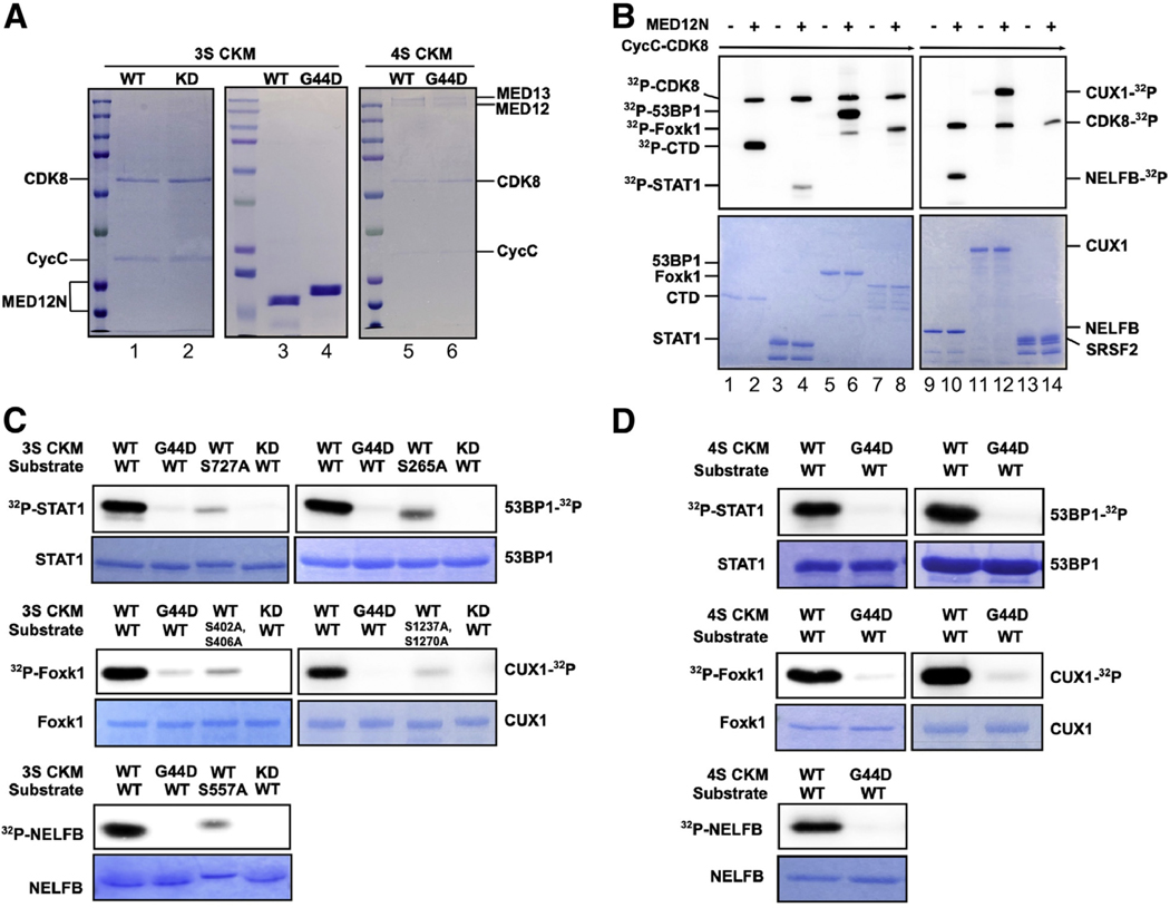
Main outcome measure(s): High-confidence Mediator kinase substrates identified by SILAC-based quantitative phosphoproteomics were determined using an empirical Bayes analysis and validated orthogonally by in vitro kinase assay featuring reconstituted Mediator kinase modules comprising wild-type or G44D mutant MED12 corresponding to the most frequent uterine fibroid driver mutation in MED12. Mediator kinase-regulated transcripts identified by RNA sequencing were linked to Mediator kinase substrates by computational analyses.
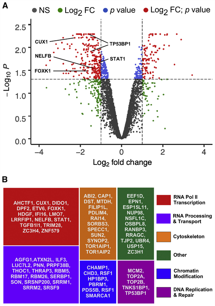
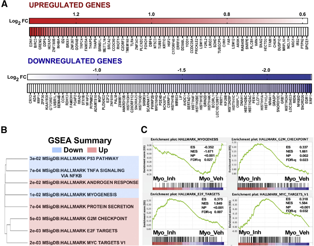
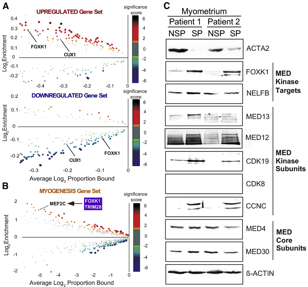
Result(s): A total of 296 unique phosphosites in 166 proteins were significantly decreased (≥ twofold) upon CDK8/19 inhibition, including 118 phosphosites in 71 nuclear proteins representing high-confidence Mediator kinase substrates linked to RNA polymerase II transcription, RNA processing and transport, chromatin modification, cytoskeletal architecture, and DNA replication and repair. Orthogonal validation confirmed a subset of these proteins, including Cut Like Homeobox 1 (CUX1) and Forkhead Box K1 (FOXK1), to be direct targets of MED12-dependent CDK8 phosphorylation in a manner abrogated by the most common uterine fibroid driver mutation (G44D) in MED12, implicating these substrates in disease pathogenesis. Transcriptome-wide profiling of Mediator kinase-inhibited myometrial stem/progenitor cells revealed alterations in cell cycle and myogenic gene expression programs to which Mediator kinase substrates could be linked directly. Among these, CUX1 is an established transcriptional regulator of the cell cycle whose corresponding gene on chromosome 7q is the locus for a recurrent breakpoint in uterine fibroids, linking MED12 and Mediator kinase with CUX1 for the first time in uterine fibroid pathogenesis. FOXK1, a transcriptional regulator of myogenic stem cell fate, was found to be coordinately enriched along with kinase, but not core, Mediator subunits in myometrial stem/progenitor cells compared with differentiated uterine smooth muscle cells.
Conclusion(s): These studies identify a new catalog of pathologically and biologically relevant Mediator kinase substrates implicated in the pathogenesis of MED12 mutation-positive uterine fibroids, and further uncover a biochemical basis to link Mediator kinase activity with CUX1 and FOXK1 in the regulation of myometrial stem/progenitor cell fate.
Keywords: MED12 mutations; mediator kinase; myometrial stem/progenitor cells; phosphoproteomics; uterine fibroids.
Copyright © 2021 American Society for Reproductive Medicine. Published by Elsevier Inc. All rights reserved.
Figures
Similar articles
-
Mediator kinase inhibition drives myometrial stem cell differentiation and the uterine fibroid phenotype through super-enhancer reprogramming.J Mol Med (Berl). 2025 Mar;103(3):311-326. doi: 10.1007/s00109-025-02517-0. Epub 2025 Feb 4. J Mol Med (Berl). 2025. PMID: 39904883 Free PMC article.
-
Oncogenic exon 2 mutations in Mediator subunit MED12 disrupt allosteric activation of cyclin C-CDK8/19.J Biol Chem. 2018 Mar 30;293(13):4870-4882. doi: 10.1074/jbc.RA118.001725. Epub 2018 Feb 13. J Biol Chem. 2018. PMID: 29440396 Free PMC article.
-
A mediator complex subunit 12 gain-of-function mutation induces partial leiomyoma cell properties in human uterine smooth muscle cells.F S Sci. 2022 Aug;3(3):288-298. doi: 10.1016/j.xfss.2022.04.002. Epub 2022 Apr 15. F S Sci. 2022. PMID: 35643626
-
Uterine fibroids at single-cell resolution: unveiling cellular heterogeneity to improve understanding of pathogenesis and guide future therapies.Am J Obstet Gynecol. 2025 Apr;232(4S):S124-S134. doi: 10.1016/j.ajog.2024.08.037. Am J Obstet Gynecol. 2025. PMID: 40253076 Review.
-
UTERINE FIBROIDS.Physiol Rev. 2025 Oct 1;105(4):1947-1988. doi: 10.1152/physrev.00010.2024. Epub 2025 Apr 11. Physiol Rev. 2025. PMID: 40214304 Review.
Cited by
-
Mediator kinase inhibition drives myometrial stem cell differentiation and the uterine fibroid phenotype through super-enhancer reprogramming.J Mol Med (Berl). 2025 Mar;103(3):311-326. doi: 10.1007/s00109-025-02517-0. Epub 2025 Feb 4. J Mol Med (Berl). 2025. PMID: 39904883 Free PMC article.
-
Structures and compositional dynamics of Mediator in transcription regulation.Curr Opin Struct Biol. 2024 Oct;88:102892. doi: 10.1016/j.sbi.2024.102892. Epub 2024 Jul 26. Curr Opin Struct Biol. 2024. PMID: 39067114 Review.
-
Aberrant R-loop-induced replication stress in MED12-mutant uterine fibroids.Sci Rep. 2022 Apr 13;12(1):6169. doi: 10.1038/s41598-022-10188-x. Sci Rep. 2022. PMID: 35418189 Free PMC article.
-
Unveiling the noncanonical activation mechanism of CDKs: insights from recent structural studies.Front Mol Biosci. 2023 Nov 9;10:1290631. doi: 10.3389/fmolb.2023.1290631. eCollection 2023. Front Mol Biosci. 2023. PMID: 38028546 Free PMC article. Review.
-
Mediator kinase inhibition drives myometrial stem cell differentiation and the uterine fibroid phenotype through super-enhancer reprogramming.Res Sq [Preprint]. 2024 Dec 16:rs.3.rs-5125876. doi: 10.21203/rs.3.rs-5125876/v1. Res Sq. 2024. Update in: J Mol Med (Berl). 2025 Mar;103(3):311-326. doi: 10.1007/s00109-025-02517-0. PMID: 39764110 Free PMC article. Updated. Preprint.
References
-
- Doherty L, Mutlu L, Sinclair D, Taylor H. Uterine fibroids: clinical manifestations and contemporary management. Reprod Sci 2014;21:1067–92. - PubMed
-
- Bulun SE. Uterine fibroids. N Engl J Med 2013;369:1344–55. - PubMed
-
- Stewart EA, Laughlin–Tommaso SK, Catherino WH, Lalitkumar S, Gupta D, Vollenhoven B. Uterine fibroids. Nat Rev Dis Primers 2016;2:16043. - PubMed
-
- Bartels CB, Cayton KC, Chuong FS, Holthouser K, Arian SE, Abraham T, et al. An evidence-based approach to the medical management of fibroids: a systematic review. Clin Obstet Gynecol 2016;59:30–52. - PubMed
Publication types
MeSH terms
Substances
Grants and funding
LinkOut - more resources
Full Text Sources
Research Materials
