

Cytotoxicity of 2D engineered nanomaterials in pulmonary and corneal epithelium
- PMID: 35560287
- PMCID: PMC9205178
- DOI: 10.1016/j.impact.2022.100404
Cytotoxicity of 2D engineered nanomaterials in pulmonary and corneal epithelium
Abstract
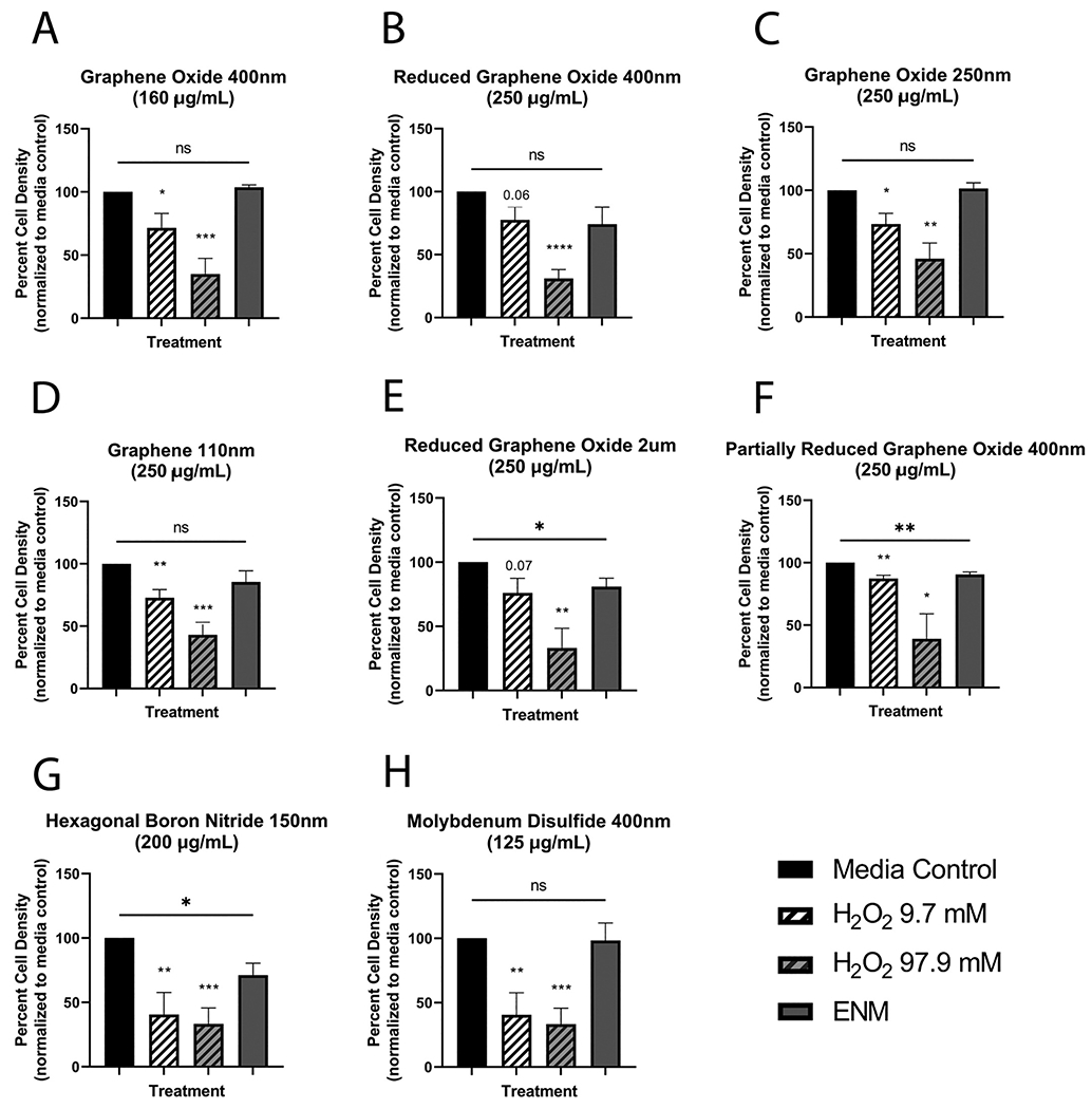
Two-dimensional (2D) engineered nanomaterials are widely used in consumer and industrial goods due to their unique chemical and physical characteristics. Engineered nanomaterials are incredibly small and capable of being aerosolized during manufacturing, with the potential for biological interaction at first-contact sites such as the eye and lung. The unique properties of 2D nanomaterials that make them of interest to many industries may also cause toxicity towards epithelial cells. Using murine and human respiratory epithelial cell culture models, we tested the cytotoxicity of eight 2D engineered nanomaterials: graphene (110 nm), graphene oxide (2 um), graphene oxide (400 nm), reduced graphene oxide (2 um), reduced graphene oxide (400 nm), partially reduced graphene oxide (400 nm), molybdenum disulfide (400 nm), and hexagonal boron nitride (150 nm). Non-graphene nanomaterials were also tested in human corneal epithelial cells for ocular epithelial cytotoxicity. Hexagonal boron nitride was found to be cytotoxic in mouse tracheal, human alveolar, and human corneal epithelial cells. Hexagonal boron nitride was also tested for inhibition of wound healing in alveolar epithelial cells; no inhibition was seen at sub-cytotoxic doses. Nanomaterials should be considered with care before use, due to specific regional cytotoxicity that also varies by cell type. Supported by U01ES027288 and T32HL007013 and T32ES007059.
Keywords: 2D engineered nanomaterials; Airway epithelial cells; Corneal epithelial cells; Hexagonal boron nitride; Nanotoxicity.
Copyright © 2022 The Authors. Published by Elsevier B.V. All rights reserved.
Conflict of interest statement
Declaration of Competing Interest
The authors declare the following financial interests/personal relationships which may be considered as potential competing interests:collaborator who supplied research consortium with nanomaterials is also editor-in-chief of Nanoimpact - Dr. Philip Demokritou.
Figures


Similar articles
-
Biotransformations and cytotoxicity of eleven graphene and inorganic two-dimensional nanomaterials using simulated digestions coupled with a triculture in vitro model of the human gastrointestinal epithelium.Environ Sci Nano. 2021 Nov 1;8(11):3233-3249. doi: 10.1039/d1en00594d. Epub 2021 Oct 8. Environ Sci Nano. 2021. PMID: 37465590 Free PMC article.
-
Effect of graphene-based nanomaterials on corneal wound healing in vitro.Exp Eye Res. 2023 Apr;229:109419. doi: 10.1016/j.exer.2023.109419. Epub 2023 Feb 16. Exp Eye Res. 2023. PMID: 36806671 Free PMC article.
-
When 2D nanomaterials meet biomolecules: design strategies and hybrid nanostructures for bone tissue engineering.J Mater Chem B. 2022 Nov 16;10(44):9040-9053. doi: 10.1039/d2tb01489k. J Mater Chem B. 2022. PMID: 36317564 Review.
-
Assessment of Physico-Chemical and Toxicological Properties of Commercial 2D Boron Nitride Nanopowder and Nanoplatelets.Int J Mol Sci. 2021 Jan 8;22(2):567. doi: 10.3390/ijms22020567. Int J Mol Sci. 2021. PMID: 33430016 Free PMC article.
-
2D nanomaterials based electrochemical biosensors for cancer diagnosis.Biosens Bioelectron. 2017 Mar 15;89(Pt 1):136-151. doi: 10.1016/j.bios.2016.06.011. Epub 2016 Jun 7. Biosens Bioelectron. 2017. PMID: 27318880 Review.
Cited by
-
2D Hexagonal Boron Nitride (h-BN) and 1D Boron Nitride Nanotubes (BNNTs): Distinct Effects at the Cellular Level in Fish Cell Lines.J Xenobiot. 2025 Jun 24;15(4):97. doi: 10.3390/jox15040097. J Xenobiot. 2025. PMID: 40700144 Free PMC article.
-
Metabolomics of V2O5 nanoparticles and V2O5 nanofibers in human airway epithelial BEAS-2B cells.Toxicol Appl Pharmacol. 2023 Jan 15;459:116327. doi: 10.1016/j.taap.2022.116327. Epub 2022 Nov 30. Toxicol Appl Pharmacol. 2023. PMID: 36460058 Free PMC article.
-
Recent Development of Nanomaterials for Transdermal Drug Delivery.Biomedicines. 2023 Apr 7;11(4):1124. doi: 10.3390/biomedicines11041124. Biomedicines. 2023. PMID: 37189742 Free PMC article. Review.
-
Environmental and Health Impacts of Graphene and Other Two-Dimensional Materials: A Graphene Flagship Perspective.ACS Nano. 2024 Feb 27;18(8):6038-6094. doi: 10.1021/acsnano.3c09699. Epub 2024 Feb 13. ACS Nano. 2024. PMID: 38350010 Free PMC article. Review.
-
Novel ultrathin ferrous sulfide nanosheets: Towards replacing black phosphorus in anticancer nanotheranostics.Bioact Mater. 2024 Oct 14;43:564-578. doi: 10.1016/j.bioactmat.2024.09.035. eCollection 2025 Jan. Bioact Mater. 2024. PMID: 40115876 Free PMC article.
References
-
- Arvidsson R, Molander S, Sandén BA, 2013. Review of potential environmental and health risks of the nanomaterial graphene. Hum. Ecol. Risk Assess. Int. J 19 (4), 873–887. 10.1080/10807039.2012.702039. - DOI
Web references
-
- ???Jain Prakhar & Prasad Eswara. (2021, July). Nanotechnology in Energy Market - 2030. Allied Market Research. URL: https://www.alliedmarketresearch.com/nanotechnology-in-energy-market.
