

Cardiac involvement in patients recovering from Delta Variant of COVID-19: a prospective multi-parametric MRI study
- PMID: 35560820
- PMCID: PMC9288765
- DOI: 10.1002/ehf2.13971
Cardiac involvement in patients recovering from Delta Variant of COVID-19: a prospective multi-parametric MRI study
Abstract
Aims: The cardiac injury and sequelae of Delta Variant of coronavirus disease 2019 (COVID-19) remain unknown. This study aimed to evaluate the presence of cardiac involvement in patients recovering from Delta Variant of COVID-19 based on multi-parametric cardiac magnetic resonance imaging (MRI).
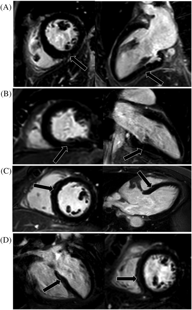
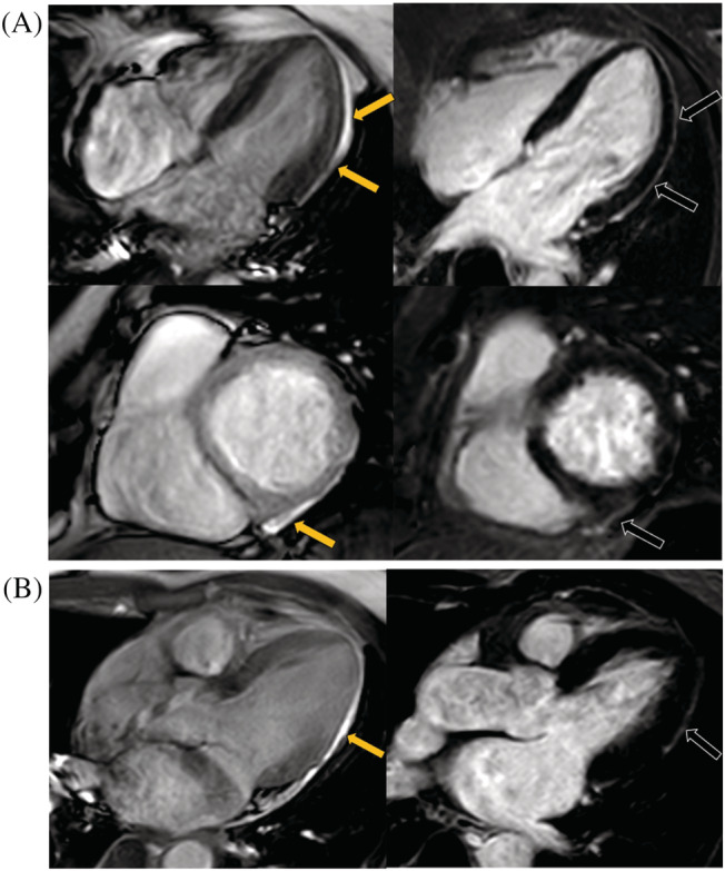
Methods and results: We prospectively assessed patients recovering from Delta Variant of COVID-19 using multi-parametric cardiac magnetic resonance imaging (MRI) between June 2021 and July 2021. Comparison was made with 25 healthy controls. Forty-four patients (median age 51 years, 28 women) recovering from Delta Variant were recruited and had a median time of 35 days between diagnosis and cardiac MRI. There were no patients with chest pain (0/44, 0%) and high sensitivity cardiac troponin T troponin elevation (median levels 2.20 pg/mL, IQR levels 0.85-4.40 pg/mL). Regarding the cardiac imaging findings, a total of 14 (32%) patients presented cardiac tissue feature abnormalities, and a total of 9 (20%) patients had a myocarditis-like injury based on cardiac MRI 2018 Lake Louise criteria. When we further assessed the T1 and T2 mapping values for of patients' individual, abnormal raised global native T1, T2, and extracellular volume were seen in 6 (14%), 6 (14%), and 4 (9%) patients, respectively. Comparing with controls, the patients had lower LV global longitudinal strain and (-22.2 ± 2.8% vs. -24.6 ± 2.0%, P < 0.001) and global circumferential strain (-20.7 ± 6.8% vs. -24.3 ± 2.9%, P = 0.014), but higher global native T1 (1318.8 ± 55.5 ms vs. 1282.9 ± 38.1 ms, P = 0.006). Four (9%) patients presented myocardial late gadolinium enhancement with subepicardial pattern mostly common seen, and two (5%) patients presented pericardial enhancement.
Conclusions: The cardiac MRI could detect subclinical functional and myocardial tissue characteristic abnormalities in individuals who were recovering from Delta Variant without cardiac-related clinical findings. The native T1 mapping and strain imaging may be a sensitive tool for the noninvasive detection of a subset of patients who are at risk for cardiac sequelae and more prone to myocardial damage in survivors with Delta Variant.
Keywords: Delta Variant; T1 mapping; T2 mapping; coronavirus disease 2019; feature tracking.
© 2022 The Authors. ESC Heart Failure published by John Wiley & Sons Ltd on behalf of European Society of Cardiology.
Conflict of interest statement
None declared.
Figures

Similar articles
-
Outcomes of Cardiovascular Magnetic Resonance Imaging in Patients Recently Recovered From Coronavirus Disease 2019 (COVID-19).JAMA Cardiol. 2020 Nov 1;5(11):1265-1273. doi: 10.1001/jamacardio.2020.3557. JAMA Cardiol. 2020. PMID: 32730619 Free PMC article.
-
Evaluation for Myocarditis in Competitive Student Athletes Recovering From Coronavirus Disease 2019 With Cardiac Magnetic Resonance Imaging.JAMA Cardiol. 2021 Aug 1;6(8):945-950. doi: 10.1001/jamacardio.2020.7444. JAMA Cardiol. 2021. PMID: 33443537 Free PMC article.
-
Cardiac magnetic resonance in recovering COVID-19 patients. Feature tracking and mapping analysis to detect persistent myocardial involvement.Int J Cardiol Heart Vasc. 2021 Oct;36:100854. doi: 10.1016/j.ijcha.2021.100854. Epub 2021 Aug 3. Int J Cardiol Heart Vasc. 2021. PMID: 34368419 Free PMC article.
-
Cardiac Magnetic Resonance Imaging Findings in 2954 COVID-19 Adult Survivors: A Comprehensive Systematic Review.J Magn Reson Imaging. 2022 Mar;55(3):866-880. doi: 10.1002/jmri.27852. Epub 2021 Jul 26. J Magn Reson Imaging. 2022. PMID: 34309139 Free PMC article.
-
Recent Advances in T1 and T2 Mapping in the Assessment of Fulminant Myocarditis by Cardiac Magnetic Resonance.Curr Cardiol Rep. 2020 May 29;22(7):47. doi: 10.1007/s11886-020-01295-0. Curr Cardiol Rep. 2020. PMID: 32472218 Review.
Cited by
-
Myocardial Injuries in COVID-19: More Questions Than Answers.J Clin Med. 2022 Aug 3;11(15):4527. doi: 10.3390/jcm11154527. J Clin Med. 2022. PMID: 35956141 Free PMC article.
-
Myocardial Tissue Characterization in Cardiac Magnetic Resonance Studies of Patients Recovering From COVID-19: A Meta-Analysis.J Am Heart Assoc. 2023 Mar 21;12(6):e027801. doi: 10.1161/JAHA.122.027801. Epub 2023 Mar 9. J Am Heart Assoc. 2023. PMID: 36892052 Free PMC article.
-
CMR Findings in COVID-19 Recovered Patients: A Review on Parametric Mapping, Feature-Tracking, and LGE.Rev Cardiovasc Med. 2022 Oct 21;23(11):355. doi: 10.31083/j.rcm2311355. eCollection 2022 Nov. Rev Cardiovasc Med. 2022. PMID: 39076192 Free PMC article. Review.
-
COVID-19, Myocarditis and Pericarditis.Circ Res. 2023 May 12;132(10):1302-1319. doi: 10.1161/CIRCRESAHA.123.321878. Epub 2023 May 11. Circ Res. 2023. PMID: 37167363 Free PMC article. Review.
-
The Impact of Vaccinations Against Respiratory Infections on the Prognosis in Heart Failure Patients.Vaccines (Basel). 2024 Nov 26;12(12):1321. doi: 10.3390/vaccines12121321. Vaccines (Basel). 2024. PMID: 39771983 Free PMC article. Review.
References
-
- Del Rio C, Malani PN, Omer SB. Confronting the Delta Variant of SARS‐CoV‐2, summer 2021. JAMA. 2021. Online ahead of print. - PubMed
-
- Dyer O. Covid‐19: Indonesia becomes Asia's new pandemic epicentre as delta variant spreads. BMJ. 2021; 374: n1815. - PubMed
-
- CDC . COVID‐19: SARS‐CoV‐2 variant classifications and definitions. Atlanta, GA: US Department of Health and Human Services. https://www.cdc.gov/coronavirus/2019‐ncov/cases‐updates/variant‐surveill... (2021; date last accessed).
Publication types
MeSH terms
Substances
Supplementary concepts
LinkOut - more resources
Full Text Sources
Medical
