. 2022 May 17;119(20):e2118129119.
doi: 10.1073/pnas.2118129119. Epub 2022 May 13.

Targeted ubiquitination of sensory neuron calcium channels reduces the development of neuropathic pain

Affiliations

Targeted ubiquitination of sensory neuron calcium channels reduces the development of neuropathic pain

Linlin Sun et al. Proc Natl Acad Sci U S A. .

Abstract

Neuropathic pain caused by lesions to somatosensory neurons due to injury or disease is a widespread public health problem that is inadequately managed by small-molecule therapeutics due to incomplete pain relief and devastating side effects. Genetically encoded molecules capable of interrupting nociception have the potential to confer long-lasting analgesia with minimal off-target effects. Here, we utilize a targeted ubiquitination approach to achieve a unique posttranslational functional knockdown of high-voltage-activated calcium channels (HVACCs) that are obligatory for neurotransmission in dorsal root ganglion (DRG) neurons. CaV-aβlator comprises a nanobody targeted to CaV channel cytosolic auxiliary β subunits fused to the catalytic HECT domain of the Nedd4-2 E3 ubiquitin ligase. Subcutaneous injection of adeno-associated virus serotype 9 encoding CaV-aβlator in the hind paw of mice resulted in the expression of the protein in a subset of DRG neurons that displayed a concomitant ablation of CaV currents and also led to an increase in the frequency of spontaneous inhibitory postsynaptic currents in the dorsal horn of the spinal cord. Mice subjected to spare nerve injury displayed a characteristic long-lasting mechanical, thermal, and cold hyperalgesia underlain by a dramatic increase in coordinated phasic firing of DRG neurons as reported by in vivo Ca2+ spike recordings. CaV-aβlator significantly dampened the integrated Ca2+ spike activity and the hyperalgesia in response to nerve injury. The results advance the principle of targeting HVACCs as a gene therapy for neuropathic pain and demonstrate the therapeutic potential of posttranslational functional knockdown of ion channels achieved by exploiting the ubiquitin-proteasome system.

Keywords: calcium channel; neuropathic pain; ubiquitin.


Conflict of interest statement

Competing interest statement: Columbia University filed a patent related to the use of engineered ubiquitin ligases as genetically encoded inhibitors for voltage-gated calcium channels.

Figures

Fig. 1.
Fig. 1.
Distinct paradigms and targets for neuropathic pain gene therapy. The circuitry for neuropathic pain involves signals from primary somatosensory neurons that synapse on secondary neurons in the spinal cord dorsal horn. Relay neurons convey signals to various parts of the brain for nociception. Key signaling molecules and proteins involved in the relaying of nociceptive signals are candidate targets for neuropathic pain gene therapy. Previously described knockdown approaches include CRISPR-dCas9-mediated epigenetic knockdown of Nav1.7 and down-regulation of NMDA receptors with siRNA. Overexpression of glutamate decarboxylase has been used to produce the inhibitory neurotransmitter GABA; enkephalin overexpression suppresses neurotransmission by activating the µ-opioid receptor to release Gβγ subunits that inhibit presynaptic CaV2.2 channels. Ca2+ influx through CaV2 channels is necessary for neurotransmitter release, and inhibiting these channels is therapeutic for chronic pain. Here, we use a targeted ubiquitination strategy to achieve direct posttranslational functional knockdown of CaV2 channels in DRG neurons using CaV-aβlator, a genetically encoded molecule featuring a CaVβ-targeted nanobody fused to the catalytic HECT domain of Nedd4 E3 ligase. µOR, µ-opioid receptors.
Fig. 2.
Fig. 2.
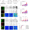
CaV-aβlator expressed in vivo ablates calcium currents in DRG neurons. (A) Experimental paradigm for CaV-aβlator injection in newborn mice. (B) Confocal images of cultured DRG neurons from Thy1-GCaMP6s mouse injected at 3 to 5 d old in the hind paw with AAV9-expressing CaV-aβlator-P2A-tdTomato. (C) Top, exemplar family of whole-cell Ca2+ channel currents obtained from young mice DRG neurons either negative (tdTomato) or positive (tdTomato +) for CaV-aβlator expression. Bottom, population Ca2+ current I-V curves from young mice DRG neurons negative (●, n = 6) or positive (red square, n = 5) for CaV-aβlator expression. (D) Experimental paradigm for CaV-aβlator subcutaneous injection in adult mice. (E) Confocal image of DRG from a CaV-aβlator-injected mouse. Autofluorescence in the green channel was exploited to isolate red signals due to CaV-aβlator-P2a-tdTomato expression from background red autofluorescence. (F) Top, exemplar family of whole-cell Ca2+ channel currents obtained from adult mice DRG neurons either negative (tdTomato) or positive (tdTomato +) for CaV-aβlator expression. Bottom, population Ca2+ current I-V curves from adult mice DRG neurons negative (●, n = 19) or positive (red square, n = 10) for CaV-aβlator expression.
Fig. 3.
Fig. 3.
Impact of CaV-aβlator expression in DRG neurons on spontaneous EPSCs and IPSCs in the spinal cord dorsal horn. (A) Exemplar sEPSC recorded in the dorsal horn from control (black traces) and CaV-aβlator-injected (red traces) sides. (B) Cumulative distribution histograms of exemplar sEPSC amplitudes for control (black line) and CaV-aβlator-injected (red line) sides. (C) Population mean sEPSC amplitudes from control and CaV-aβlator-injected sides. (D) Summary of sEPSC frequency. N.S., not significant. (E) Exemplar sIPSC recorded in the dorsal horn from control (black traces) and CaV-aβlator-injected (red traces) sides. (F) Cumulative distribution histograms of exemplar sIPSC amplitudes for control (black line) and CaV-aβlator-injected (red line) sides. (G) Population mean sIPSC amplitudes from control and CaV-aβlator-injected sides. (H) Summary of sIPSC frequency.
Fig. 4.
Fig. 4.
Impact of CaV-aβlator on nerve-injury-induced enhancement of spontaneous calcium spikes in DRG neurons in vivo. (A) Experimental design. (B) Spontaneous Ca2+ spikes recorded in vivo in DRG neurons from a control Thy1-GCaMP6s mouse (injected with AAV9-tdTomato) pre-SNI and 3 and 7 d post-SNI. Left, images of DRG neurons expressing GCaMP6s and/or tdTomato. Right, representative Ca2+ traces (Top), deconvoluted somatic Ca2+ spikes from 30 DRG neurons (Middle), and representative correlation coefficient matrix of Ca2+ transients from all active neuron pairs in DRG (Bottom). Ca2+ traces 1 to 6 correspond to individual neurons 1 to 6 indicated by arrows in the DRG image. (C) In vivo spontaneous Ca2+ transient data for Thy1-GCaMP6s mouse injected subcutaneously with AAV9-encoding tdTomato-CaV-aβlator, with the same format as B. (D) Summary of mean peak amplitude of spontaneous Ca2+ spikes between tdTomato- and CaV-aβlator-injected mice pre- and post-SNI. (E) Summary of mean frequency of spontaneous Ca2+ spikes. (F) Average integrated Ca2+ activity of DRG neurons (n = 140 neurons in tdTomato group, n = 52 neurons in CaV-aβlator group). (G) Cumulative curve of the correlation coefficients (c.c.) of all active DRG neuron pairs (n = 140 neurons in tdTomato group, n = 52 neurons in CaV-aβlator group). Summary data are presented as mean ± SEM *P < 0.05, **P < 0.01, ***P < 0.001; by unpaired t test.
Fig. 5.
Fig. 5.
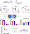
CaV-aβlator reduces hyperalgesia after SNI with no apparent adverse effects. (A) Top, shows experimental design. Mice were tested 4 weeks after viral injection. Bottom, shows behavior tests for nocifensive responses. SNI triggered the development of mechanical, thermal, and cold hypersensitivity in the ipsilateral (ipsi) but not contralateral (contra) side of the mice, and the hypersensitivity was significantly reduced in CaV-aβlator injected mice. (n = 6 to 9 mice per group). (B) Open field test showed no difference between tdTomato- and CaV-aβlator-injected mice. (C) Rotarod performance test. Each dots represent data from a single animal. (D) Scores for placing, grasping, and righting reflexes were based on counts of each normal reflex exhibited in five trials. All values are mean (SEM). n = 6 mice per group. There was no difference in locomotor functions between tdTomato- and CaV-aβlator-injected mice. Summary data are presented as mean ± SEM *P < 0.05, **P < 0.01, ***P < 0.001, ****P < 0.0001; #P < 0.05, ##P < 0.01, ####P < 0.0001; by two-way RM ANOVA with Dunnett’s multiple comparisons test (A), or unpaired t test (B, C). Asterisks (*) denote post-SNI versus pre-SNI (day 0) comparisons, and the pound signs (#) denote the comparison CaV-aβlator versus control.

Comment in

Similar articles

Cited by

References

    1. Toth C., Lander J., Wiebe S., The prevalence and impact of chronic pain with neuropathic pain symptoms in the general population. Pain Med. 10, 918–929 (2009). - PubMed
    1. van Hecke O., Austin S. K., Khan R. A., Smith B. H., Torrance N., Neuropathic pain in the general population: A systematic review of epidemiological studies. Pain 155, 654–662 (2014). - PubMed
    1. Colloca L., et al. , Neuropathic pain. Nat. Rev. Dis. Primers 3, 17002 (2017). - PMC - PubMed
    1. Bates D., et al. , A comprehensive algorithm for management of neuropathic pain. Pain Med. 20 (suppl. 1), S2–S12 (2019). - PMC - PubMed
    1. Cavalli E., Mammana S., Nicoletti F., Bramanti P., Mazzon E., The neuropathic pain: An overview of the current treatment and future therapeutic approaches. Int. J. Immunopathol. Pharmacol. 33, 2058738419838383 (2019). - PMC - PubMed

Publication types

Substances

LinkOut - more resources