

Periventricular gradient of T1 tissue alterations in multiple sclerosis
- PMID: 35561554
- PMCID: PMC9112026
- DOI: 10.1016/j.nicl.2022.103009
Periventricular gradient of T1 tissue alterations in multiple sclerosis
Abstract
Objective: Pathology in multiple sclerosis is not homogenously distributed. Recently, it has been shown that structures adjacent to CSF are more severely affected. A gradient of brain tissue involvement was shown with more severe pathology in periventricular areas and in proximity to brain surfaces such as the subarachnoid spaces and ependyma, and hence termed the "surface-in" gradient. Here, we study whether (i) the surface-in gradient of periventricular tissue alteration measured by T1 relaxometry is already present in early multiple sclerosis patients, (ii) how it differs between early and progressive multiple sclerosis patients, and (iii) whether the gradient-derived metrics in normal-appearing white matter and lesions correlate better with physical disability than conventional MRI-based metrics.
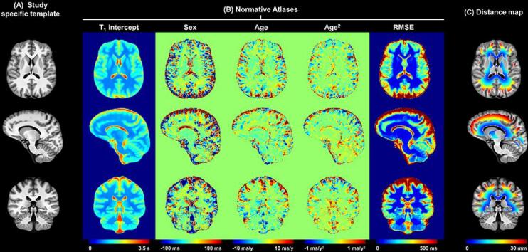
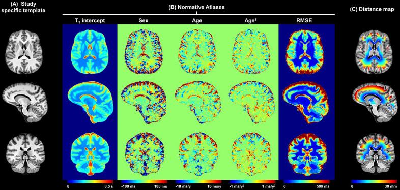
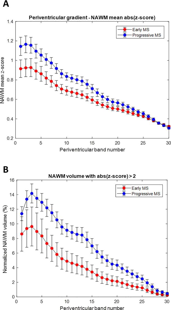
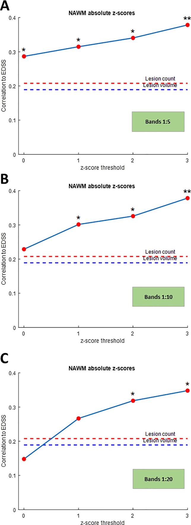
Methods: Forty-seven patients with early multiple sclerosis, 52 with progressive multiple sclerosis, and 92 healthy controls were included in the study. Isotropic 3D T1 relaxometry maps were obtained using the Magnetization-Prepared 2 Rapid Acquisition Gradient Echoes sequence at 3 T. After spatially normalizing the T1 maps into a study-specific common space, T1 inter-subject variability within the healthy cohort was modelled voxel-wise, yielding a normative T1 atlas. Individual comparisons of each multiple sclerosis patient against the atlas were performed by computing z-scores. Equidistant bands of voxels were defined around the ventricles in the supratentorial white matter; the z-scores in these bands were analysed and compared between the early and progressive multiple sclerosis cohorts. Correlations between both conventional and z-score-gradient-derived MRI metrics and the Expanded Disability Status Scale were assessed.
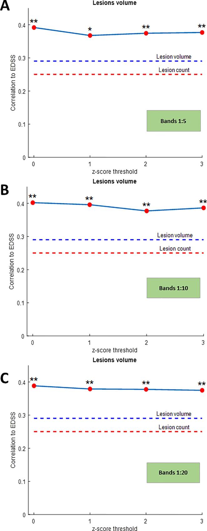
Results: Patients with early and progressive multiple sclerosis demonstrated a periventricular gradient of T1 relaxation time z-scores. In progressive multiple sclerosis, z-score-derived metrics reflecting the gradient of tissue abnormality in normal-appearing white matter were more strongly correlated with disability (maximal rho = 0.374) than the conventional lesion volume and count (maximal rho = 0.189 and 0.21 respectively). In early multiple sclerosis, the gradient of normal-appearing white matter volume with z-scores > 2 at baseline correlated with clinical disability assessed at two years follow-up.
Conclusion: Our results suggest that the surface-in white matter gradient of tissue alteration is detectable with T1 relaxometry and is already present at clinical disease onset. The periventricular gradients correlate with clinical disability. The periventricular gradient in normal-appearing white matter may thus qualify as a promising biomarker for monitoring of disease activity from an early stage in all phenotypes of multiple sclerosis.
Trial registration: ClinicalTrials.gov NCT03706118.
Keywords: Atlas-based assessment; Gradient of tissue damage; MP2RAGE; Multiple sclerosis; T(1)-relaxometry.
Copyright © 2022 The Author(s). Published by Elsevier Inc. All rights reserved.
Conflict of interest statement
M. Vaneckova received speaker honoraria, consultant fees, and travel expenses from Biogen Idec, Novartis, Roche, Genzyme, and Teva, as well as support for research activities from Biogen Idec. G.F. Piredda, T. Hilbert, B. Marechal and T. Kober are employees of Siemens Healthcare AG, Switzerland. M. Andelova received financial support for conference travel from Novartis, Genzyme, Merck Serono, Biogen Idec, and Roche. J. Krasensky received financial support for research activities from Biogen Idec. T. Uher received financial support for conference travel and honoraria from Biogen Idec, Novartis, Roche, Genzyme, and Merck Serono, as well as support for research activities from Biogen Idec and Sanofi.B. Srpova received financial support for conference travel from Novartis, Genzyme, Merck Serono, Biogen Idec, and Roche. E. Kubala Havrdova received speaker honoraria and consultant fees from Biogen Idec, Merck Serono, Novartis, Genzyme, Teva, Actelion, and Receptos, as well as support for research activities from Biogen Idec and Merck Serono. K. Vodehnalova received compensation for travel, conference fees, and consulting fees from Merck, Sanofi Genzyme, Biogen Idec, and Novartis. D. Horakova received compensation for travel, speaker honoraria and consultant fees from Biogen Idec, Novartis, Merck Serono, Bayer Shering, and Teva, as well as support for research activities from Biogen Idec.
Figures
References
-
- Bakshi R., Neema M., Healy B.C., Liptak Z., Betensky R.A., Buckle G.J., Gauthier S.A., Stankiewicz J., Meier D., Egorova S., Arora A., Guss Z.D., Glanz B., Khoury S.J., Guttmann C.R.G., Weiner H.L. Predicting clinical progression in multiple sclerosis with the magnetic resonance disease severity scale. Arch. Neurol. 2008;65(11):1449–1453. doi: 10.1001/archneur.65.11.1449. - DOI - PMC - PubMed
-
- Bodini B., Poirion E., Tonietto M., Benoit C., Palladino R., Maillart E., Portera E., Battaglini M., Bera G., Kuhnast B., Louapre C., Bottlaender M., Stankoff B. Individual mapping of innate immune cell activation is a candidate marker of patient-specific trajectories of worsening disability in multiple sclerosis. J. Nucl. Med. 2020;61(7):1043–1049. doi: 10.2967/jnumed.119.231340. - DOI - PMC - PubMed
-
- Brown J.W.L., Pardini M., Brownlee W.J., Fernando K., Samson R.S., Prados Carrasco F., Ourselin S., Gandini Wheeler-Kingshott C.A.M., Miller D.H., Chard D.T. An abnormal periventricular magnetization transfer ratio gradient occurs early in multiple sclerosis. Brain. 2017;140(2):387–398. - PMC - PubMed
Publication types
MeSH terms
Associated data
LinkOut - more resources
Full Text Sources
Other Literature Sources
Medical
