

Medulla oblongata volume as a promising predictor of survival in amyotrophic lateral sclerosis
- PMID: 35561555
- PMCID: PMC9111981
- DOI: 10.1016/j.nicl.2022.103015
Medulla oblongata volume as a promising predictor of survival in amyotrophic lateral sclerosis
Abstract
Background: Unconventional magnetic resonance imaging studies of the brainstem have recently acquired a growing interest in amyotrophic lateral sclerosis (ALS) pathology since they provide a unique opportunity to evaluate motor tract degeneration and bulbar lower motor neuron involvement. The aim of this study was to investigate the role of brainstem structures as accurate biomarkers of disease severity and predictors of survival.
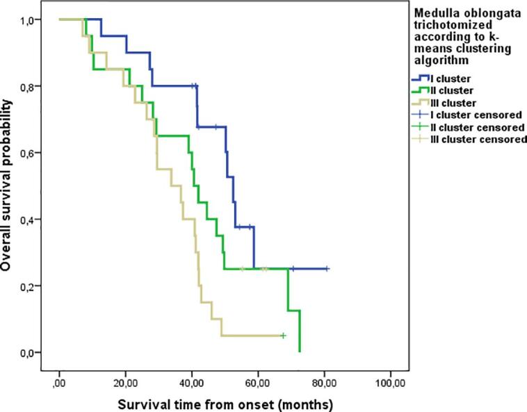
Materials and methods: A total of 60 ALS patients and 30 healthy controls subjects (CS) were recruited in this study. Patients were divided in two subgroups according to the onset of the disease: 42 spinal (S-ALS) and 18 bulbar (B-ALS). All subjects underwent 3D-structural MRI. Brainstem volume both of the entire cohort of ALS patients and S-ALS and B-ALS onset were compared with those of CS. In addition the two ALS subgroups were tested for differences in brainstem volumes. Volumetric, vertex-wise, and voxel-based approaches were implemented to assess correlations between MR structural features and clinical characteristics expressed as ALSFRS-r and its bulbar (ALSFSR-r-B) and spinal subscores (ALSFSR-r-S). ROC curves were performed to test the accuracy of midbrain, pons, and medulla oblongata volumes able to discriminate patients dichotomized into long and short survivors by using Two-Steps cluster analysis. Univariate and multivariate survival analyses were carried out to test the prognostic role of brainstem structures' volume, trichotomized by applying a k-means clustering algorithm.
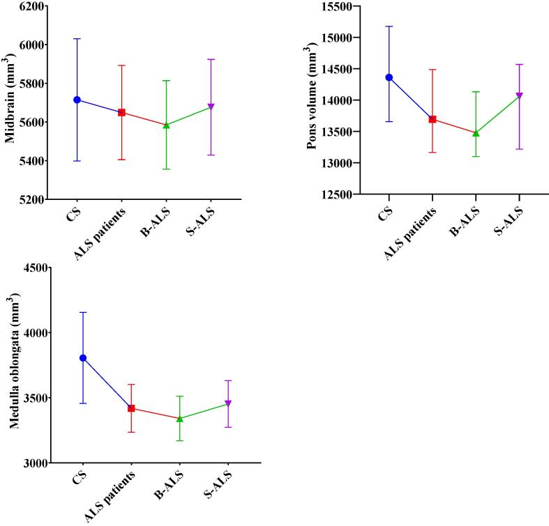
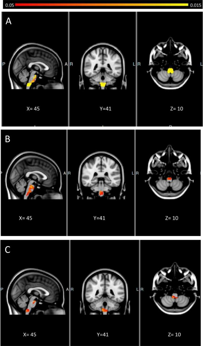
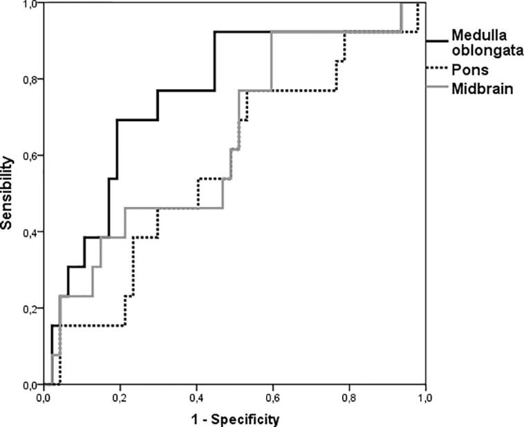
Results: Both the entire cohort of ALS patients and B-ALS and S-ALS showed significant lower volumes of both medulla oblongata and pons compared to CS. Furthermore, B-ALS showed a significant lower volume of medulla oblongata, compared to S-ALS. Lower score of ALSFRS-r correlated to atrophy in the anterior compartment of midbrain, pons, and medulla oblongata, as well as in the posterior portion of only this latter region. ALSFSR-r-S positively correlated with shape deformation and density reduction of the anterior portion of the entire brainstem, along the corticospinal tracts. ALSFSR-r-B instead showed a positive correlation with shape deformation of the floor of the fourth ventricle in the medulla oblongata and the crus cerebri in the midbrain. Only medulla oblongata volume demonstrated a significant accuracy to discriminate long and short survivors ALS patients (ROC AUC 0.76, p < 0.001). Univariate and multivariate analysis confirmed the survival predictive role of the medulla oblongata (log rank test p: 0.003).
Discussions: Our findings suggest that brainstem volume may reflect the impairment of corticospinal and corticobulbar tracts as well as lower bulbar motor neurons. Furthermore, medulla oblongata could be used as an early predictor of survival in ALS patients.
Keywords: ALS; Brainstem; MRI; Medulla oblongata; Survival.
Copyright © 2022 The Author(s). Published by Elsevier Inc. All rights reserved.
Conflict of interest statement
The authors declare that they have no known competing financial interests or personal relationships that could have appeared to influence the work reported in this paper.
Figures
References
-
- Bede P., Bokde A., Elamin M., Byrne S., McLaughlin R.L., Jordan N., Hampel H., Gallagher L., Lynch C., Fagan A.J., Pender N., Hardiman O. Grey matter correlates of clinical variables in amyotrophic lateral sclerosis (ALS): a neuroimaging study of ALS motor phenotype heterogeneity and cortical focality. J. Neurol. Neurosurg. Psychiatry. 2013;84:766–773. doi: 10.1136/jnnp-2012-302674. - DOI - PubMed
-
- P. Bede, R.H. Chipika, E. Finegan, S. Li Hi Shing, K.M. Chang, M.A. Doherty, J.C. Hengeveld, A. Vajda, S. Hutchinson, C. Donaghy, R.L. McLaughlin, O. Hardiman, 2020. Progressive brainstem pathology in motor neuron diseases: Imaging data from amyotrophic lateral sclerosis and primary lateral sclerosis. Data Brief 29, 105229. https://doi.org/10.1016/j.dib.2020.105229. - PMC - PubMed
-
- P. Bede, R.H. Chipika, E. Finegan, S. Li Hi Shing, M.A. Doherty, J.C. Hengeveld, A. Vajda, S. Hutchinson, C. Donaghy, R.L. McLaughlin, O. Hardiman, 2019, Brainstem pathology in amyotrophic lateral sclerosis and primary lateral sclerosis: A longitudinal neuroimaging study. NeuroImage Clin. 24, 102054. https://doi.org/10.1016/j.nicl.2019.102054. - PMC - PubMed
MeSH terms
LinkOut - more resources
Full Text Sources
Medical
Miscellaneous
