. 2022 Jun 14;17(6):1428-1441.
doi: 10.1016/j.stemcr.2022.04.009. Epub 2022 May 12.

Ripk3 signaling regulates HSCs during stress and represses radiation-induced leukemia in mice

Affiliations

Ripk3 signaling regulates HSCs during stress and represses radiation-induced leukemia in mice

Lei Zhang et al. Stem Cell Reports. .

Abstract

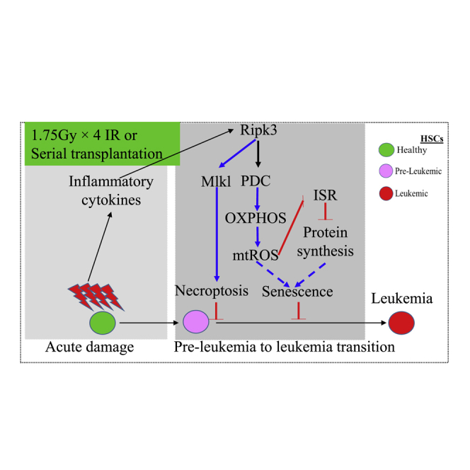
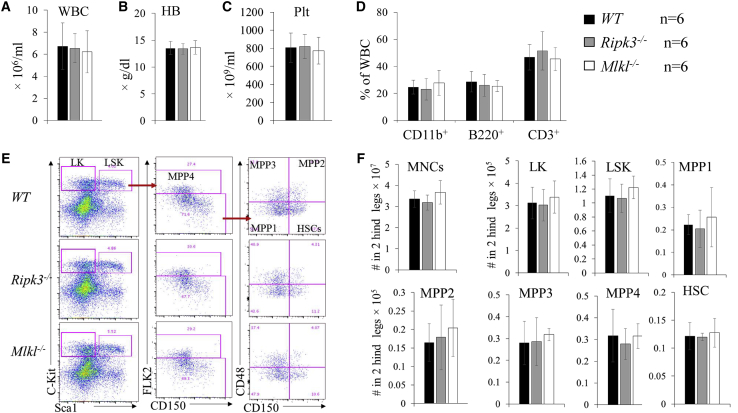
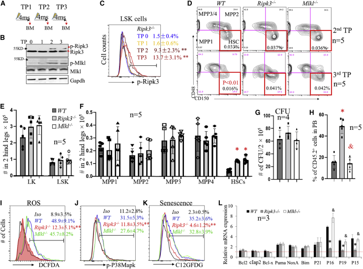
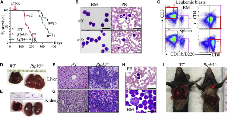
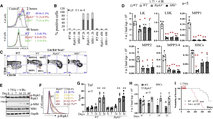
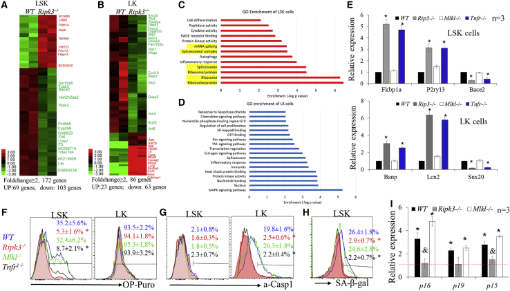
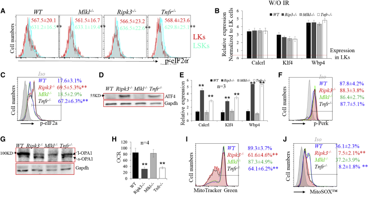
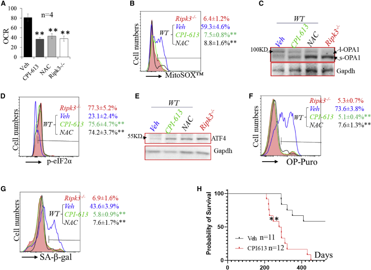
Receptor-interacting protein kinase 3 (Ripk3) is one of the critical mediators of inflammatory cytokine-stimulated signaling. Here we show that Ripk3 signaling selectively regulates both the number and the function of hematopoietic stem cells (HSCs) during stress conditions. Ripk3 signaling is not required for normal homeostatic hematopoiesis. However, in response to serial transplantation, inactivation of Ripk3 signaling prevents stress-induced HSC exhaustion and functional HSC attenuation, while in response to fractionated low doses of ionizing radiation (IR), inactivation of Ripk3 signaling accelerates leukemia/lymphoma development. In both situations, Ripk3 signaling is primarily stimulated by tumor necrosis factor-α. Activated Ripk3 signaling promotes the elimination of HSCs during serial transplantation and pre-leukemia stem cells (pre-LSCs) during fractionated IR by inducing Mlkl-dependent necroptosis. Activated Ripk3 signaling also attenuates HSC functioning and represses a pre-LSC-to-LSC transformation by promoting Mlkl-independent senescence. Furthermore, we demonstrate that Ripk3 signaling induces senescence in HSCs and pre-LSCs by attenuating ISR-mediated mitochondrial quality control.

Keywords: HSCs; Mlkl; Ripk3; ionizing radiation; leukemia; necroptosis.


Figures

None
Graphical abstract
Figure 1
Figure 1
Normal hematopoiesis in Ripk3−/− and Mlkl−/− mice under normal homeostatic conditions (A–F) PB (A–D) and BM (E and F) were collected from WT, Ripk3−/−, and Mlkl−/− mice. Six mice per genotype were studied (three males and three females). All mice were analyzed at 6 months of age. WBC counts (A), Hb concentrations (B), and Plt numbers (C) were analyzed using a Hemavet 950FS. The percentages of CD11b+ granulocytes/monocytes, B220+ B lymphocytes, and CD3+ T lymphocytes were analyzed by flow cytometry (D). Representative flow cytometric data (E) and absolute numbers (F) of HSCs and HPCs in the BM of WT, Ripk3−/−, and Mlkl−/− mice are shown.
Figure 2
Figure 2
Ripk3 selectively regulates number and function of HSCs during serial transplantation in Mlkl-dependent and -independent manners, respectively (A) Serial transplantation and analysis schedule. TP0 represents non-transplanted control. TP1, TP2, and TP3 indicate first, second, and third transplantations, respectively. “4ms” represents 4 months post-transplantation. (B and C) BM MNCs were collected from recipient mice 4 months post-transplantation for each transplantation cycle. Activation of Ripk3-Mlkl signaling in BM MNCs (B) and LSK cells (C) was examined by western blotting and flow cytometric assays, respectively. For flow cytometric analysis, Ripk3−/− LSK cells were always used as a negative control to set up the flow cytometer in order to make the data consistent in all of the experiments. (D) Representative flow cytometric data for HSCs and MPPs among CD45.2+ BM MNCs from second and third transplantation recipients of the indicated genotypes of donors. (E and F) Absolute numbers of LK cells and LSK cells (E) as well as HSCs and MPPs (F) in the BM from third transplantation recipients of the indicated donor genotypes. (G–L) BM MNCs were collected from third transplantation recipients of the indicated genotypes of donors. The cells were seeded into methylcellulose medium for CFU-C assay. The numbers of colonies were counted after 7 days of culturing (G). Cells were mixed with equal numbers of competitor BM cells for competitive transplantation study. The CHRC of the donor cells was analyzed 4 months after transplantation by examining the percentage of donor-derived cells (CD45.2+) in PB (H). ROS levels (I), p-p38 Mapk levels (J), and senescence (K) in LSK cells were examined by dichlorofluores cin diacetate (DCFDA), p-p38 antibody, and 5-Dodecanoylaminofluorescein Di-β-D-Galactopyranoside (C12GFDG) staining, respectively, followed by flow cytometric analysis. Data in (C), (I), (J), and (K) show one of the three biological triplicate experiments. “Iso” stands for isoform control. The expression of the indicated genes in LSK cells of the indicated genotypes was examined by qRT-PCR assay and normalized to the levels of the same gene in LSK cells isolated from non-transplanted WT mice (L). p < 0.05 and ∗∗p < 0.01, compared with WT group or TP0 group. &p < 0.05, compared with Ripk3−/− group.
Figure 3
Figure 3
Inactivation of Ripk3 signaling promotes IR-induced leukemia in mice WT, Ripk3−/−, and Mlkl−/− mice were X-irradiated at 1.75 Gy weekly × 4. The mice were monitored for leukemia development. (A–C) Survival curves for the mice were plotted by Kaplan-Meier graphing (A). Leukemia in the mice was diagnosed by examination for leukemic blasts in BM and PB by morphology (B) and flow cytometry (C). (D–G) Leukemia was further verified by liver and kidney infiltration of leukemic blasts. (H) Mice transplanted with 1 × 106 leukemic cells from Ripk3−/− mice developed the same types of leukemia as donor mice as demonstrated by morphologic analysis of leukemic blasts in the PB and BM. (I) Thymoma (indicated by arrow) developed in Ripk3−/− mice but not in WT mice by 200–250 days post-IR. Scale bars in (B), (F), (G), and (H) represent 50 μm.
Figure 4
Figure 4
Ripk3 signaling promotes low-dose IR-induced HSC elimination in an Mlkl-dependent manner (A and B) WT, Ripk3−/−, and Mlkl−/− mice were irradiated with 6 Gy X-ray. BM MNCs were collected 2 and 48 h after IR. DNA damage was examined in LSK cells by γ-H2A.X staining followed by flow cytometry (A) and microscopic analysis (B). (C and D) WT, Ripk3−/−, and Mlkl−/− mice were irradiated with 1.75 Gy X-ray weekly × 4. BM MNCs were collected from the mice 1 month following the last IR. HSCs and HPCs were analyzed by flow cytometry gating on LSK and LK populations. Representative flow cytometric data (C) and absolute numbers of HSCs and HPCs (D) in the BM of the indicated genotypes of mice are presented. Five mice were studied in each group. $p < 0.05 compared with Ripk3−/− or Mlkjl−/− groups. (E–H) WT and Ripk3−/− mice were irradiated with X-rays, 1.75 Gy, every week for a maximum of 4 weeks. BM and PB were collected from the mice at the indicated times after the first IR. Ripk3-Mlkl signaling in BM MNC and LSK populations from WT mice was examined at the indicated times after the first IR by western blotting (E) and flow cytometry (F), respectively. Data in (F) show one of the three biological triplicate experiments, and LSK cells from Ripk3−/− mice were always studied in parallel as controls. Levels of Tnf-α in PB were examined by ELISA (G). HSC numbers/two hindlegs were examined by flow cytometric analysis (H). (I) WT and Tnfr−/− mice were irradiated with 1.75 Gy X-ray weekly × 4. Mice were monitored for leukemia development. Survival curves for the mice were plotted by Kaplan-Meier graphing. p < 0.05 and ∗∗p < 0.01, compared with non-irradiated WT mice or D0 group. &p < 0.05, compared with irradiated Ripk3−/− or Mlkl−/− mice. In (G), ∗∗p < 0.01 compared with days 0, 1, and 7.
Figure 5
Figure 5
Ripk3 signaling induces protein synthesis and cellular senescence in HSCs in an Mlkl-independent fashion (A–D) WT and Ripk3−/− mice were irradiated with X-rays, 1.75 Gy weekly × 4. LK and LSK cells were collected from mouse BM 1 month after the final IR. Gene expression was examined by RNA-seq. Genes up- or downregulated in LK and LSK cells of Ripk3−/− mice compared with WT mice are presented in the heatmaps (A and B). Signaling pathways specifically altered in the LSK and LK populations of Ripk3−/− mice compared with WT mice were analyzed by GO enrichment analysis (C and D). (E–I) WT, Ripk3−/−, Mlkl−/−, and Tnfr−/− mice were irradiated with X-rays, 1.75 Gy weekly × 4. LK and LSK cells were collected from mouse BM 1 month after the last IR. The expression of selected genes from RNA-seq data was verified in LSK and LK cells by qRT-PCR (E). Protein synthesis (F), inflammasome activity (G), and senescence (H and I) were examined by OP-puro, a-Casp1, and C12GFDG staining, as well as p16, p19, and p15 expression (I). Data in (F), (G), and (H) show one of the three biological triplicate experiments. p < 0.01, compared with non-irradiated controls. &p < 0.05, compared with WT and Mlkl−/− mice.
Figure 6
Figure 6
Ripk3 signaling induces protein synthesis and cellular senescence in HSCs by stimulating PDC-mediated OXPHOS and attenuating mitochondrial ISR (A and B) The basal activity of eIF2a-Atf4 signaling in WT, Ripk3−/−, Mlkl−/−, and Tnfr−/− HSCs was examined by flow cytometry for p-eIF2a levels (mean fluorescence intensity, MFI) (A) and qRT-PCR for the expression of Atf4 target genes (B). (C–J) WT, Ripk3−/−, Mlkl−/−, and Tnfr−/− mice were irradiated with X-rays, 1.75 Gy weekly × 4. LSK cells were collected from mouse BM 1 month after the final IR. The activity of eIF2a-Atf4 signaling was examined by flow cytometry for p-eIF2a levels (C), western blotting for ATF4 expression (D), and qRT-PCR for the expression of Atf4 target genes (E). The activity of Perk-ER stress signaling was examined by p-Perk levels (F). The activity of OMA1 was examined by western blotting for OPA cleavage (G); OXPHOS was examined by OCR (H); and mitochondrial mass and mtROS were examined by MitoTracker green staining (I) and MitoSOX red staining (J). Data in (A), (C), (F), (I), and (J) show one of the three biological triplicate experiments. ∗∗p < 0.01 compared with LK in (A) or WT and Mlkl−/− mice.
Figure 7
Figure 7
Inhibition of PDH or ROS restores eIF2α-Atf4 ISR signaling in HSCs of irradiated WT mice (A–G) WT mice were irradiated with X-rays, 1.75 Gy weekly × 4, and treated with vehicle (Veh), CPI-613, or NAC. LSK cells were collected from mouse BM 1 month after the last IR. LSKs isolated from irradiated Ripk3−/− mice were studied as controls. OCR (A), mtROS (B), OPA cleavage (C), p-eIF2a (D), ATF4 expression (E), rate of protein synthesis (F), and senescence (G) were examined. (H) BM MNCs were collected 1 week after the last IR and transplanted into lethally irradiated mice. Recipient mice were monitored for leukemia development. Survival curves for the mice were plotted by Kaplan-Meier graphing. Data in (B), (D), (F), and (G) show one of the three biological triplicate experiments. ∗∗p < 0.01, compared with respective vehicle controls.

References

    1. Aird K.M., Worth A.J., Snyder N.W., Lee J.V., Sivanand S., Liu Q., Blair I.A., Wellen K.E., Zhang R. ATM couples replication stress and metabolic reprogramming during cellular senescence. Cell Rep. 2015;11:893–901. doi: 10.1016/j.celrep.2015.04.014. - DOI - PMC - PubMed
    1. Boniver J., Humblet C., Defresne M.P. Tumor necrosis factor and interferon gamma inhibit the development of radiation-induced thymic lymphomas in C57BL/Ka mice. Leukemia. 1989;3:611–613. - PubMed
    1. Brault M., Olsen T.M., Martinez J., Stetson D.B., Oberst A. Intracellular nucleic acid sensing triggers necroptosis through synergistic type I IFN and TNF signaling. J. Immunol. 2018;200:2748–2756. doi: 10.4049/jimmunol.1701492. - DOI - PMC - PubMed
    1. Cadet J., Davies K.J.A. Oxidative DNA damage & repair: an introduction. Free Radic. Biol. Med. 2017;107:2–12. doi: 10.1016/j.freeradbiomed.2017.03.030. - DOI - PMC - PubMed
    1. Campisi J., d'Adda di Fagagna F. Cellular senescence: when bad things happen to good cells. Nat. Rev. Mol. Cell Biol. 2007;8:729–740. doi: 10.1038/nrm2233. - DOI - PubMed

Publication types

MeSH terms

Substances