. 2022 Jun 9;185(12):2071-2085.e12.
doi: 10.1016/j.cell.2022.04.025. Epub 2022 May 12.

Topical therapy for regression and melanoma prevention of congenital giant nevi

Affiliations

Topical therapy for regression and melanoma prevention of congenital giant nevi

Yeon Sook Choi et al. Cell. .

Abstract

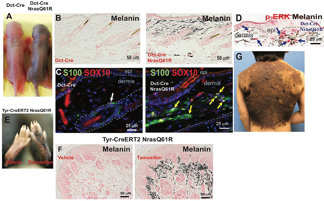
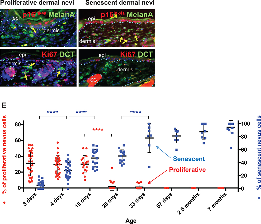
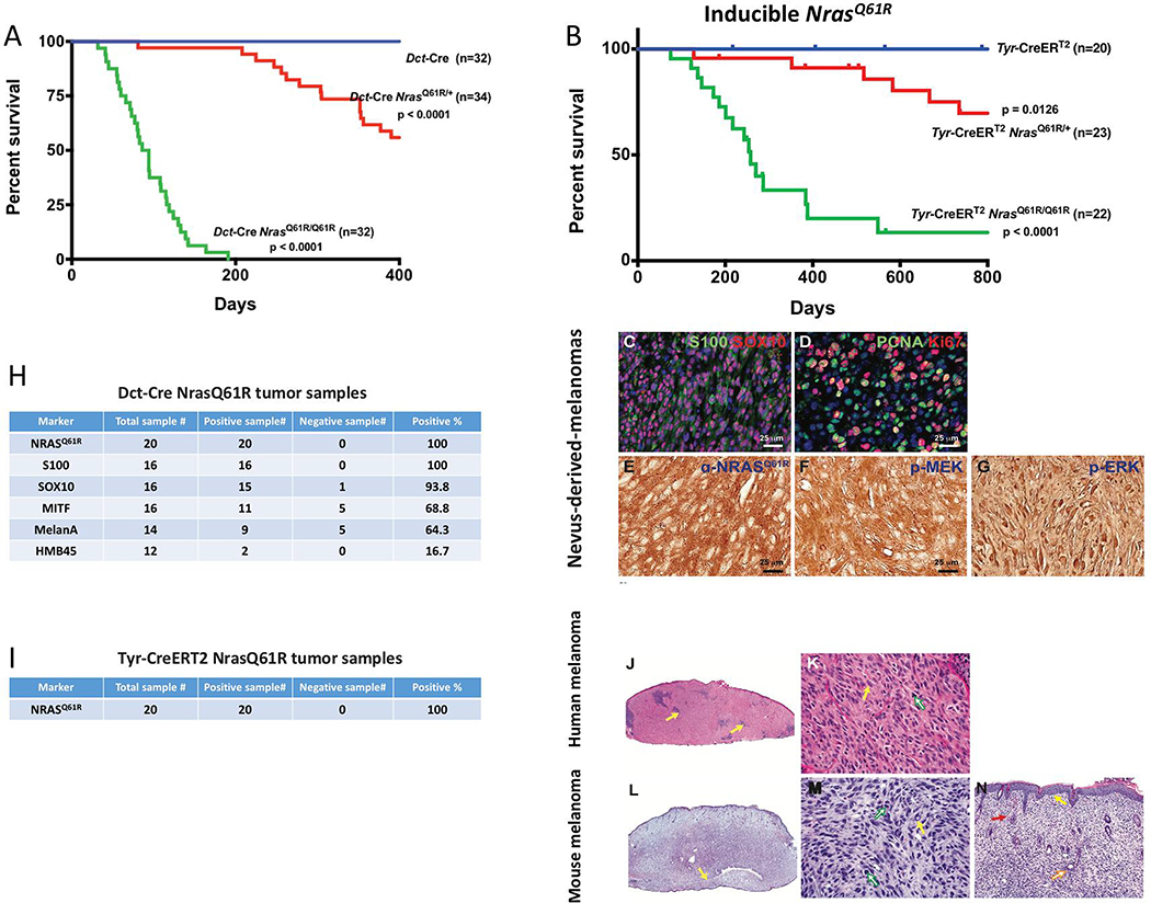
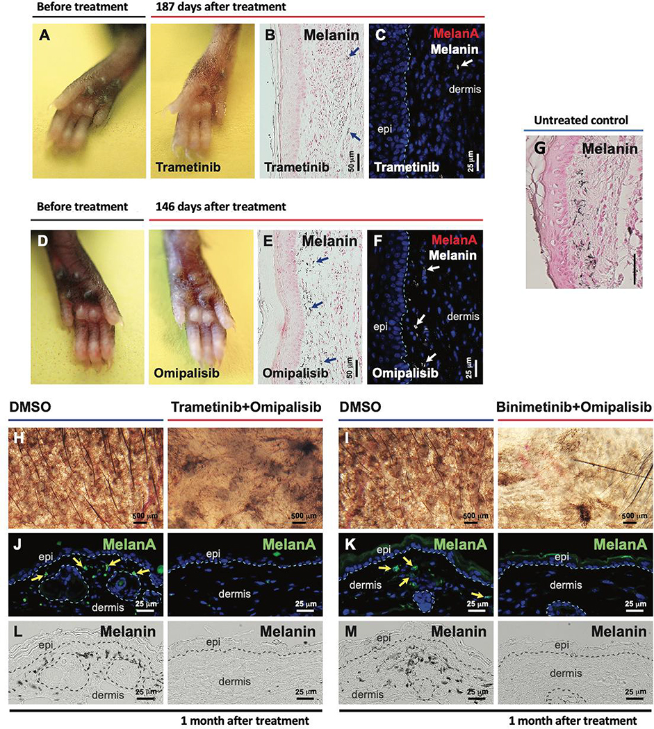
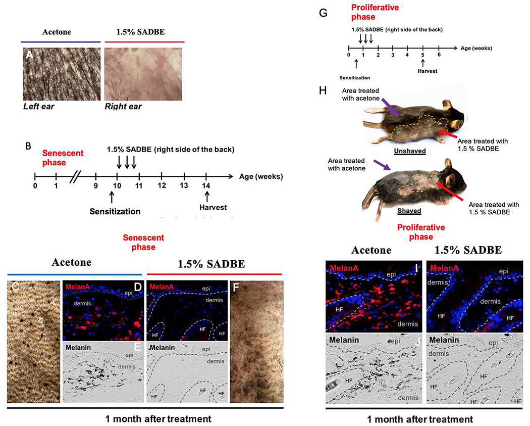
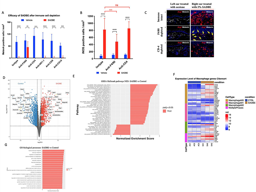
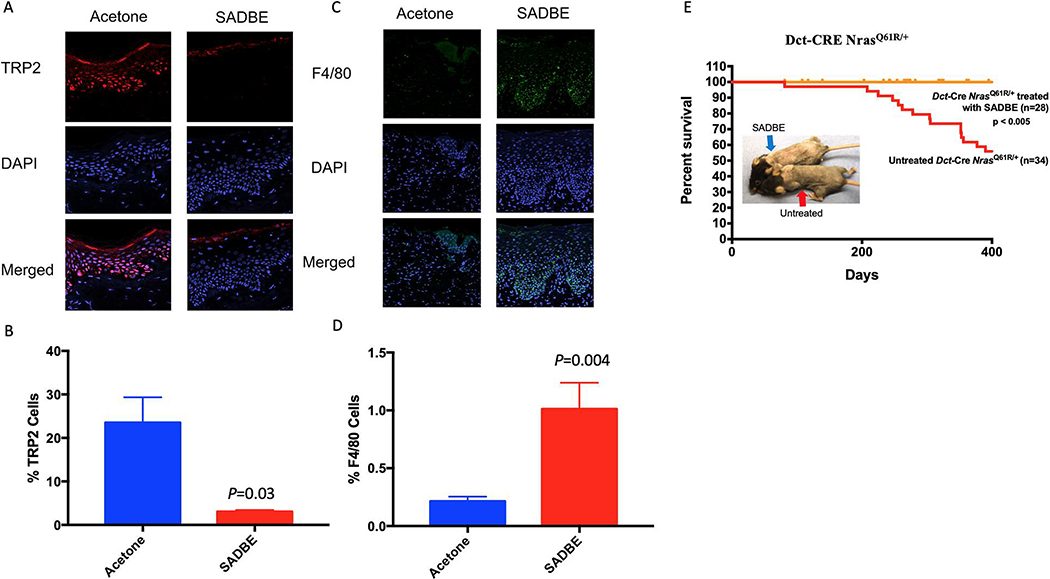
Giant congenital melanocytic nevi are NRAS-driven proliferations that may cover up to 80% of the body surface. Their most dangerous consequence is progression to melanoma. This risk often triggers preemptive extensive surgical excisions in childhood, producing severe lifelong challenges. We have presented preclinical models, including multiple genetically engineered mice and xenografted human lesions, which enabled testing locally applied pharmacologic agents to avoid surgery. The murine models permitted the identification of proliferative versus senescent nevus phases and treatments targeting both. These nevi recapitulated the histologic and molecular features of human giant congenital nevi, including the risk of melanoma transformation. Cutaneously delivered MEK, PI3K, and c-KIT inhibitors or proinflammatory squaric acid dibutylester (SADBE) achieved major regressions. SADBE triggered innate immunity that ablated detectable nevocytes, fully prevented melanoma, and regressed human giant nevus xenografts. These findings reveal nevus mechanistic vulnerabilities and suggest opportunities for topical interventions that may alter the therapeutic options for children with congenital giant nevi.

Keywords: Nras; congenital melanocytic nevus; hapten; melanoma; mole; prevention; topical.


Conflict of interest statement

Declaration of interests D.E.F. has a financial interest in Soltego, a company developing salt inducible kinase inhibitors for topical skin-darkening treatments that might be used for a broad set of human applications. The interests of D.E.F. were reviewed and are managed by Massachusetts General Hospital and Partners HealthCare in accordance with their conflict-of-interest policies. C.L.C. has a financial interest in 4Immune, a company developing cell therapy treatments that can be used for a broad set of human applications. The interests of C.L.C were reviewed and are managed by Mass General Brigham in accordance with their conflict-of-interest policies. X.S.L. is a cofounder, board member, SAB member, and consultant of GV20 Oncotherapy and its subsidiaries; stockholder of BMY, TMO, WBA, ABT, ABBV, and JNJ; and received research funding from Takeda, Sanofi, Bristol Myers Squibb, and Novartis. M.C.M. discloses consulting relationship with Novartis, Advisory Board with BioCoz and Caliber ID, and author royalties with Wiley & Sons.

Figures

Figure 1.
Figure 1.. Melanocyte-specific NrasQ61R mutant mice recapitulates histologic and molecular features of human giant congenital melanocytic nevi.
(A) Features of melanocytic nevi in 3-day-old Dct-Cre NrasQ61R. (B) Melanin of adult Dct-Cre NrasQ61R mice was detected by Fontana-Masson staining. (C-D) Immunostains. Paraffin sections were immunostained for S100 (C, green), SOX10 (C, red), or p-ERK (D, red). (E) Features of melanocytic nevi in tamoxifen-induced Tyr-CreERT2 NrasQ61R mice. (F) Melanin in tamoxifen-induced Tyr-CreERT2 NrasQ61R mice was detected by Fontana-Masson staining. (G) The clinical appearance of a human giant congenital nevus is shown in. White arrow in (C) indicates normal signal in hair follicle. Yellow arrows in (C) indicate ectopic signals in the dermis of nevus skin. Blue arrows in (D) indicate positive signals. See also supplementary Figures 1 and 2.
Figure 2.
Figure 2.. NrasQ61R mutation-driven congenital nevi are initially proliferative and subsequently become senescent.
(A, B) Representative images of proliferative nevi. (C, D) Representative images of senescent nevi. Paraffin sections were immunostained for p16Ink4a (A and C, red), MelanA/MART1 (A and C, green), Ki67 (B and D, red), or DCT (B and D, green). Yellow arrows indicate double-positive cells, and white arrows indicate single-positive cells. Dorsal skin samples from Dct-Cre NrasQ61R/Q61R mutant mice were harvested at the ages indicated in E. Samples from three mice of each age group, with five to ten of non-adjacent samples per mouse, were quantified. Nevus cell proliferation and senescence were measured by immunofluorescence staining for Ki67 and p16Ink4a, respectively. Quantification of the above staining revealed a statistically significant decrease in proliferative nevus cell between 10 and 20 days of age (E, red). The percentage of senescent nevus cells gradually increases between age groups until mice reach approximately 1 month of age (E, blue). Red and blue colored dots indicate mean percentages of nevus cells that are proliferative (left Y-axis) or senescent (right Y-axis), respectively. The bars represent the standard error of the mean (SEM). P-values were calculated using one-way ANOVA with Tukey’s multiple comparison test (****P<0.0001).
Figure 3.
Figure 3.. Melanocyte-specific NrasQ61R activating mutation in mice results in high incidence of melanoma with shared histologic features of human melanoma.
(A) Dct-Cre NrasQ61R/Q61R mutant (green, n=35), Dct-Cre NrasQ61R/+ mutant (red, n=34), and Dct-Cre control (blue, n=32) mice were examined to compare the incidence of NrasQ61R mutation-driven melanoma formation. Survival curves were plotted using the Kaplan-Meier method and analyzed by the log-rank test. The tumor-free survival rates were significantly different between the control and Dct-Cre NrasQ61R/Q61R groups (P<0.0001), and between the control and Dct-Cre NrasQ61R/+ groups (P<0.0001). (B) The incidence of NrasQ61R mutation-driven melanomas was measured in tamoxifen-induced Tyr-CreERT2 NrasQ61R/Q61R mutant (green, n=22), Tyr-CreERT2 NrasQ61R/+ mutant (red, n=23), and Tyr-CreERT2 control (blue, n=20) mice. Statistical significance was defined as P<0.05. (C-G) Dct-Cre NrasQ61R tumor sections were immunostained for S100 (C, green), SOX10 (C, red), PCNA (D, green), Ki67 (D, red), NrasQ61R (E, brown), p-MEK (F, brown), or p-ERK (G, brown). (H) Melanocytic markers, including S100, SOX10, MITF, MelanA/MART-1, and HMB45, were assessed by immunostaining in Dct-Cre NrasQ61R tumor samples, and the percentage of positive samples for each marker was calculated. (I) Tyr-CreERT2 NrasQ61R tumor samples were examined using an antibody against NrasQ61R and the percentage of NrasQ61R-positive samples was calculated. (J-K) H&E stained histological images of human melanomas. (L-N) H&E stained histological images of NrasQ61R mutation-driven murine melanomas. are compared with those of. Histologic images at low and high magnifications are shown (J, L, 20x) (K, M, 400x; N, 100x). The infiltrate of human primary malignant melanomas replaces the entire dermis as a sheet, punctuated by nidi of inflammation (J, yellow arrows). The nuclear-to-cytoplasmic ratios, hyperchromatic nuclei, and sometimes multiple nucleoli are shown (K, yellow arrow). The atypical mitotic figure is visible (K, green arrow). The infiltration of murine melanoma can be observed in the ear as a sheet of tumor cells, encircling the cartilage (L, yellow arrow). Severe pleomorphism of the murine melanoma cells, with large nuclear-to-cytoplasmic ratios, hyperchromatic nuclei, and intranuclear vacuoles (M, yellow arrow), and mitotic activity, (M, green arrows) is observed. The murine melanoma abuts the epidermis, with no grenz zone (N, yellow arrow), and invades the hair follicle adventitia (N, orange arrow). The extravasation of red blood cells (N, red arrow), a common finding in human melanoma, is also detected. See also supplemental Figures 3 and 4.
Figure 4.
Figure 4.. Local therapy with small molecule inhibitors of the NRAS signaling pathway regress NrasQ61R-driven melanocytic nevi.
(A-F) NrasQ61R mutation-driven nevi were induced in Tyr-CreERT2 LSL-NrasQ61R newborn mice with tamoxifen over the first postnatal week, and 2 months later local treatments of senescent nevi were initiated with MEK and/or PI3K inhibitors or DMSO vehicle control. Trametinib (A-C, 0.1 μg /μl) or omipalisib (D-F, 0.03 μg /μl) was subcutaneously injected into the pigmented paws of tamoxifen-induced Tyr-CreERT2 NrasQ61R/+ heterozygous mutants in 10 μl of 10% DMSO, three times per week for two weeks. Paws were visualized 187 days after treatment with Trametinib (A) or 146 days after treatment with omipalisib (D). Melanin (B, and E, blue arrows) was detected by Fontana-Masson staining. Tissue sections were stained for MelanA/MART1 (C, F). White arrows (in C and F) indicate dermal melanin detected by bright-field microscopy. (G) Representative image of melanin detected by Fontana-Masson staining of non-treated tamoxifen-induced Tyr-CreERT2 NrasQ61R/+ mice from postnatal day 16, 2 weeks after tamoxifen induction. (H-M) Topical combinatorial therapies. Topical combinatorial therapies with the PI3K inhibitor, omipalisib (10 μg/ μl), plus the MEK inhibitor, trametinib (H, J, L, 2 μg/ μl) or binimetinib (I, K, M, 10 μg/ μl), were applied to the pigmented ears of tamoxifen-induced Tyr-CreERT2 NrasQ61R/Q61R mutant mice. All topical agents were administered in 5 μl DMSO, five times per week for 3 weeks (‘Trametinib+Omipalisib’ and their vehicle control treatments) or for 6 weeks (‘Binimetinib+Omipalisib’ and their vehicle control treatments). Ear skin tissues were harvested 1 month after the final drug treatment, and nevus regression was examined. Images were obtained with a stereomicroscope, and the dark brown particles indicate melanin (H, I). Tissue sections were stained for MelanA/MART1 (J, K, green). Yellow arrows indicate positive signals. Dermal melanin detected by bright-field microscopy is shown (L, M, black). We used five mice per treatment group, and representative images are shown. See also supplemental Figure 5.
Figure 5.
Figure 5.. Single-agent local immunotherapy with topical SADBE results in significant regression of both neonatal proliferative and postnatal senescent congenital nevi.
(A) SADBE (1.5%, topical) was applied to the pigmented right ears of tamoxifen-induced Tyr-CreERT2 NrasQ61R/Q61R mutant mice 2 months after tamoxifen induction, whereas the pigmented left ears of the same animals were treated with acetone vehicle. Ear tissue images were obtained with a stereomicroscope, and dark brown particles indicate melanin. (B) Treatment scheme of adult Dct-Cre NrasQ61R/Q61R. Single-drug local therapy with 1.5% SADBE was administered topically to the right back. Acetone was applied topically to the left back of each SADBE-treated mouse as a vehicle control. All treatments were carried out three times per week (every other day), and the treated skin tissues were harvested 1 month after the first treatment to assess regression of melanocytic nevi (C, F). Tissue sections were immunostained for MelanA/MART1 (D, I). Melanin (black) detected by bright-field microscopy is shown in (E, J). (G) Treatment scheme of neonatal Dct-Cre NrasQ61R/Q61R mice. (H) Representative images of treated Dct-Cre NrasQ61R/Q61R mice unshaved (upper panel) and shaved (lower panel) are shown. White hairs were detectable in treated areas (H, upper panel red arrow). Regression of melanocytic nevi was detectable in treated areas (H, lower panel red arrow). The purple arrows in (H) indicate unchanged nevi in the nevus lesion treated with acetone vehicle. Yellow dashed lines in H demarcate the region treated with 1.5% SADBE. Stippled white lines in (D, E, I, J) separate the epidermis (epi) and hair follicles (HF) from the dermis.
Figure 6.
Figure 6.. Antibody mediated depletion of inflammatory cell lineages in mice and RNAseq reveal macrophage recruitment by SADBE.
(A) Quantification of MelanA positive cells per mm2 in Dct-Cre NrasQ61R mice after immunodepletion by IP injection with 750 μg of anti F4/80, anti-CD-4, anti-NK1.1, anti-CD-8 and anti-CD-19. (B) Quantification of INOS positive cells per mm2 in Dct-Cre NrasQ61R mice after immunodepletion by IP injection with 750 μg of anti F4/80 and anti-CD-4. (C) Arg1 staining (red) of ear skin sections following depletion with anti-CD-4, anti-F4/80, isotype control and 2% SADBE treatment for 1 week. Yellow arrows indicate negative signals. Melanin detected by bright-field microscopy is shown in white. (D) Differential gene expression analysis (E) GSEA hallmark pathways analysis (F) Heat map of macrophage marker groups according to macrophage phase and (G) GO biological processes analysis. Dct-Cre NrasQ61R mice were treated with SADBE (2%) or vehicle control (acetone) on dorsal skin (3 mice per treatment or control group). Whole skin RNA-Seq was performed 72h after treatment. Macrophage-associated genes for M0, M1, and M2 phases were collected from the hematopoietic gene signature set LM22.
Figure 7.
Figure 7.. SADBE decreases melanocyte numbers in CMN xenografts and prevents melanoma formation in mice.
(A) Immunofluorescent staining of CMN tissue xenografts treated with topical application of 1% SADBE or acetone control for 8 months: DAPI (blue) TRP2 (red) Merged (purple). (B) Quantification of the percentage of TRP2 positive cells out of total cells in the epidermis. (C) Immunofluorescent staining of CMN tissue xenografts treated with topical application of 1% SADBE or acetone control for 2 weeks: DAPI (blue) F4/80 (green) Merged (cyan). (D) Quantification of F4/80 positive cells as percent of total cells. (E) Effect of SADBE on melanoma-genesis and mouse survival in Dct-Cre NrasQ61R/+ mice. Mice were sensitized with a 2% SADBE in acetone treatment of the right side of the shaved abdomen. Three days later, sensitized mice were treated with 1.5% SADBE over the shaved dorsal (back) skin (treated and untreated controls indicated by arrows). Treatments with SADBE were carried out every other day (total of three treatments), and tumor formation was monitored. Untreated control tumor incidences are the same (historical) control Dct-CRE NrasQ61R/+ mice in Figure 3A. In a population where anatomic location was tracked, dorsal back specific melanomas occurred in 13 of 21 (61.9%) control mice. See also supplemental Figure 6.

Comment in

Similar articles

Cited by

References

    1. Ackermann J, Frutschi M, Kaloulis K, McKee T, Trumpp A, and Beermann F (2005). Metastasizing melanoma formation caused by expression of activated N-RasQ61K on an INK4adeficient background. Cancer Res 65, 4005–4011. 10.1158/0008-5472.CAN-04-2970. - DOI - PubMed
    1. Bajaj MS, Khuraijam N, Sen S, and Pushker N (2009). Congenital melanocytoma manifesting as proptosis with multiple cutaneous melanocytic nevi and oculodermal melanosis. Arch Ophthalmol 127, 937–939. 10.1001/archophthalmol.2009.146. - DOI - PubMed
    1. Bancroft GJ, and Kelly JP (1994). Macrophage activation and innate resistance to infection in SCID mice. Immunobiology 191, 424–431. 10.1016/S0171-2985(11)80448-1. - DOI - PubMed
    1. Bauer J, Curtin JA, Pinkel D, and Bastian BC (2007). Congenital melanocytic nevi frequently harbor NRAS mutations but no BRAF mutations. J Invest Dermatol 127, 179–182. 10.1038/sj.jid.5700490. - DOI - PubMed
    1. Bett BJ (2005). Large or multiple congenital melanocytic nevi: occurrence of cutaneous melanoma in 1008 persons. J Am Acad Dermatol 52, 793–797. 10.1016/j.jaad.2005.02.024. - DOI - PubMed

Publication types

Supplementary concepts