

doi: 10.1055/a-1838-4311.
Epub 2022 May 13.
Underwater endoscopic mucosal resection after endoscopic ultrasound examination for safe and reliable complete resection of a deeply invasive submucosal cecal cancer
Affiliations
- PMID: 35561982
- PMCID: PMC9767750
- DOI: 10.1055/a-1838-4311
Item in Clipboard
Underwater endoscopic mucosal resection after endoscopic ultrasound examination for safe and reliable complete resection of a deeply invasive submucosal cecal cancer
Endoscopy.
2023 Jan.
No abstract available
Conflict of interest statement
H. Yamamoto is a consultant for Fujifilm Corporation and has received honoraria, a grant, and royalties from the company. The remaining authors declare that they have no conflict of interest.
Figures
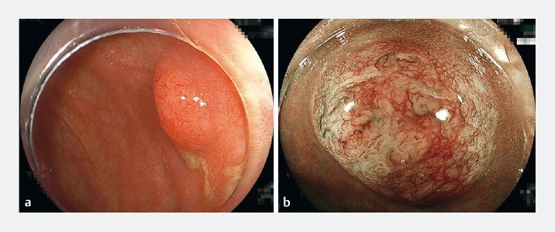
Images from magnifying narrow-band imaging (NBI) of a cecal lesion showing:
a
a 7-mm sessile tumor without a demarcated depressed area, which initially resembled a sessile serrated lesion and was suspected to be an adenocarcinoma on a previous biopsy;
b
under blue light, a vascular pattern with both interruption of thick vessels and loose vascular areas, and a surface pattern with amorphous areas, which was classified as type 3 (the Japan NBI Expert Team classification), consistent with a diagnosis of deep submucosal invasive cancer.
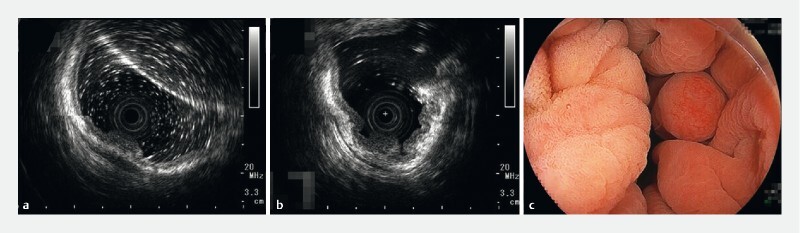
Endoscopic ultrasound using a 20-MHz miniature probe showing:
a
a high echoic submucosal layer between the tumor and the muscularis during insufflation;
b
when the cecum contracted while still under water, the muscularis becoming circumferentially recessed and the submucosa becoming thicker, with the tumor appearing to float on the submucosa.
c
On underwater endoscopic view, the tumor was transformed from a sessile tumor to a floating subpedunculated tumor.
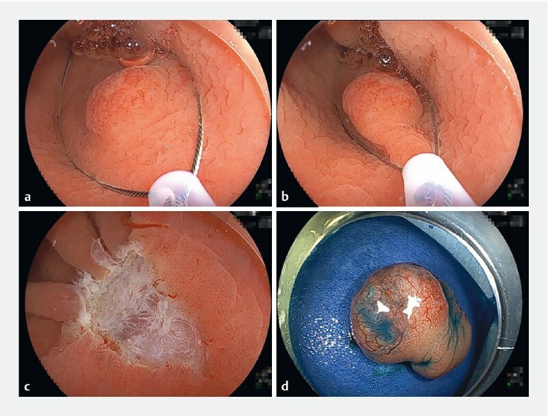
Sequential pictures of the underwater endoscopic mucosal resection of the tumor showing:
a
the tip of the snare (15-mm Rota snare; Medi-Globe GmbH, Achenmühle, Germany) securely placed on the normal mucosa beyond the tumor with a sufficient proximal margin;
b
the snare being gradually closed, while confirming it was capturing the entire tumor with its surrounding normal mucosa and aspirating the water, with the completely captured tumor then cut with pure cut mode diathermy (ESG-100; Olympus);
c
no residual lesions around the mucosal defect, meaning endoscopic en bloc resection had been achieved, and the mucosal defect was closed with a reopenable clip (Sureclip Plus; Micro-Tech Co. Ltd., NanJing, China) and endoclips (EZ-clip; Olympus);
d
the resected specimen.
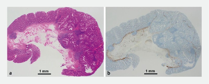
Histopathological views (× 40 magnification) showing:
a
on hematoxylin and eosin staining, a well-to-moderately differentiated deeply invasive submucosal adenocarcinoma, with no lymphovascular invasion and a negative vertical margin;
b
on desmin staining, absence of the muscularis mucosa, with submucosal invasion to a depth of 3 mm.
Comment in
-
Commentary.Endoscopy. 2023 Jan;55(1):99. doi: 10.1055/a-1966-9046. Epub 2022 Dec 20. Endoscopy. 2023. PMID: 36538921 No abstract available.
References
-
- Fukuda H, Takeuchi Y, Shoji A et al. Curative value of underwater endoscopic mucosal resection for submucosally invasive colorectal cancer. J Gastroenterol Hepatol. 2021;36:2471–2478. - PubMed
MeSH terms
LinkOut - more resources
Full Text Sources
Medical