. 2022 May 11;12(1):7753.
doi: 10.1038/s41598-022-11843-z.

Translational development of a tumor junction opening technology

Affiliations

Translational development of a tumor junction opening technology

Jiho Kim et al. Sci Rep. .

Abstract

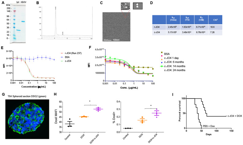
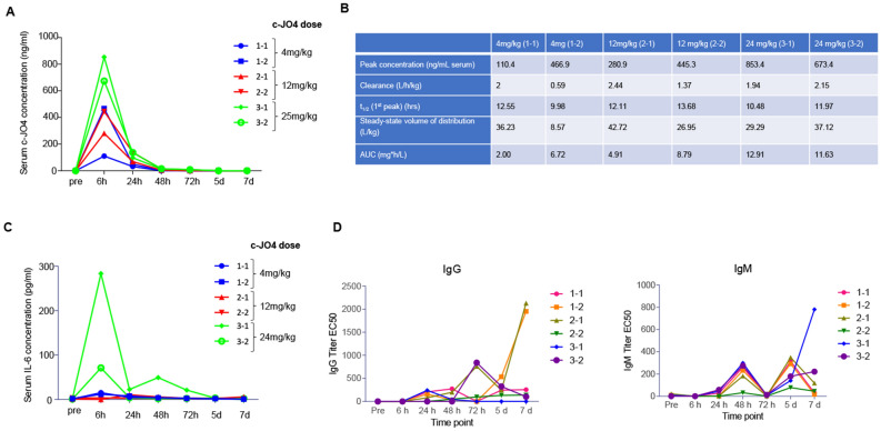
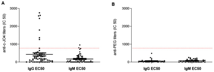
Our goal is to overcome treatment resistance in ovarian cancer patients which occurs in most cases after an initial positive response to chemotherapy. A central resistance mechanism is the maintenance of desmoglein-2 (DSG2) positive tight junctions between malignant cells that prevents drug penetration into the tumor. We have generated JO4, a recombinant protein that binds to DSG2 resulting in the transient opening of junctions in epithelial tumors. Here we present studies toward the clinical translation of c-JO4 in combination with PEGylated liposomal doxorubicin/Doxil for ovarian cancer therapy. A manufacturing process for cGMP compliant production of JO4 was developed resulting in c-JO4. GLP toxicology studies using material from this process in DSG2 transgenic mice and cynomolgus macaques showed no treatment-related toxicities after intravenous injection at doses reaching 24 mg/kg. Multiple cycles of intravenous c-JO4 plus Doxil (four cycles, 4 weeks apart, simulating the treatment regimen in the clinical trial) elicited antibodies against c-JO4 that increased with each cycle and were accompanied by elevation of pro-inflammatory cytokines IL-6 and TNFα. Pretreatment with steroids and cyclophosphamide reduced anti-c-JO4 antibody response and blunted cytokine release. Our data indicate acceptable safety of our new treatment approach if immune reactions are monitored and counteracted with appropriate immune suppression.


Conflict of interest statement

D.C. is founder of and holds an equity interest in HDT Bio Corp. a company that licensed the JO4 technology and is assisting in its clinical development.

Figures

Figure 1
Figure 1
Characterization of c-JO4. (A) Polyacrylamide gel electrophoresis. Gennova’s preparation of JO4 (c-JO4) was treated as indicated. b/r—boiled and reduced; nb/nr—not boiled and not reduced. 1 μg protein was run on the gel. (B) Size exclusion chromatography (SEC) profile of c-JO4 run through a Superdex 200 increase 10/300 column with TBS. (C) Electron microscopy of c-JO4 negative staining with sodium silicotungstate. The zoomed in image showed a dimer of trimeric fiber knob domains. A schematic illustration of this structure is shown on the right. (D) Surface Plasmon Resonance analysis of JO4 binding to recombinant DSG2. DSG2 was immobilized on sensorchips, and background was automatically subtracted from the control flow cell. r-JO4 and c-JO4 were injected for 3 min at 2.5 μg/ml, followed by a 2.5-min dissociation period. Shown is the summary of SPR data. Parameters were evaluated using BIAeval software. kon- association rate; koff—dissociation rate; KD—equilibrium dissociation constant; chi2—area of the squared difference between the measured data point and the fit. (EI) Potency assays. (E) Ad3-GFP Infection competition. Research r-JO4 run (#237) was compared with c-JO4. 293 cells were incubated in 96-well plates with JO4 proteins at indicated concentrations for 1 h before addition of Ad3-GFP virus for competition. GFP expression was measured 18 h after virus addition to measure extent of JO4 competition with Ad3-GFP. (F) c-JO4 was stored at 4 °C for 1 day (red dot), 6 months (orange triangle), 14 months (blue triangle), and 24 months (empty green triangle) and then tested in infection competition assay. (G,H) c-JO4-enhanced Doxil penetration in epithelial tumor spheroids. (G) Confocal immunofluorescence images of T84 spheroids stained for DSG2 (Green) and DAPI (blue). (H) Left panel: Uptake of liposomal doxorubicin (Doxil) (DOX) in T84 cell spheroids after treatment with c-JO4. The amount of Doxil (mean autofluorescence intensity—MAFI) in tumor cells was measured 1 h after adding it to tumor sphere cultures. Right panel: Doxil released from spheroids into the supernatant after trypsin digestion of spheroids. *p < 0.05. (I) c-JO4-enhanced anti-tumor activity. CB17 immunodeficient mice bearing epithelial tumors derived from T84 human colon adenocarcinoma cells were used for testing. When tumors reached a volume of 100 mm3, mice were intravenously injected with either PBS or c-JO4 (2 mg/kg) followed by Doxil (1 mg/kg) one hour later. Mice received three injection cycles 3 days apart. Tumor volumes were measured twice a week until reaching a tumor volume of 1000 mm3 when they were sacrificed. The day of sacrifice served as the endpoint in Kaplan–Meier survival studies. In the c-JO4/Doxil group, tumor growth was significantly delayed (p < 0.0001) and 45% of the animals were still tumor-free at day 140 after tumor cell inoculation. N = 10. p < 0.001 for Doxil vs c-JO4 + Doxil.
Figure 2
Figure 2
Summary of animal studies. (A) Four-week intravenous GLP-toxicology study of c-JO4 in female hDSG2-transgenic mice. The objectives of this study were to evaluate the potential toxicity of c-JO4 when administered intravenously together with Doxil to hDSG2-transgenic mice weekly for 4 consecutive weeks with 2 weeks of recovery. c-JO4 was injected intravenously one hour before intravenous injection of Doxil. The animals were approximately 5–11 weeks old and weighed 19.9–26.0 g at the time of the first dose administration. The study consisted of one control and five treated groups. Each group had 6 Core female mice (undergoing standard evaluations such as clinical observations, body weight, clinical pathology, organ weights and histopathology). In addition, 4 female mice per group in groups 1, 3, and 6 were designated as Recovery animals. The recovery animals were handled exactly as the Core mice but were held for at least 14 days of rest after the last dose to determine the reversibility of potential treatment-related effects. (B) Intravenous dose range finding study in female cynomolgus monkeys. Dose range study plan of c-JO4 in female cynomolgus monkeys. 3 groups of monkeys (N = 2) were administered with escalating doses of c-JO4 (4, 12, 24 mg/kg). Group 3 was dosed after no toxicity was observed at the previous dose level. Necropsy was performed at day 7 after administration of the c-JO4 dose to collect organ and tissue samples. (C) Non-GLP four-cycle toxicity /pharmacokinetics /immunogenicity study with c-JO4 + Doxil in cynomolgus monkeys. One animal received c-JO4 and, one hour later, Doxil as indicated (“Tx”). The second animal additionally received immunosuppressive drugs (“IS + Tx”). Immunosuppression consisted of Cyclophosphamide given 2 days before c-JO4/Doxil) and methylprednisolone + dexamethasone (0.5 hrs before c-JO4 injection). Timing and dosage of drug injection is shown on the right side.
Figure 3
Figure 3
Intravenous dose range finding study in female cynomolgus monkeys. (A,B) Pharmacokinetics parameters of c-JO4 in cynomolgus monkeys. (A) c-JO4 concentrations in serum collected at indicated time points subsequent to c-JO4 administration were measured by ELISA. Standard curve used to quantify c-JO4 is shown on the right side. (B) Pharmacokinetic data calculated by using serum concentration curves of each animal. Units are as indicated. (C) Cytokines IL-2, IL-4, IL-5, IL-6, TNF-alpha, and IFN-gamma were measured by cytometric bead array (CBA) (NHP th1/Th2 cytokine kit from BD Biosciences). Only IL-6 was detectable. (D) Antibody titers present in serum against c-JO4 were measured by ELISA, using recombinant c-JO4 protein for capture and anti-NHP-IgG-HRP and anti-NHP-IgM-HRP for detection. Shown are IgM and IgG titers as IC50.
Figure 4
Figure 4
Non-GLP four-cycle toxicity /pharmacokinetics /immunogenicity study with c-JO4 + Doxil in cynomolgus monkeys. (A,B) c-JO4 pharmacokinetics. (A) c-JO4 concentration in serum collected at indicated time points as measured by ELISA. (B) Pharmacokinetics parameters. (C) c-JO4 concentration in it issues. Tissues were ground with QiaShredder and resulting tissue lysates were measured by ELISA for c-JO4. c-JO4 levels in tissue lysates were normalized for mg tissue. (D) Immunofluorescence with anti-CD163 and anti-cJO4 antibodies on liver sections of the “Tx” animal. Nuclei are stained blue with DAPI. The scale bar is 20 μm. In the composite panel (right) co-staining is marked by arrows.
Figure 5
Figure 5
Non-GLP four-cycle toxicity/immunogenicity study with c-JO4 + Doxil in cynomolgus monkeys. (A,B) Serum anti-c-JO4 antibody titers. Anti-c-JO4 IgG and IgM titers were measured in at indicated time points by ELISA. (C) Serum cytokines. Serum samples were run on a Th1/Th2 inflammatory cytokine array using the CBA flow panel (BD Biosciences). The only cytokines above limit of detection were IL-6 and TNFα. Other cytokines tested included IL-2, IL-4, IL-10 and IFNγ. Inlet shows the values at and around each injection time point.
Figure 6
Figure 6
Pharmacokinetics of Doxil in cynomolgus monkeys. Doxil was detected in serum using anti-PEG antibodies and a HRP-conjugated secondary antibody. (A) Pharmacokinetics after each cycle. (B) Pharmacokinetics parameters after the first cycle.
Figure 7
Figure 7
Serum antibodies against c-JO4 and PEG/Doxil in ovarian cancer patients A titer below 1:800 is not neutralizing in in vitro infection studies. Serum samples from the Fred Hutchinson Cancer Research Center was taken for analysis of antibodies against (A) c-JO4 (B) PEG/Doxil using anti-Ad3 and anti-PEG antibodies respectively in an ELISA. EC50 values are shown.

References

    1. Tannock IF, Lee CM, Tunggal JK, Cowan DS, Egorin MJ. Limited penetration of anticancer drugs through tumor tissue: A potential cause of resistance of solid tumors to chemotherapy. Clin. Cancer Res. 2002;8:878–884. - PubMed
    1. Minchinton AI, Tannock IF. Drug penetration in solid tumours. Nat. Rev. Cancer. 2006;6:583–592. doi: 10.1038/nrc1893. - DOI - PubMed
    1. Choi IK, Strauss R, Richter M, Yun CO, Lieber A. Strategies to increase drug penetration in solid tumors. Front. Oncol. 2013;3:193. doi: 10.3389/fonc.2013.00193. - DOI - PMC - PubMed
    1. Kim J, et al. Desmoglein-2 as a prognostic and biomarker in ovarian cancer. Cancer Biol. Ther. 2020;21:1154–1162. doi: 10.1080/15384047.2020.1843323. - DOI - PMC - PubMed
    1. Wang H, et al. Desmoglein 2 is a receptor for adenovirus serotypes 3, 7, 11 and 14. Nat. Med. 2011;17:96–104. doi: 10.1038/nm.2270. - DOI - PMC - PubMed

Publication types