Review
. 2022 Sep;23(9):623-640.
doi: 10.1038/s41580-022-00483-w. Epub 2022 May 13.

Establishment of H3K9-methylated heterochromatin and its functions in tissue differentiation and maintenance

Affiliations
Review

Establishment of H3K9-methylated heterochromatin and its functions in tissue differentiation and maintenance

Jan Padeken et al. Nat Rev Mol Cell Biol. 2022 Sep.

Abstract

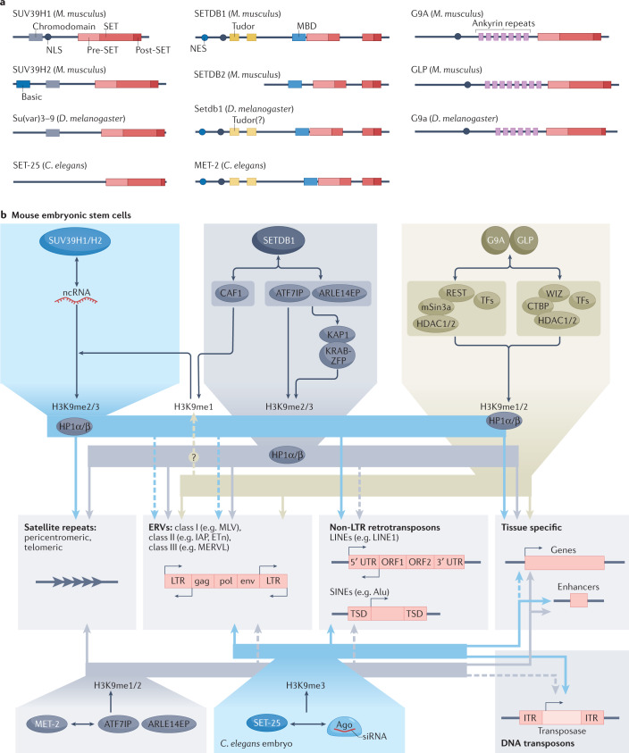
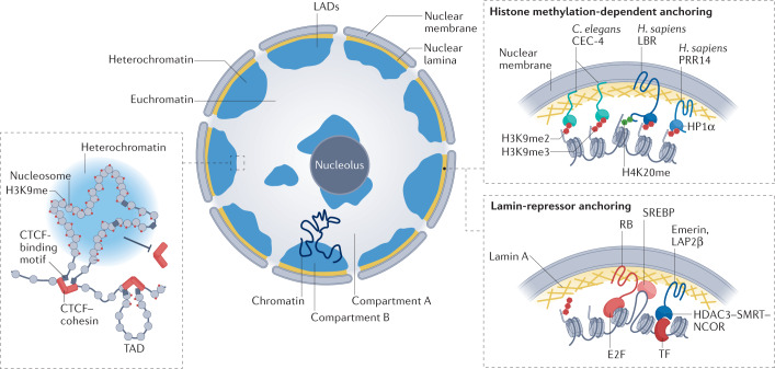
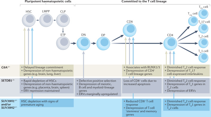
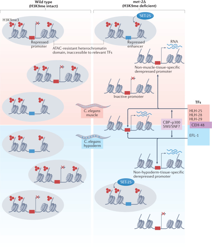
Heterochromatin is characterized by dimethylated or trimethylated histone H3 Lys9 (H3K9me2 or H3K9me3, respectively) and is found at transposable elements, satellite repeats and genes, where it ensures their transcriptional silencing. The histone methyltransferases (HMTs) that methylate H3K9 - in mammals Suppressor of variegation 3-9 homologue 1 (SUV39H1), SUV39H2, SET domain bifurcated 1 (SETDB1), SETDB2, G9A and G9A-like protein (GLP) - and the 'readers' of H3K9me2 or H3K9me3 are highly conserved and show considerable redundancy. Despite their redundancy, genetic ablation or mistargeting of an individual H3K9 methyltransferase can correlate with impaired cell differentiation, loss of tissue identity, premature aging and/or cancer. In this Review, we discuss recent advances in understanding the roles of the known H3K9-specific HMTs in ensuring transcriptional homeostasis during tissue differentiation in mammals. We examine the effects of H3K9-methylation-dependent gene repression in haematopoiesis, muscle differentiation and neurogenesis in mammals, and compare them with mechanistic insights obtained from the study of model organisms, notably Caenorhabditis elegans and Drosophila melanogaster. In all these organisms, H3K9-specific HMTs have both unique and redundant roles that ensure the maintenance of tissue integrity by restricting the binding of transcription factors to lineage-specific promoters and enhancer elements.


Conflict of interest statement

The authors declare no competing interests.

Figures

Fig. 1
Fig. 1. The major H3K9 methyltransferases, cofactors and targets.
a | The structural domains of the major histone H3 Lys9 (H3K9) methyltransferases in Mus musculus, Drosophila melanogaster and Caenorhabditis elegans. The SET domain contains the catalytic site. The enzymes are grouped by structural relatedness. One outlier is C. elegans SET-25, which has a SUV39-like SET and pre-SET domain, but does not have the methylated-lysine-binding chromodomain like other SUV39 enzymes. The amino-terminal basic domain in SUV39H2 is found uniquely in the mouse, and not the human, enzyme. SET domain bifurcated 1 (SETDB1), Setdb1 and MET-2 are structurally closely related and also share conserved essential cofactors. Tudor domains, ankyrin repeats and methyl-CpG-binding domains (MBDs) have roles in targeting the histone methyltransferases (HMTs) to specific sites of action. b | The three families of H3K9 methyltransferases are shown with cofactors and interacting proteins that enable site-specific HMT recruitment. Suppressor of variegation 3–9 homologue 1 (SUV39H1) and SUV39H2 are often recruited to chromatin by non-coding RNA (ncRNA), or in worms by endogenous siRNA bound to Argonaut (Ago) proteins. SUV39 enzymes bind both to heterochromatin protein 1 (HP1) proteins and to dimethylated or trimethylated H3K9 (H3K9me2/3), which help to further recruit the HMT. SETDB1 class enzymes form a complex with activating transcription factor 7-interacting protein 1 (ATF7IP) (LIN-65 in worms) and ARLE14EP (adenosine 5′-diphosphate-ribosylation factor-like 14 (ARLE-14) in worms) or with chromatin assembly factor 1 (CAF1). With CAF1, SETDB1 monomethylates H3K9, whereas with ATF7IP, it binds either Krüppel-associated box (KRAB)-interacting protein 1 (KAP1) together with KRAB domain-containing zinc-finger proteins (KRAB-ZFPs) at endogenous retroviruses (ERVs) or is directly recruited by HP1 proteins to produce H3K9me2/3. In worms, MET-2 binds LIN-65 and ARLE-14. The enzymes G9A and G9A-like protein (GLP) bind a variety of transcription factors (TFs), together with histone deacetylases (HDACs), mSin3a and other chromatin modifiers. They also bind monomethylated H3K9 (H3K9me1) or H3K9me2 with their ankyrin repeats. The repeat elements targeted by these HMTs belong to three classes: centromeric or telomeric satellites (simple repeats), ERVs, which are long terminal repeat (LTR)-containing retrotransposons, or non-LTR-containing transposons such as long interspersed nuclear elements (LINEs) and short interspersed nuclear elements (SINEs). All HMTs are also recruited to genes and enhancer elements. CTBP, C-terminal-binding protein 1; ETn, early transposon; IAP, intracisternal A-particle; MERVL, murine endogenous retrovirus type L; MLV, murine leukaemia virus; NES, nuclear export signal; NLS, nuclear localization signal; ORF, open reading frame; TSD, target site duplication; UTR, untranslated region; WIZ, widely interspaced zinc-finger-containing protein.
Fig. 2
Fig. 2. Roles of H3K9 methylation in long-range chromatin interactions and nuclear organization.
An interphase nucleus in a differentiated vertebrate tissue is shown with heterochromatin (blue) sequestered against the nucleolus and against the nuclear lamina.The separation of active and inactive chromatin domains can manifest itself as lamin-associated domains (LADs) or as A and B compartments (Box 2). B compartments correspond to gene-inactive chromatin (heterochromatin) and largely include LADs. A compartments correspond to gene-active chromatin, which is further organized into topologically associating domains (TADs; left), which enable enhancer–promoter interaction that stimulates gene expression (not shown). In differentiated tissues, methylated histone H3 Lys9 (H3K9me) blocks TAD formation by preventing the binding of CCCTC-binding factor (CTCF) and cohesin complexes, which delineate TAD borders. On the right are shown several mechanisms for heterochromatin anchorage at the nuclear periphery. In Caenorhabditis elegans, the nuclear envelope-associated protein CEC-4 recognizes all forms of H3K9me (monomethylated H3K9 (H3K9me1), dimethylated H3K9 (H3K9me2) and trimethylated H3K9 (H3K9me3)) through its chromodomain, with affinity roughly equal to that of heterochromatin protein 1 (HP1). CEC-4 contributes directly to heterochromatin anchoring in worm embryos, but upon tissue differentiation, as in mammals, additional anchoring pathways are induced, which in worms are independent of H3K9me. Anchoring mechanisms present in differentiated mammalian cells include a histone methylation-dependent pathway with at least two potential anchors: proline-rich protein 14 (PRR14; potentially a functional homologue of CEC-4) is a non-transmembrane protein that anchors heterochromatin through H3K9me and HP1, and lamin B receptor (LBR), which is a transmembrane factor that binds both HP1α and dimethylated histone H4 Lys20 (H4K20me2) through its Tudor domain. Another mechanism is lamin A dependent and likely involves its interaction with RB and/or with transcription factors (TFs), such as SREBP. Emerin and lamina-associated polypeptide 2, isoform-β (LAP2β) are additional lamin-associated factors that bind chromatin through the histone deacetylase 3 (HDAC3)–nuclear receptor co-repressor 1 (NCOR)–silencing mediator of retinoic acid and thyroid hormone receptor (SMRT; also known as NCOR2) complex and potentially through tissue-specific transcription factors, (Box 2). H. sapiens, Homo sapiens.
Fig. 3
Fig. 3. Roles of H3K9 methyltransferases in T cell lineage determination.
The well-characterized differentiation steps from haematopoietic stem cells (HSCs) into terminally differentiated T helper cells (T helper 1 (TH1) cells, TH2 cells, T follicular helper (TFH) cells and TH17 cells) are shown. The effects of deletion of SUV39H1 and/or SUV39H2, SETDB1 or G9A are listed. Although not shown, histone H3 Lys9 (H3K9) methyltransferases also have a role in the maturation of B cell progenitors (pro-B cells). In this case, the loss of SETDB1 leads to derepression of both specific endogenous retroviruses (ERVs), and non-lineage-specific genes, including genes specific to innate immunity. The selection of genes and repeats that are derepressed likely depends on the variable presence of lineage-specific transcription factors, as described in Caenorhabditis elegans (Box 4). CLP, common lymphoid progenitor; DN, double negative (CD4CD8); DP, double positive (CD4+CD8+); ETP, early T cell progenitor; LMPP, lymphoid primed multipotent progenitor; Treg cell, regulatory T cell.
Fig. 4
Fig. 4. Transcription factors control tissue-specific transcription derepression upon H3K9me loss.
A major question is whether histone H3 Lys9 (H3K9) methylation contributes to gene repression in differentiated tissues by blocking transcription factor (TF) binding to chromatin. A recent study addressed this question in Caenorhabditis elegans by comparing the effects of loss of the SET domain bifurcated 1 (SETDB1)-like histone methyltransferase MET-2 on gene expression in embryos and in isolated muscle and hypoderm tissues. As depicted schematically, the genes upregulated by MET-2 loss constitute only a fraction (12% in embryos and 13% in muscle) of the genes marked by trimethylated H3K9 (H3K9me3), and the subset of genes affected is both tissue specific and developmental stage specific. In muscle and hypoderm, H3K9 methylation by MET-2 represses tissue-specific genes expressed in other cell lineages, including, but not restricted to, germline-specific genes,,. In addition, MET-2 and SET-25 redundantly repress a large number of enhancer elements in muscle. Methylated H3K9 (H3K9me) heterochromatin ensures transcription inactivity by restricting the binding of a defined set of transcription factors at promoters and enhancers, which are either tissue specific — for example, HLH25, HLH-28 and HLH-29 in muscle and EFL-1 in hypoderm — or more widely expressed, for example CEH-48 (ref.). As measured by assay for transposase-accessible chromatin with high-throughput sequencing (ATAC–seq), increased accessibility upon loss of H3K9me is neither sufficient nor necessary to drive transcription, but a subset of β-helix–turn–helix and bZip-containing factors that can recruit chromatin remodelling complexes such as SWI/SNF or the acetyltransferase complex CBP–p300 drive transcription at the genes that become both accessible and upregulated. These results suggest that H3K9 methylation confers tissue-specific gene expression by restricting transcription factor access. The met-2 mutation compromises both muscle ultrastructure and worm mobility.

Similar articles

Cited by

References

    1. Győry I, Wu J, Fejér G, Seto E, Wright KL. PRDI-BF1 recruits the histone H3 methyltransferase G9a in transcriptional silencing. Nat. Immunol. 2004;5:299–308. doi: 10.1038/ni1046. - DOI - PubMed
    1. Tachibana M, et al. G9a histone methyltransferase plays a dominant role in euchromatic histone H3 lysine 9 methylation and is essential for early embryogenesis. Genes Dev. 2002;16:1779–1791. doi: 10.1101/gad.989402. - DOI - PMC - PubMed
    1. Kubicek S, et al. Reversal of H3K9me2 by a small-molecule inhibitor for the G9a histone methyltransferase. Mol. Cell. 2007;25:473–481. doi: 10.1016/j.molcel.2007.01.017. - DOI - PubMed
    1. Becker JS, et al. Genomic and proteomic resolution of heterochromatin and its restriction of alternate fate genes. Mol. Cell. 2017;68:1023–1037.e15. doi: 10.1016/j.molcel.2017.11.030. - DOI - PMC - PubMed
    1. Garrigues JM, Sidoli S, Garcia BA, Strome S. Defining heterochromatin in C. elegans through genome-wide analysis of the heterochromatin protein 1 homolog HPL-2. Genome Res. 2015;25:76–88. doi: 10.1101/gr.180489.114. - DOI - PMC - PubMed

Publication types

MeSH terms