. 2022 May 13;28(1):54.
doi: 10.1186/s10020-022-00484-7.

MAIT cell compartment characteristics are associated with the immune response magnitude to the BNT162b2 mRNA anti-SARS-CoV-2 vaccine

Collaborators, Affiliations

MAIT cell compartment characteristics are associated with the immune response magnitude to the BNT162b2 mRNA anti-SARS-CoV-2 vaccine

Caroline Boulouis et al. Mol Med. .

Abstract

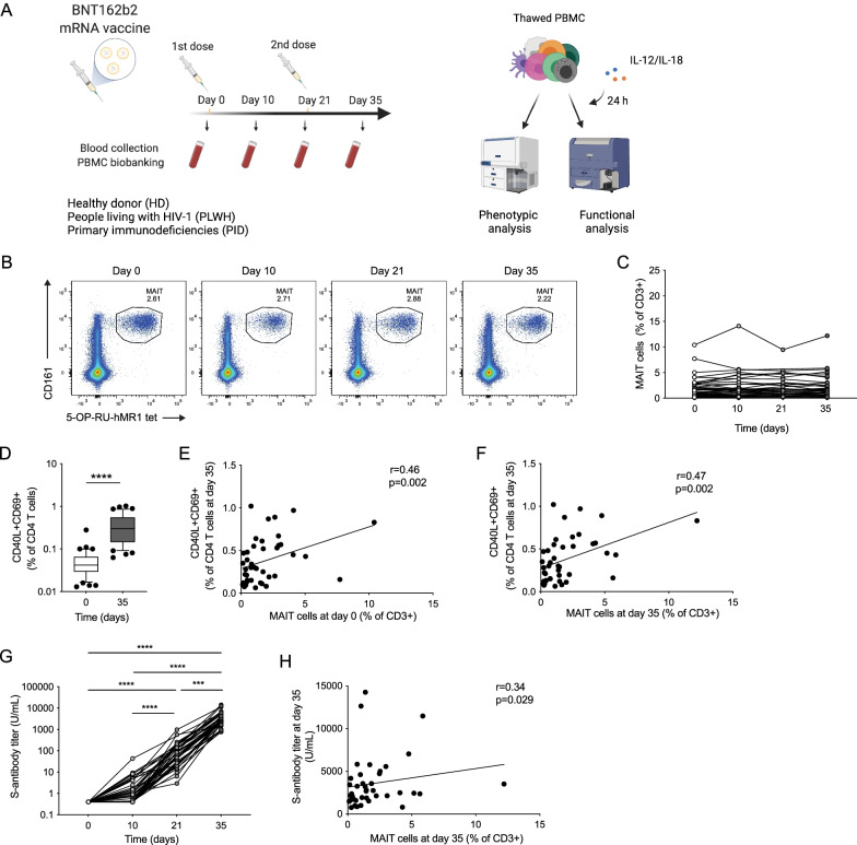
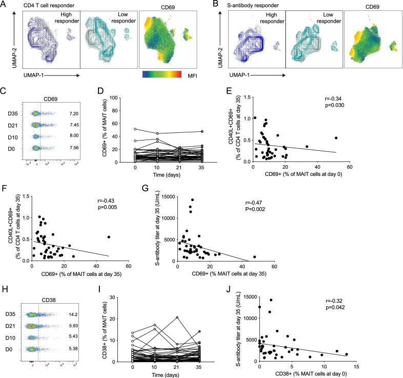
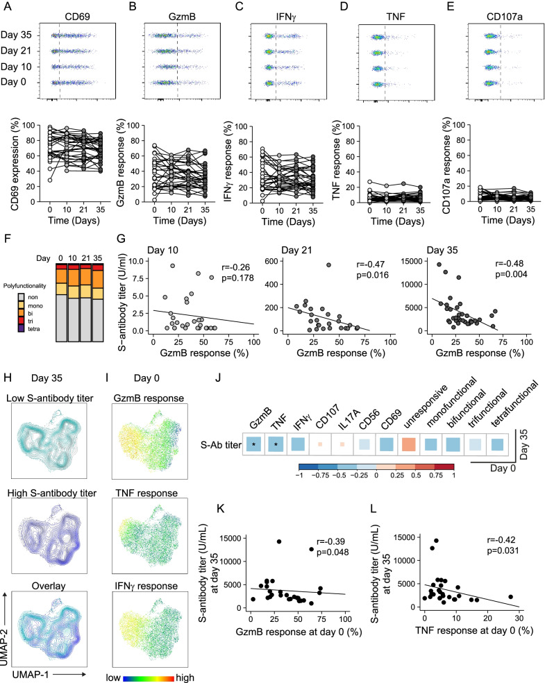
Mucosa-associated invariant T (MAIT) cells are unconventional T cells with innate-like capacity to rapidly respond to microbial infection via MR1-restricted antigen recognition. Emerging evidence indicate that they can also act as rapid sensors of viral infection via innate cytokine activation. However, their possible role in the immune response to mRNA vaccination is unknown. Here, we evaluated the involvement of MAIT cells in individuals vaccinated with the BNT162b2 mRNA SARS-CoV-2 vaccine. MAIT cell levels, phenotype and function in circulation were preserved and unperturbed through day 35 post-vaccination in healthy donor (HD) vaccinees, as well as people living with HIV (PLWH) or with primary immunodeficiency (PID). Unexpectedly, pre-vaccination and post-vaccination levels of MAIT cells correlated positively with the magnitude of the SARS-CoV-2 spike protein-specific CD4 T cell and antibody responses in the HD vaccinees. This pattern was largely preserved in the PID group, but less so in the PLWH group. Furthermore, in the HD vaccinees levels of MAIT cell activation and cytolytic potential correlated negatively to the adaptive antigen-specific immune responses. These findings indicate an unexpected association between MAIT cell compartment characteristics and the immune response magnitude to the BNT162b2 mRNA vaccine.

Keywords: Antibodies; BNT162b2; CD4 T cells; COVID-19; MAIT cells; SARS-CoV-2; mRNA vaccine.


Conflict of interest statement

The authors declare that they have no competing financial interests, patents, patent applications, or material transfer agreements associated with this study. M.B. is a consultant for Oxford Immunotec. S.A. has received honoraria for lectures and educational events, not related to this work, from Gilead, AbbVie, MSD, Biogen and Netdoktor.

Figures

Fig. 1
Fig. 1
MAIT cells are positively associated with adaptive immune responses to BNT162b2 mRNA vaccination. A Schematic description of the vaccine study. Representative flow cytometry plots (B) and combined data of MAIT cell percentage (C) in the healthy donor (HD) group at consecutive time points (n = 36–42). D CD40L+CD69+CD4 T cells in the HD group after SARS-CoV-2 spike peptide pool stimulation (n = 42). Correlation between the percentage of CD40L+CD69+CD4 T cells at day 35 (n = 42) and the MAIT cell percentage at day 0 (n = 42) (E) and day 35 (n = 42) (F). G Spike antibody titer at different time points in the HD group (n = 40–42). H Correlation between the MAIT cell percentage at day 35 (n = 42) and the spike antibody titer at day 35 (n = 42). Box plot indicates the median and interquartile range and whisker bars the 10th and 90th percentile. Mann–Whitney test was performed in D. Kruskal–Wallis test followed by Dunn’s multiple comparison test was performed in G. Correlation was assessed using the Spearman rank correlation test in E, F, and H. ***p < 0.005, ****p < 0.0001. A Was created using Biorender
Fig. 2
Fig. 2
Negative association between MAIT cell activation markers and vaccine responses. Uniform Manifold Approximation and Projection (UMAP) plots of the total MAIT cell pool with overlay color indicating data from high or low spike-specific CD4 T cell responses (A, left) or S-antibody titer (B, left) and showing MAIT cell expression of the activation marker CD69 (A, right; B, right). Concatenated flow cytometry plots (C) and combined data (D) of MAIT cell CD69 expression (n = 36–42) at the different time points in the healthy donor group. Correlation of the percentage of CD40L+CD69+CD4 T cells at day 35 (n = 42) with the percentage of CD69 + MAIT cells at day 0 (n = 42) (E) or day 35 (n = 42) (F). G Correlation between the S-antibody titer at day 35 (n = 41) and expression of CD69 on MAIT cells at day 35 (n = 42). Concatenated flow cytometry plot (H) and combined data (I) of MAIT cell CD38 expression (n = 36–42) at the different time points in the healthy donor group. J Correlation between the S-antibody titer at day 35 (n = 42) and the CD38 expression on MAIT cells at day 0 (n = 42). Associations were assessed using the Spearman rank correlation in EG and J
Fig. 3
Fig. 3
MAIT cell cytolytic arming in response to innate cytokine stimulation is negatively associated with antibody titers against SARS-CoV-2 spike protein. AE Expression of CD69, granzyme B (GzmB), IFNγ, TNF and CD107a in MAIT cells from the healthy donor (HD) group stimulated for 24 h with IL-12 and IL-18, shown as representative concatenated flow cytometry plots (upper panel) and combined data from all donors at the different time points (lower panel, n = 26–35). Significance was assessed by Kruskal–Wallis test followed by Dunn’s multiple comparison with Benjamini–Hochberg correction of p-values. F Comparison of polyfunctionality defined by co-expression of combinations of GzmB, TNF, IFNɣ and CD107a indicating mono-, bi-, tri- or tetra-functionality or cells expressing none of the tested effector molecules. G Correlation between S-antibody titers and GzmB response of stimulated MAIT cells at day 10 (n = 28), day 21 (n = 26) or day 35 (n = 34, from left to right). H, I Uniform Manifold Approximation and Projection (UMAP) plot of concatenated MAIT cells from the healthy donor group (n = 27) at day 0. Color indicates pooled samples from individuals with (< 3000 U/mL) or high (≥ 3000 U/mL) spike-antibody titers at day 35 (H), or I expression of GzmB, TNF or IFNɣ pre-vaccination following IL-12 and IL-18 stimulation. J Correlation analysis of expression of selected functional markers in stimulated MAIT cells before vaccination (day 0) and S-antibody titer at the end of the study (day 35, n = 27). Significant correlations (p < 0.05) against GzmB or TNF response levels are depicted in K or L, respectively. Correlations were assessed using the Spearman rank correlation in G, JL

References

    1. Baden LR, El Sahly HM, Essink B, Kotloff K, Frey S, Novak R, et al. Efficacy and safety of the mRNA-1273 SARS-CoV-2 vaccine. N Engl J Med. 2021;384(5):403–416. doi: 10.1056/NEJMoa2035389. - DOI - PMC - PubMed
    1. Bennett MS, Trivedi S, Iyer AS, Hale JS, Leung DT. Human mucosal-associated invariant T (MAIT) cells possess capacity for B cell help. J Leukoc Biol. 2017;102(5):1261–1269. doi: 10.1189/jlb.4A0317-116R. - DOI - PMC - PubMed
    1. Bergman P, Blennow O, Hansson L, Mielke S, Nowak P, Chen P, et al. Safety and efficacy of the mRNA BNT162b2 vaccine against SARS-CoV-2 in five groups of immunocompromised patients and healthy controls in a prospective open-label clinical trial. EBioMedicine. 2021;74:103705. doi: 10.1016/j.ebiom.2021.103705. - DOI - PMC - PubMed
    1. Boulouis C, Gorin JB, Dias J, Bergman P, Leeansyah E, Sandberg JK. Opsonization-enhanced antigen presentation by MR1 activates rapid polyfunctional MAIT cell responses acting as an effector arm of humoral antibacterial immunity. J Immunol. 2020;205(1):67–77. doi: 10.4049/jimmunol.2000003. - DOI - PMC - PubMed
    1. Boulouis C, Sia WR, Gulam MY, Teo JQM, Png YT, Phan TK, et al. Human MAIT cell cytolytic effector proteins synergize to overcome carbapenem resistance in Escherichia coli. PLoS Biol. 2020;18(6):e3000644. doi: 10.1371/journal.pbio.3000644. - DOI - PMC - PubMed

Publication types